
ISSN: 1885-5857 Impact factor 2025 4.2
Corrected proofs Journal pre-proofs

Original article
Exercise-induced ventricular ectopy with RBBB morphology suggests underlying myocardial fibrosis in inherited left ventricular cardiomyopathy

La extrasistolia ventricular con morfología de bloqueo de rama derecha durante el ejercicio sugiere fibrosis miocárdica en la miocardiopatía hereditaria del ventrículo izquierdo

Elena Sola-Garcíaabc◊Francisco J. Bermúdez-Jiménezabc◊Carlos Gómez-NavarrodJosé Ángel Urbano-MoraleEva Cabrera-BorregoabLuis Tercedor-SánchezabPablo Javier Sánchez-MillánabRosa Macías-RuízabJuan Jiménez-Jáimezabc
https://doi.org/10.1016/j.rec.2026.08.005
La versión en español de este artículo estará disponible en breve

Options

Supplementary data
Imagen extra
10.1016/j.rec.2026.08.005
Abstract
Introduction and objectives

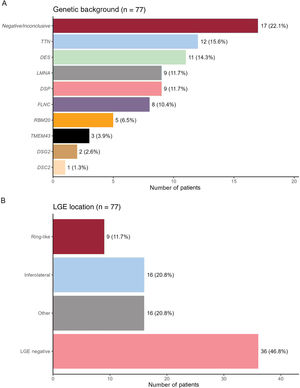
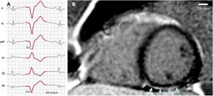
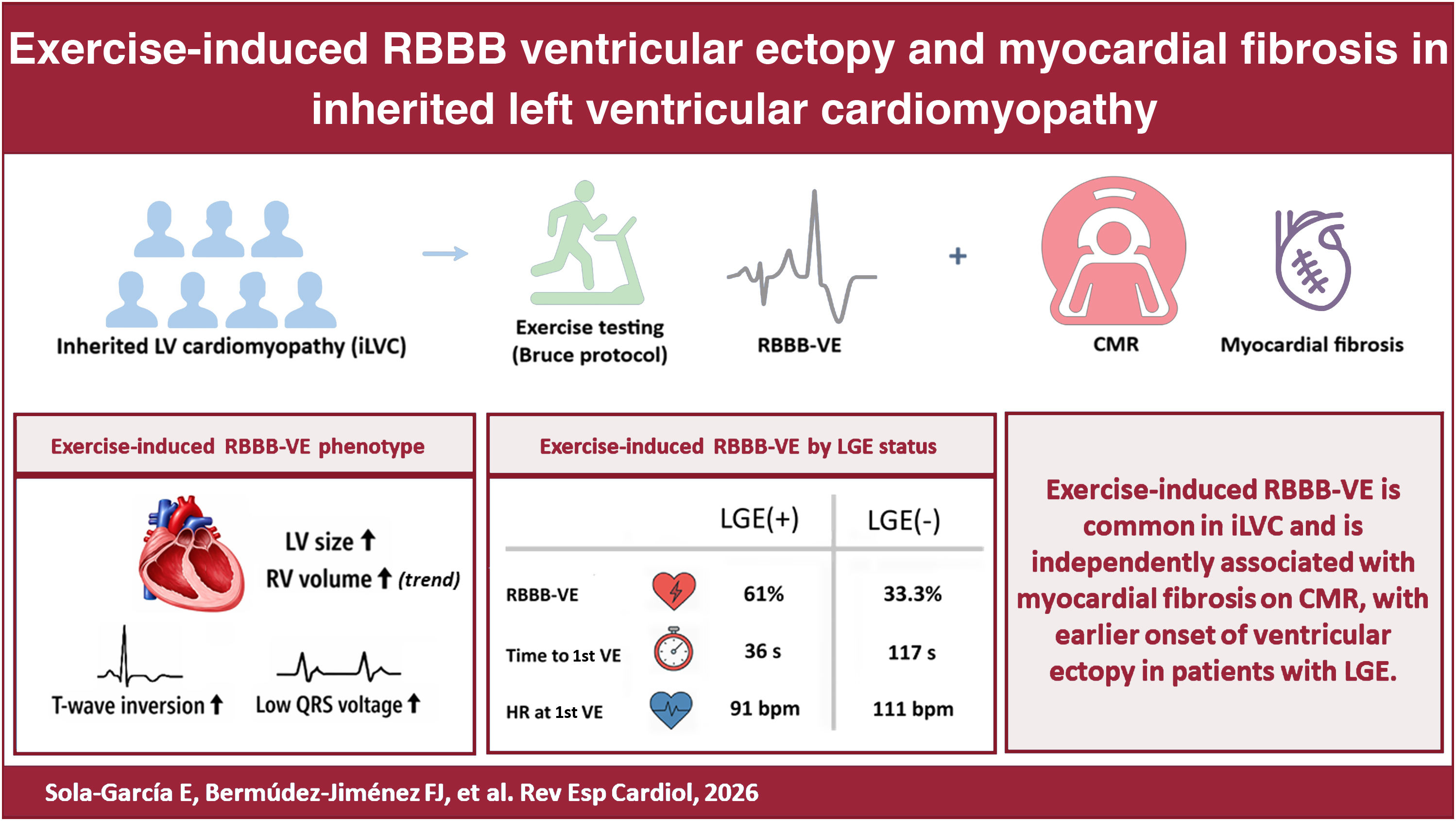
Inherited left ventricular cardiomyopathies (iLVC) are genetic disorders characterized by myocardial remodeling and fibrosis, predisposing patients to ventricular arrhythmias. The prevalence of exercise-induced right bundle branch block-pattern ventricular ectopy (EI-RBBB-VE) and its relationship with myocardial fibrosis in iLVC remain poorly characterized. We aimed to assess the prevalence and characteristics of EI-RBBB-VE in iLVC and its association with myocardial fibrosis detected by late gadolinium enhancement (LGE) on cardiac magnetic resonance (CMR).

Methods

In this multicenter, cross-sectional study, 77 patients with genetically characterized iLVC underwent CMR and treadmill exercise testing. The presence, morphology, and timing of EI-RBBB-VE were assessed.

Results

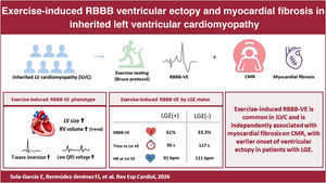
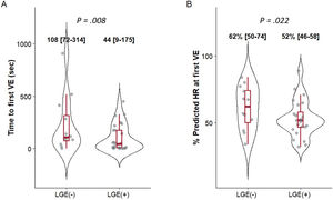
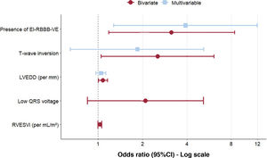
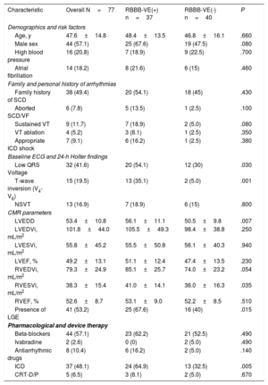
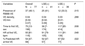
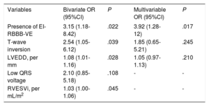
EI-RBBB-VE was present in 48.1% of patients and was significantly more frequent in those with LGE (LGE(+)) than in those without LGE (LGE(-)) (61.0% vs 33.3%; P=.015). Patients with LGE exhibited a lower arrhythmic threshold, with earlier ectopy onset (median 36 [IQR, 7-174] vs 117 [85-414] seconds; P=.008) and ectopy occurring at a lower percentage of predicted maximal heart rate (52% vs 67%; P=.022). The prevalence of LGE did not differ between low- and high-burden ectopy groups (62.5% vs 66.7%; P=1.0). On multivariable analysis, EI-RBBB-VE was independently associated with myocardial fibrosis (OR, 3.92; 95%CI, 1.28-12; P=.017), whereas resting Holter ectopic burden and genotype positivity were not.

Conclusions

EI-RBBB-VE is common in iLVC and is independently associated with myocardial fibrosis on CMR. Earlier exercise-induced ectopy also identifies patients with fibrosis, supporting exercise testing as an accessible tool for detecting an underlying fibrotic substrate beyond resting ECG, Holter monitoring, and genotype.

Keywords

Cardiomyopathies
Exercise test
Myocardial fibrosis
Ventricular premature complexes

Abbreviations

CMR
EI-RBBB-VE
iLVC
LGE
LV
PVC

Identify yourself

Not yet a subscriber to the journal?
Purchase access to the article

By purchasing the article, the PDF of the same can be downloaded

Price: 19,34 €

Phone for incidents

Monday to Friday from 9am to 6pm (GMT+1) except for the months of July and August, which will be from 9am to 3pm

Calls from Spain 932 415 960
Calls from outside Spain +34 932 415 960
References
[1]
E. Arbelo, A. Protonotarios, J.R. Gimeno, et al.
2023 ESC Guidelines for the management of cardiomyopathies.
Eur Heart J., (2023), 44 pp. 3503-3626
[2]
J.A. Towbin, W.J. McKenna, D.J. Abrams, et al.
2019 HRS expert consensus statement on evaluation, risk stratification, and management of arrhythmogenic cardiomyopathy: Executive summary.
Heart Rhythm., (2019), 16 pp. e373-e407
[3]
D. Corrado, M. Perazzolo Marra, A. Zorzi, et al.
Diagnosis of arrhythmogenic cardiomyopathy: The Padua criteria.
Int J Cardiol., (2020), 319 pp. 106-114
[4]
D. Corrado, C. Basso.
Arrhythmogenic left ventricular cardiomyopathy.
[5]
D. Corrado, A. Anastasakis, C. Basso, et al.
Proposed diagnostic criteria for arrhythmogenic cardiomyopathy: European Task Force consensus report.
Int J Cardiol., (2024), 395
[6]
D.J. Hammersley, A. Zegard, E. Androulakis, et al.
Arrhythmic risk stratification by cardiovascular magnetic resonance imaging in patients with nonischemic cardiomyopathy.
J Am Coll Cardiol., (2024), 84 pp. 1407-1420
[7]
C. Eichhorn, D. Koeckerling, R.K. Reddy, et al.
Risk stratification in nonischemic dilated cardiomyopathy using CMR imaging: a systematic review and meta-analysis.
JAMA., (2024), 332 pp. 1535-1540
[8]
D. Segura-Rodríguez, F.J. Bermúdez-Jiménez, V. Carriel, et al.
Myocardial fibrosis in arrhythmogenic cardiomyopathy: a genotype-phenotype correlation study.
Eur Heart J Cardiovasc Imaging., (2020), 21 pp. 378-386
[9]
A. Zorzi, T. Vessella, M. De Lazzari, et al.
Screening young athletes for diseases at risk of sudden cardiac death: role of stress testing for ventricular arrhythmias.
Eur J Prev Cardiol., (2020), 27 pp. 311-320
[10]
C.R. di Gioia, C. Giordano, B. Cerbelli, et al.
Nonischemic left ventricular scar and cardiac sudden death in the young.
Hum Pathol., (2016), 58 pp. 78-89
[11]
A. Zorzi, M. Perazzolo Marra, et al.
Nonischemic Left Ventricular Scar as a Substrate of Life-Threatening Ventricular Arrhythmias and Sudden Cardiac Death in Competitive Athletes.
Circ Arrhythm Electrophysiol., (2016), 9
[12]
A. Cipriani, A. Zorzi, P. Sarto, et al.
Predictive value of exercise testing in athletes with ventricular ectopy evaluated by cardiac magnetic resonance.
Heart Rhythm., (2019), 16 pp. 239-248
[13]
C. Crescenzi, A. Zorzi, T. Vessella, et al.
Predictors of Left Ventricular Scar Using Cardiac Magnetic Resonance in Athletes With Apparently Idiopathic Ventricular Arrhythmias.
J Am Heart Assoc., (2021), 10
[14]
L. Calò, G. Panattoni, M. Tatangelo, et al.
Electrocardiographic characteristics of right-bundle-branch-block premature ventricular complexes predicting absence of left ventricular scar in athletes with apparently structural normal heart.
Europace., (2023), 25
[15]
F. Schnell, G. Claessen, A. La Gerche, et al.
Subepicardial delayed gadolinium enhancement in asymptomatic athletes: let sleeping dogs lie?.
Br J Sports Med., (2016), 50 pp. 111-117
[16]
G. Brunetti, F. Graziano, L. Cavigli, et al.
Reproducibility of ventricular arrhythmias at exercise testing for prediction of non-ischaemic left ventricular scar in athletes.
Eur J Prev Cardiol., (2023), 30 pp. 107-116
[17]
S. Richards, N. Aziz, S. Bale, et al.
Standards and guidelines for the interpretation of sequence variants: a joint consensus recommendation of the American College of Medical Genetics and Genomics and the Association for Molecular Pathology.
Genet Med., (2015), 17 pp. 405-424
[18]
G.F. Fletcher, P.A. Ades, P. Kligfield, et al.
Exercise standards for testing and training: a scientific statement from the American Heart Association.
Circulation., (2013), 12 pp. 873-934
[19]
B.J. Petek, J.A. Drezner, T.W. Churchill.
The International Criteria for Electrocardiogram Interpretation in Athletes: Common Pitfalls and Future Directions.
Cardiol Clin., (2023), 41 pp. 35-49
[20]
D.F. Marchlinski, C.M. Tschabrunn, E.S. Zado, P. Santangeli, F.E. Marchlinski.
Right bundle branch block ventricular tachycardia in arrhythmogenic right ventricular cardiomyopathy more commonly originates from the right ventricle: criteria for identifying chamber of origin.
Heart Rhythm., (2021), 18 pp. 163-171
[21]
M. Laredo, O. Tovia-Brodie, A. Milman, et al.
Electrocardiographic findings in patients with arrhythmogenic cardiomyopathy and right bundle branch block ventricular tachycardia.
Europace., (2023), 25 pp. 1025-1034
[22]
B. Belhassen, M. Laredo, R.W. Roudijk, et al.
The prevalence of left and right bundle branch block morphology ventricular tachycardia amongst patients with arrhythmogenic cardiomyopathy and sustained ventricular tachycardia: insights from the European Survey on Arrhythmogenic Cardiomyopathy.
Europace., (2022), 24 pp. 285-295
[23]
E. Cabrera-Borrego, F.J. Bermúdez-Jiménez, A. Gasperetti, et al.
Electrophysiological phenotype-genotype study of sustained monomorphic ventricular tachycardia in inherited, high arrhythmic risk, left ventricular cardiomyopathy.
Circ Arrhythm Electrophysiol., (2024), 17
[24]
A. Di Marco, I. Anguera, M. Schmitt, et al.
Late gadolinium enhancement and the risk for ventricular arrhythmias or sudden death in dilated cardiomyopathy: systematic review and meta-analysis.
JACC Heart Fail., (2017), 5 pp. 28-38
[25]
E. Feliu, R. Moscicki, L. Carrillo, A. García-Fernández, J.G. Martínez Martínez, J.M. Ruiz-Nodar.
Importance of cardiac magnetic resonance findings in the diagnosis of left dominant arrhythmogenic cardiomyopathy.
Rev Esp Cardiol., (2020), 73 pp. 885-892
[26]
D.J. Tester, J.P. Ackerman, J.R. Giudicessi, et al.
Plakophilin-2 Truncation Variants in Patients Clinically Diagnosed With Catecholaminergic Polymorphic Ventricular Tachycardia and Decedents With Exercise-Associated Autopsy Negative Sudden Unexplained Death in the Young.
JACC Clin Electrophysiol., (2019), 5 pp. 120-127
[27]
J. Ramos-Maqueda, J.H. Migueles, M. Molina-Jiménez, et al.
Lifestyle physical activity and rapid-rate non-sustained ventricular tachycardia in arrhythmogenic cardiomyopathy.
[28]
S. Verheule, U. Schotten.
Electrophysiological Consequences of Cardiac Fibrosis.
Cells., (2021), 10 pp. 3220
[29]
T.P. Nguyen, Z. Qu, J.N. Weiss.
Cardiac fibrosis and arrhythmogenesis: the road to repair is paved with perils.
J Mol Cell Cardiol., (2014), 70 pp. 83-91
[30]
B.B. Lerman, S.M. Markowitz, J.W. Cheung, G. Thomas, J.E. Ip.
Ventricular Tachycardia Due to Triggered Activity: Role of Early and Delayed Afterdepolarizations.
JACC Clin Electrophysiol., (2024), 10 pp. 379-401
[31]
V.Y.H. van Weperen, M.A. Vos, O.A. Ajijola.
Autonomic modulation of ventricular electrical activity: recent developments and clinical implications.
Clin Auton Res., (2021), 31 pp. 659-676
[32]
S.G. Priori, C. Napolitano, N. Tiso, et al.
Mutations in the cardiac ryanodine receptor gene (hRyR2) underlie catecholaminergic polymorphic ventricular tachycardia.
Circulation., (2001), 103 pp. 196-200
Copyright © 2026. Sociedad Española de Cardiología