
ISSN: 1885-5857 Impact factor 2025 4.2
Vol. 79. Num. 4.
Pages 305-315 (April 2026)

Original article
Impact of brachial-ankle pulse wave velocity on progression of diastolic dysfunction: a cohort study

Impacto de la velocidad de la onda de pulso brazo-tobillo en la progresión de la disfunción diastólica: un estudio de cohorte

Seung Hun Leea◊Ki Hong Choib◊Sung Won ChocSoo-Hee ChoicTaek Kyu ParkbJoo Myung LeebYoung Bin SongbJoo-Yong HahnbSeung-Hyuk ChoibHyeon-Cheol GwonbSoo Jin ChocDanbee KangdeJeong Hoon Yangbf
https://doi.org/10.1016/j.rec.2025.09.004

Options

Supplementary data
Imagen extra
Rev Esp Cardiol. 2026;79:305-15
Abstract
Introduction and objectives

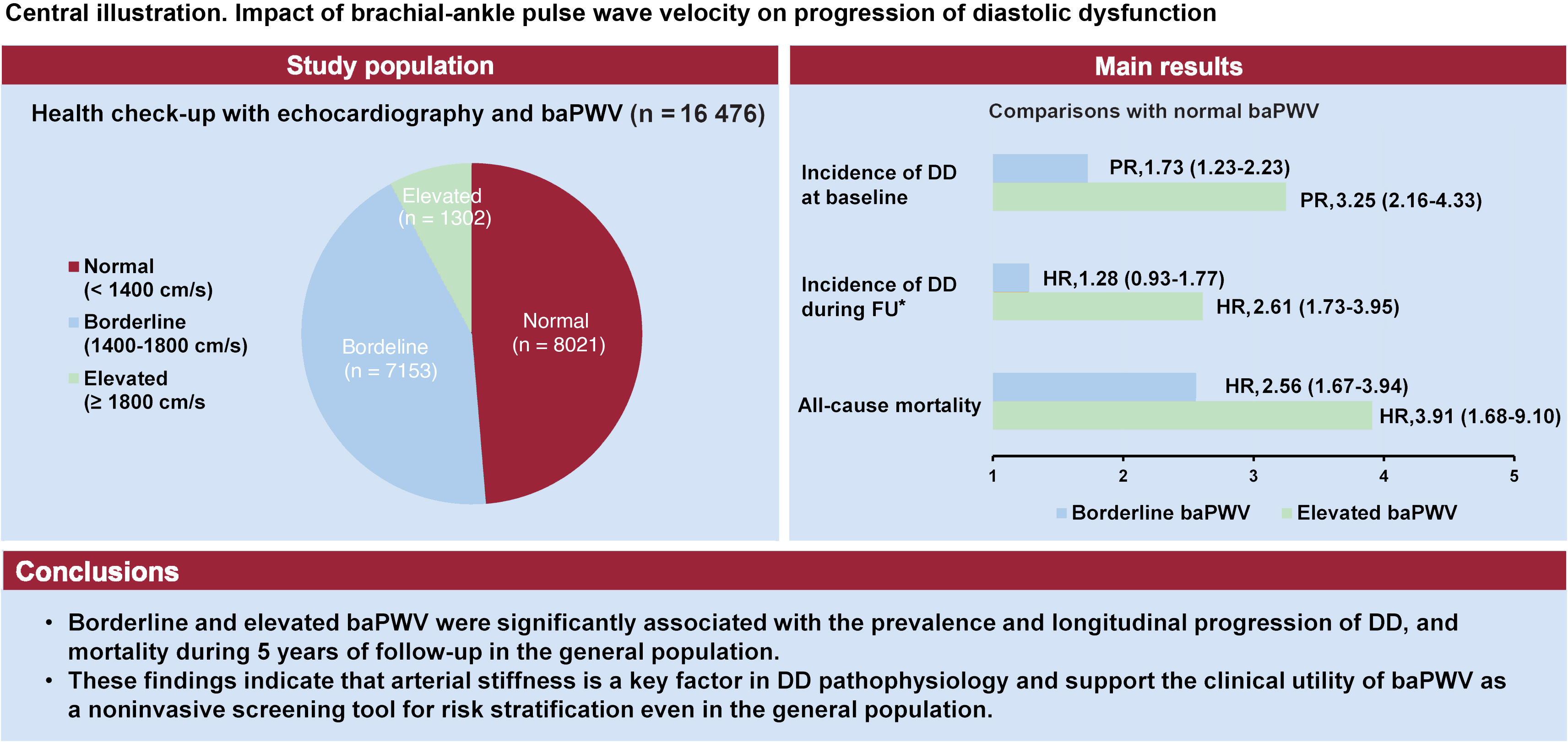
Arterial stiffness is associated with left ventricular diastolic dysfunction (DD), which can lead to heart failure with preserved ejection fraction. However, longitudinal data on their relationship in the general population are limited. This study aimed to evaluate the association between arterial stiffness, assessed by brachial-ankle pulse wave velocity (baPWV), and the prevalence and progression of DD, as well as long-term outcomes in a large, community-based cohort.

Methods

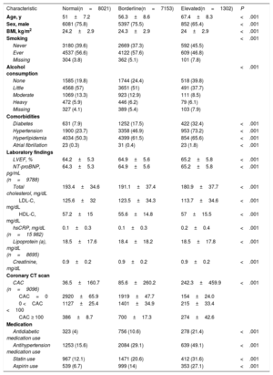
This retrospective cohort study enrolled 16 476 adults (≥ 18 years) who underwent health screening, including echocardiography and baPWV measurements, between 2010 and 2019. Participants were categorized into normal (< 1400cm/s), borderline (1400-1799cm/s), and elevated (≥ 1800cm/s) baPWV groups. Multivariable regression was conducted to analyze the association between baPWV, DD progression, and all-cause mortality during long-term follow-up.

Results

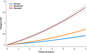
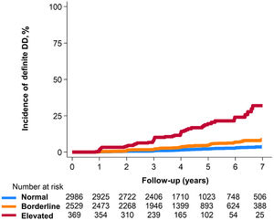
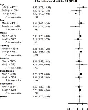
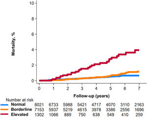
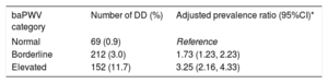
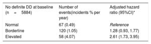
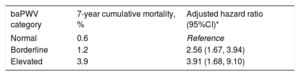
At baseline, the prevalence of definite DD was significantly higher in the borderline baPWV group (PR, 1.73; 95%CI, 1.23-2.23) and the elevated baPWV group (PR, 3.25; 95%CI, 2.16-4.33) compared with the normal group. Participants with borderline or elevated baPWV had a faster increase in left ventricular filling pressure (E/e′) over a mean follow-up of 4 years. Elevated baPWV was independently associated with an increased risk of incident DD (HR, 2.61; 95%CI, 1.73-3.95) and higher all-cause mortality during follow-up (HR, 3.91; 95%CI, 1.68-9.10), even after adjustment for traditional cardiovascular risk factors.

Conclusions

Borderline and elevated baPWV were significantly associated with a higher prevalence and faster DD progression, as well as an increased mortality risk in the general population, suggesting that baPWV is an important factor in DD pathophysiology and a useful noninvasive tool for cardiovascular risk stratification.

Keywords

Arterial stiffness
Diastolic dysfunction
Pulse wave velocity
Heart failure
Prognosis

Abbreviations

baPWV
DD
LVEF
HFpEF

Identify yourself

Not yet a subscriber to the journal?
Purchase access to the article

By purchasing the article, the PDF of the same can be downloaded

Price: 19,34 €

Phone for incidents

Monday to Friday from 9am to 6pm (GMT+1) except for the months of July and August, which will be from 9am to 3pm

Calls from Spain 932 415 960
Calls from outside Spain +34 932 415 960
References
[1]
S.F. Nagueh.
Left Ventricular Diastolic Function: Understanding Pathophysiology Diagnosis, and Prognosis With Echocardiography.
JACC Cardiovasc Imaging., (2020), 13 pp. 228-244
[2]
C. Russo, Z. Jin, S. Homma, et al.
Effect of diabetes and hypertension on left ventricular diastolic function in a high-risk population without evidence of heart disease.
Eur J Heart Fail., (2010), 12 pp. 454-461
[3]
J.A. Naser, E. Lee, C.G. Scott, et al.
Prevalence and incidence of diastolic dysfunction in atrial fibrillation: clinical implications.
Eur Heart J., (2023), 44 pp. 5049-5060
[4]
P. Ponikowski, A.A. Voors, S.D. Anker, et al.
2016 ESC Guidelines for the diagnosis and treatment of acute and chronic heart failure: The Task Force for the diagnosis and treatment of acute and chronic heart failure of the European Society of Cardiology (ESC) Developed with the special contribution of the Heart Failure Association (HFA) of the ESC.
Eur Heart J., (2016), 37 pp. 2129-2200
[5]
B. Pieske, C. Tschope, R.A. de Boer, et al.
How to diagnose heart failure with preserved ejection fraction: the HFA-PEFF diagnostic algorithm: a consensus recommendation from the Heart Failure Association (HFA) of the European Society of Cardiology (ESC).
Eur Heart J., (2019), 40 pp. 3297-3317
[6]
M. Chetrit, P.C. Cremer, A.L. Klein.
Imaging of Diastolic Dysfunction in Community-Based Epidemiological Studies and Randomized Controlled Trials of HFpEF.
JACC Cardiovasc Imaging., (2020), 13 pp. 310-326
[7]
B. Chow, S.W. Rabkin.
The relationship between arterial stiffness and heart failure with preserved ejection fraction: a systemic meta-analysis.
Heart Fail Rev., (2015), 20 pp. 291-303
[8]
H. Tomiyama, K. Shiina.
State of the Art Review: Brachial-Ankle PWV.
J Atheroscler Thromb., (2020), 27 pp. 621-636
[9]
S. Kwak, H.L. Kim, M. In, et al.
Associations of Brachial-Ankle Pulse Wave Velocity With Left Ventricular Geometry and Diastolic Function in Untreated Hypertensive Patients.
Front Cardiovasc Med., (2021), 8
[10]
M. Kim, H.L. Kim, W.H. Lim, et al.
Association between arterial stiffness and left ventricular diastolic function: A large population-based cross-sectional study.
Front Cardiovasc Med., (2022), 9
[11]
C. Mitchell, P.S. Rahko, L.A. Blauwet, et al.
Guidelines for Performing a Comprehensive Transthoracic Echocardiographic Examination in Adults: Recommendations from the American Society of Echocardiography.
J Am Soc Echocardiogr., (2019), 32 pp. 1-64
[12]
R.M. Lang, L.P. Badano, V. Mor-Avi, et al.
Recommendations for cardiac chamber quantification by echocardiography in adults: an update from the American Society of Echocardiography and the European Association of Cardiovascular Imaging.
J Am Soc Echocardiogr., (2015), 28 pp. 1-39
[13]
S.F. Nagueh, O.A. Smiseth, C.P. Appleton, et al.
Recommendations for the Evaluation of Left Ventricular Diastolic Function by Echocardiography: An Update from the American Society of Echocardiography and the European Association of Cardiovascular Imaging.
J Am Soc Echocardiogr., (2016), 29 pp. 277-314
[14]
S.Y. Jae, K.H. Lee, H.J. Kim, et al.
Association between cardiorespiratory fitness and trend of age-related rise in arterial stiffness in individuals with and without hypertension or diabetes.
Am J Hypertens., (2024),
[15]
J.B. Park, J.E. Sharman, Y. Li, et al.
Expert Consensus on the Clinical Use of Pulse Wave Velocity in Asia.
Pulse (Basel)., (2022), 10 pp. 1-18
[16]
S. Wu, L. Xu, M. Wu, et al.
Association between triglyceride-glucose index and risk of arterial stiffness: a cohort study.
Cardiovasc Diabetol., (2021), 20 pp. 146
[17]
D.W. Crabb, G.Y. Im, G. Szabo, et al.
Diagnosis and Treatment of Alcohol-Associated Liver Diseases: 2019 Practice Guidance From the American Association for the Study of Liver Diseases.
Hepatology., (2020), 71 pp. 306-333
[18]
A.S. Agatston, W.R. Janowitz, F.J. Hildner, et al.
Quantification of coronary artery calcium using ultrafast computed tomography.
J Am Coll Cardiol., (1990), 15 pp. 827-832
[19]
H. Tanaka, M. Munakata, Y. Kawano, et al.
Comparison between carotid-femoral and brachial-ankle pulse wave velocity as measures of arterial stiffness.
J Hypertens., (2009), 27 pp. 2022-2027
[20]
T. Dong, F. Fan, H. Chen, et al.
Comparison between vascular age based on brachial-ankle pulse wave velocity or carotid-femoral pulse wave velocity.
Hypertens Res., (2025),
[21]
J. Sugawara, K. Hayashi, T. Yokoi, et al.
Brachial-ankle pulse wave velocity: an index of central arterial stiffness?.
J Hum Hypertens., (2005), 19 pp. 401-406
[22]
A. Yamashina, H. Tomiyama, T. Arai, et al.
Brachial-ankle pulse wave velocity as a marker of atherosclerotic vascular damage and cardiovascular risk.
Hypertens Res., (2003), 26 pp. 615-622
[23]
I. Sequí-Domínguez, I. Cavero-Redondo, C. Álvarez-Bueno, et al.
Accuracy of Pulse Wave Velocity Predicting Cardiovascular and All-Cause Mortality. A Systematic Review and Meta-Analysis.
J Clin Med., (2020), pp. 9
[24]
R. Mori, M. Miyata, T. Kubozono, et al.
Cutoff Values of Brachial-Ankle Pulse Wave Velocity for Atherosclerotic Risks by Age and Sex in the Japanese General Population.
J Atheroscler Thromb., (2023), 30 pp. 481-490
[25]
K.H. Choi, D. Kang, S.H. Lee, et al.
Impact of coronary artery calcium on progression of diastolic dysfunction: a cohort study.
[26]
L. Tartière-Kesri, J.M. Tartière, D. Logeart, et al.
Increased proximal arterial stiffness and cardiac response with moderate exercise in patients with heart failure and preserved ejection fraction.
J Am Coll Cardiol., (2012), 59 pp. 455-461
[27]
J.A. Chirinos, P. Segers, T. Hughes, et al.
Large-Artery Stiffness in Health and Disease: JACC State-of-the-Art Review.
J Am Coll Cardiol., (2019), 74 pp. 1237-1263
[28]
R. Baral, B. Loudon, M.P. Frenneaux, et al.
Ventricular-vascular coupling in heart failure with preserved ejection fraction: A systematic review and meta-analysis.
Heart Lung., (2021), 50 pp. 121-128
[29]
C.S.P. Lam, A.A. Voors, R.A. de Boer, et al.
Heart failure with preserved ejection fraction: from mechanisms to therapies.
Eur Heart J., (2018), 39 pp. 2780-2792
[30]
G.C. Kane, B.L. Karon, D.W. Mahoney, et al.
Progression of left ventricular diastolic dysfunction and risk of heart failure.
JAMA., (2011), 306 pp. 856-863
[31]
H. Aisu, M. Saito, S. Inaba, et al.
Association of worsening arterial stiffness with incident heart failure in asymptomatic patients with cardiovascular risk factors.
Hypertens Res., (2017), 40 pp. 173-180
[32]
T. Weber, J.A. Chirinos.
Pulsatile arterial haemodynamics in heart failure.
Eur Heart J., (2018), 39 pp. 3847-3854
[33]
J.J. DuPont, R.M. Kenney, A.R. Patel, et al.
Sex differences in mechanisms of arterial stiffness.
Br J Pharmacol., (2019), 176 pp. 4208-4225
[34]
O.S. Andersen, O.A. Smiseth, H. Dokainish, et al.
Estimating Left Ventricular Filling Pressure by Echocardiography.
J Am Coll Cardiol., (2017), 69 pp. 1937-1948
Copyright © 2025. Sociedad Española de Cardiología