
Cardiac dysfunction in septic patients admitted to the intensive care unit (ICU)—referred to as sepsis-induced myocardial dysfunction or septic cardiomyopathy (SCM)—is usually classified as left ventricular (LV), right ventricular (RV), or biventricular systolic dysfunction (LVSD/RVSD). In addition to conventional LV functional assessment using ejection fraction, myocardial strain measured by speckle-tracking echocardiography has emerged as a robust marker of intrinsic cardiac function, detecting systolic and diastolic dysfunction up to 50% more frequently.1
The presence of harmful variants in genes associated with cardiomyopathies has been reported in forms of ventricular dysfunction or dilated cardiomyopathy (DCM) that were previously considered predominantly acquired or nongenetic. This is particularly evident for variants in titin (TTN) in the context of peripartum cardiomyopathy, cancer therapy–related cardiotoxicity, alcohol-associated cardiomyopathy,2 and myocarditis.3
Therefore, we hypothesized that a similar genetic predisposition may exist in SCM and investigated whether rare genetic variants in cardiomyopathy-associated genes could be detected in this context. Such findings may have implications for both clinical and familial management.
A single-center prospective observational study was conducted in a general ICU between October 2022 and December 2023. The study was approved by the ethics committees of Hospital Garcia de Orta and the Instituto de Medicina Molecular (T01/2022), and written informed consent was obtained from all participants. The study was registered on ClinicalTrials.gov (NCT05552521) on 15 September 2022.
Patients aged ≥18 years who met septic shock criteria according to the Surviving Sepsis Campaign and who had cardiac dysfunction were consecutively recruited. A comprehensive echocardiogram was performed within 24hours of ICU admission and analyzed offline using dedicated software (GE EchoPAC PC v206, GE Healthcare, United States). Cardiac dysfunction was classified based on current guidelines. LV systolic dysfunction was defined as an LV ejection fraction (LVEF)<50% or a global longitudinal strain (GLS) >–15%. Exclusion criteria included pregnancy, congenital heart disease, prior LV or RV dysfunction, valve prostheses, severe valvular disease, atrial fibrillation, acute coronary syndrome, acute pulmonary embolism, and inadequate image quality. All patients had previous echocardiograms from routine ambulatory health check-ups within the year prior to admission, none of which showing an LVEF <50%.
Whole-exome sequencing was performed at the Institute of Molecular Medicine, Lisbon. Analysis focused on a panel of 281 genes associated with cardiomyopathies, including genes linked to metabolic conditions and all genes with definitive, strong, or moderate evidence for association with DCM, as classified by ClinGen. These included: definitive: BAG3, DES, FLNC, LMNA, MYH7, PLN, RBM20, SCN5A, TNNC1, TNNT2, TTN; strong: DSP; moderate: ACTC1, ACTN2, JPH2, NEXN, TNNI3, TPM1, VCL. Variants were filtered for a minor allele frequency <0.0001 and classified according to the American College of Medical Genetics and Genomics (ACMG) criteria.4 Copy-number variation was also analysed. Additionally, we analyzed a group of genes recently suggested to be associated with septic cardiomyopathy: ADGRG1, BCL2A1, BCL6, CD44, FTH1, GAB2, IL1R1, IL6ST, ING3, JAK3, KLRG1, MMP9, PRF1, SERPINE1, TGIF1, and TPT1.5
All demographic, clinical, hemodynamic, respiratory, fluid balance, vasopressor use, and laboratory data were prospectively collected, along with 30-day mortality.
Continuous variables are presented as medians with interquartile ranges and compared between 2 groups using the Mann-Whitney U test, as appropriate. Categorical variables are expressed as proportions and compared using the Fisher exact test.
Of the initial 151 patients assessed, 71 met exclusion criteria, 4 had insufficient DNA quality for genetic analysis, and 6 had isolated diastolic dysfunction. The remaining 70 patients had systolic dysfunction, as defined by either LVEF or LV-GLS criteria; among them, 45 patients (64%) had an LVEF<50%.
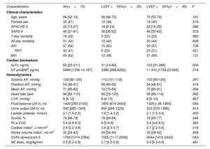
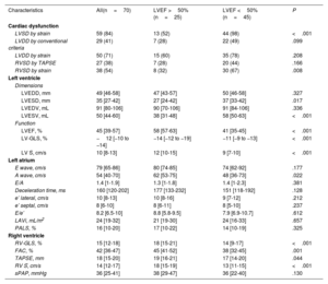
The baseline clinical and echocardiographic characteristics of the 70 patients with LVSD, obtained within the first 24hours of ICU admission, are presented in table 1 and table 2, respectively.
Baseline clinical characteristics
| Characteristics | All(n=70) | LVEF >50%(n=25) | LVEF <50%(n=45) | P |
|---|---|---|---|---|
| Clinical characteristics | ||||
| Age, years | 66 [52-74] | 56 [48-72] | 70 [55-74] | .181 |
| Female sex | 30 (41) | 12 (48) | 18 (40) | .516 |
| APACHE II | 20 [13-27] | 18 [9-24] | 22 [16-29] | .086 |
| SAPS II | 46 [31-61] | 38 [26-52] | 48 [35-62] | .033 |
| 7-day mortality | 16 (22) | 5 (25) | 10 (22) | .860 |
| 30-day mortality | 31 (42) | 10 (40) | 20 (44) | .609 |
| AKI | 47 (64) | 15 (60) | 30 (67) | .708 |
| RRT | 30 (41) | 5 (20) | 23 (51) | .021 |
| MVi | 45 (62) | 12 (48) | 31 (69) | .131 |
| Cardiac biomarkers | ||||
| hsTn, ng/mL | 62 [22-211] | 31 [14-80] | 103 [31-286] | .004 |
| NT-proBNP, pg/mL | 5899 [1336-14 187] | 3389 [998-8282] | 11 910 [1709-23 626] | .018 |
| Hemodynamics | ||||
| Systolic AP, mmHg | 108 [95-120] | 110 [101-119] | 103 [95-120] | .297 |
| Diastolic AP, mmHg | 54 [48-61] | 56 [49-62] | 54 [48-61] | .416 |
| Mean AP, mmHg | 71 [65-82] | 72 [70-84] | 70 [65-81] | .099 |
| Heart rate, bpm | 98 [82-110] | 90 [78-103] | 99 [89-114] | .062 |
| CVP, mmHg | 8 [4-12] | 8 [4-13] | 8 [5-12] | .867 |
| Fluid balance (24-h), mL | 1400 [293-2100] | 1800 [674-2443] | 1329 [–38-1950] | .084 |
| Urine output (24-h), mL | 590 [285-1300] | 850 [265-1220] | 532 [276-1366] | .810 |
| Lactate (arterial), mmol/L | 2.1 [1.5-3.3] | 1.8 [1.4-3.2] | 2.1 [1.5-3.4] | .403 |
| ScvO2, % | 74 [68-79] | 78 [69-84] | 72 [65-77] | .046 |
| Pv-a CO2 | 5.4 [4.8-8.3] | 6 [5-8.9] | 5.4 [4.6-8.5] | .482 |
| Cardiac index*, L/min/m2 | 2.9 [2.2-3.9] | 3.6 [2.5-4.7] | 2.7 [2.2-3.4] | .016 |
| Stroke volume index*, mL/m2 | 32 [28-42] | 34 [24-40] | 32 [28-42] | .849 |
| SVRI, dynes/s/cm-5 | 1794 [1314-2364] | 1505 [1117-2058] | 2044 [1412-2442] | .043 |
| NE dose, mcg/kg/min | 0.5 [0.2-0.9] | 0.7 [0.2-0.9] | 0.4 [0.2-0.9] | .481 |
AKI, acute kidney injury; AP, arterial pressure; APACHE II, Acute Physiology and Chronic Health Evaluation II; CKD, chronic kidney disease; CVP, central venous pressure; DM, diabetes mellitus; ESR, erythrocyte sedimentation rate; ICU, intensive care unit; LOS, length of stay; MVi, invasive mechanical ventilation; NE, norepinephrine; hsTn, high-sensitivity troponin; NT-proBNP, N-terminal pro B-type natriuretic peptide; Pv-aCO2, central venous-to-arterial carbon dioxide difference; RRT, continuous renal replacement therapy; SAPS II, Simplified Acute Physiology Score II; SOFA, Sequential Organ Failure Assessment; SVRI, systemic vascular resistance index.
Data are presented as No. (%) or median [interquartile range]. The Mann-Whitney U test was used for continuous variables and the Fisher exact test for categorical variables.
Baseline echocardiographic characteristics
| Characteristics | All(n=70) | LVEF >50%(n=25) | LVEF <50%(n=45) | P |
|---|---|---|---|---|
| Cardiac dysfunction | ||||
| LVSD by strain | 59 (84) | 13 (52) | 44 (98) | <.001 |
| LVDD by conventional criteria | 29 (41) | 7 (28) | 22 (49) | .099 |
| LVDD by strain | 50 (71) | 15 (60) | 35 (78) | .208 |
| RVSD by TAPSE | 27 (38) | 7 (28) | 20 (44) | .166 |
| RVSD by strain | 38 (54) | 8 (32) | 30 (67) | .008 |
| Left ventricle | ||||
| Dimensions | ||||
| LVEDD, mm | 49 [46-58] | 47 [43-57] | 50 [46-58] | .327 |
| LVESD, mm | 35 [27-42] | 27 [24-42] | 37 [33-42] | .017 |
| LVEDV, mL | 91 [80-106] | 90 [70-106] | 91 [84-106] | .336 |
| LVESV, mL | 50 [44-60] | 38 [31-48] | 58 [50-63] | <.001 |
| Function | ||||
| LVEF, % | 45 [39-57] | 58 [57-63] | 41 [35-45] | <.001 |
| LV-GLS, % | −12 [−10 to −14] | −14 [−12 to −19] | −11 [−9 to −13] | <.001 |
| LV Ś, cm/s | 10 [8-13] | 12 [10-15] | 9 [7-10] | <.001 |
| Left atrium | ||||
| E wave, cm/s | 79 [65-86] | 80 [74-85] | 74 [62-92] | .177 |
| A wave, cm/s | 54 [40-70] | 62 [53-75] | 48 [36-73] | .022 |
| E/A | 1.4 [1-1.9] | 1.3 [1-1.8] | 1.4 [1-2.3] | .381 |
| Deceleration time, ms | 160 [120-202] | 177 [133-232] | 151 [118-192] | .128 |
| e’ lateral, cm/s | 10 [8-13] | 10 [8-16] | 9 [7-12] | .212 |
| e’ septal, cm/s | 8 [6-10] | 8 [6-11] | 8 [5-10] | .237 |
| E/e’ | 8.2 [6.5-10] | 8.8 [5.8-9.5] | 7.9 [6.9-10.7] | .612 |
| LAVi, mL/m2 | 24 [19-32] | 21 [19-30] | 24 [16-33] | .657 |
| PALS, % | 16 [10-20] | 17 [10-22] | 14 [10-19] | .325 |
| Right ventricle | ||||
| RV-GLS, % | 15 [12-18] | 18 [15-21] | 14 [9-17] | <.001 |
| FAC, % | 42 [36-47] | 45 [41-52] | 38 [32-45] | .001 |
| TAPSE, mm | 18 [15-20] | 19 [16-21] | 17 [14-20] | .044 |
| RV Ś, cm/s | 14 [12-17] | 18 [15-19] | 13 [11-15] | <.001 |
| sPAP, mmHg | 36 [25-41] | 38 [29-47] | 36 [22-40] | .130 |
A, peak late inflow velocity; E, peak early inflow velocity; E/A, peak early inflow velocity to peak late inflow velocity; E/e’, peak early inflow velocity to peak early longitudinal diastolic myocardial velocity; e’, peak early longitudinal diastolic myocardial velocity; Ea, arterial elastance; Ees, end-systolic elastance (myocardial performance); GWI, global work index; GWE, global work efficiency; IVC, inferior vena cava; IVS, interventricular septum; LAVi, left atrial volume indexed; LV S’, LV systolic myocardial velocity; LV, left ventricle; LV-AC, left ventriculo-arterial coupling; LVEDD, LV end-diastolic diameter; LVEDV, LV end-diastolic volume; LVEF, LV ejection fraction; LVESD, LV end-systolic diameter; LVESV, LV end-systolic volume; LV-GLS, LV global longitudinal strain; LVOT VTI, LV outflow tract velocity-time integral; LVPW, LV posterior wall; LVSV, LV stroke volume; max, maximum; min, minimum; PALS, peak atrial longitudinal strain during the reservoir phase; RV, right ventricle; FAC, fractional area change; S’, peak systolic myocardial velocity; sPAP, systolic pulmonary artery pressure; TAPSE, tricuspid annular plane systolic excursion.
Data are presented as No. (%) or median [interquartile range] unless otherwise specified. The Mann-Whitney U test was used for continuous variables and the Fisher exact test for categorical variables.
The median age was 66 years (IQR: 52-74), with 41% of patients being female. The median Acute Physiology and Chronic Health Evaluation II (APACHE II) score was 20 (IQR: 13-27), and the median Simplified Acute Physiology Score II (SAPS II) was 46 (IQR: 31-61). Invasive mechanical ventilation was required in 62% of patients, 41% received renal replacement therapy, and the 30-day mortality rate was 42%.
Patients with LVEF <50% had higher SAPS II scores, a greater need for renal replacement therapy, and elevated levels of high-sensitivity troponin and N-terminal pro B-type natriuretic peptide (NT-proBNP). They also had a lower cardiac index and reduced central venous oxygen saturation. Echocardiographically, these patients showed a larger LV end-systolic diameter (37 [33-42] vs 27 [24-42] mm; P=.017) and LV end-systolic volume (58 [50-63] vs 38 [31-48] mL; P<.001), lower LV-GLS (−11 [−9 to −13] vs -14 [−12 to −19] %; P<.001) and worse right ventricle systolic function.
No pathogenic or likely pathogenic variants were identified in the analyzed DCM-associated genes, genes related to inherited metabolic conditions associated with cardiomyopathy, or genes recently proposed as candidates for an association with septic cardiomyopathy.
This is the first study to investigate the contribution of inherited cardiomyopathy-related genetic variation in a septic shock population with cardiac dysfunction. The absence of pathogenic or likely pathogenic variants in 70 patients with systolic dysfunction—including 45 with LVEF <50%—suggests a potential lack of this specific genetic predisposition in SCM. However, given the limited sample size, validation in larger, multicenter cohorts is necessary to confirm these findings.
In contrast, a genetic predisposition has been demonstrated in other forms of non-ischemic cardiomyopathy previously considered acquired. In alcoholic cardiomyopathy, heterozygous rare protein-altering variants in 9 genes associated with inherited DCM were identified in 19 of 141 patients (13.5%), a frequency significantly higher than in controls and comparable to that seen in genetic DCM. Similarly, titin truncating variants (TTNtv) were identified in 172 cases of peripartum cardiomyopathy, indicating a shared genetic predisposition with familial DCM.2 Rare TTNtv have also been associated with an increased risk of cancer therapy-induced cardiomyopathy.2 DCM- or arrhythmogenic cardiomyopathy-associated variants were identified in 8% of 336 patients with acute myocarditis,3 with TTNtv predominance in those with reduced left ventricular ejection fraction.
An association between sepsis, SCM, and immune response genes has been previously reported.5 Although SCM can be profound, it is usually reversible in survivors.6 Our findings might therefore reflect similarities with other clinical scenarios of stress-induced LV dysfunction (eg, tako-tsubo cardiomyopathy) in which the recovery of LV function and normalization of LV size can occur.6 In these entities, there is likewise no established contribution from cardiomyopathy-associated genetic variants. In summary, our results suggest that cardiomyopathy-related genetic variation does not play a significant role in the pathogenesis of SCM.
FUNDINGThis project was funded by Academia CUF (No. 01/2022), Lisbon, Portugal, and by the Centro Cardiovascular da Universidade de Lisboa (CCUL@RISE), Faculdade de Medicina, Universidade de Lisboa, Portugal.
ETHICAL CONSIDERATIONSThe study was approved by the Ethics Committees of Hospital Garcia de Orta and the Instituto de Medicina Molecular (T01/2022). Written informed consent was obtained from all participants, including consent for publication of anonymized data. This manuscript does not contain any identifiable personal data in any form.
STATEMENT ON THE USE OF ARTIFICIAL INTELLIGENCENo artificial intelligence tools were used in the preparation of this manuscript or in the generation of the data presented.
AUTHORS’ CONTRIBUTIONSF.A. Gonzalez, L.R. Lopes, and A.G. Almeida designed the study, analyzed the data, and drafted the manuscript. F.A. Gonzalez, J. Barcariza, J. Leonte, and A.R. Varudo recruited patients, collected data, and performed the echocardiographic assessments. All authors revised and approved the final manuscript. A.G. Almeida and L.R. Lopes are joint senior authors.
CONFLICTS OF INTERESTWe declare no conflicts of interest relevant to this study, including financial.