The 2019 ESC guidelines1 on the management of patients with supraventricular tachycardia (SVT) are an update of the document published in 2003.2 To make their various recommendations, the authors have thoroughly reviewed the literature and evaluated the level of evidence. This article follows the order of the new guidelines document and reviews its content.
The main changes in the 16 years since the publication of the previous guidelines are due to developments in catheter ablation techniques, which have become the primary treatment of choice in the chronic management of these arrhythmias. In addition, although drug therapy has not changed significantly, the relevant indications have been refined, with the disappearance of a considerable number of drug therapy-related recommendations. New therapeutic alternatives are mentioned, such as ivabradine and ibutilide, although the latter drug is not available here in Spain. Finally, recommendations are made for special situations, such as for pregnant or pediatric patients and for patients with congenital heart disease or tachycardia-induced cardiomyopathy.
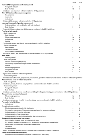
Table 3 of the guidelines,1 which contains the most representative developments and changes vs the 2003 guidelines, and Table 4, which summarizes the new recommendations, are required reading. Their contents are summarized in table 1.
Main novelties in the 2019 guidelines
| 2003 | 2019 | |
|---|---|---|
| Narrow QRS tachycardias: acute management | ||
| Verapamil, diltiazem | I | IIa |
| Beta-blockers | IIb | IIa |
| • Amiodarone and digoxin are not mentioned in the 2019 guidelines | ||
| Wide QRS tachycardias: acute management | ||
| Procainamide | I | IIa |
| Adenosine | IIb | IIa |
| Amiodarone | I | IIb |
| • Sotalol and lidocaine are not mentioned in the 2019 guidelines | ||
| Inappropriate sinus tachycardia: management | ||
| Ivabradine (alone or in combination with beta-blockers) | – | IIa |
| Beta-blockers | I | IIa |
| • Verapamil/diltiazem and catheter ablation are not mentioned in the 2019 guidelines | ||
| Focal atrial tachycardia | ||
| Acute management | ||
| Beta-blockers | I | IIa |
| Flecainide/propafenone | IIa | IIb |
| Amiodarone | IIa | IIb |
| Ibutilide | – | IIb |
| • Procainamide, sotalol, and digoxin are not mentioned in the 2019 guidelines | ||
| Chronic management | ||
| Beta-blockers | I | IIa |
| Verapamil, diltiazem | I | IIa |
| Ivabradine with beta-blockers | – | IIb |
| • Amiodarone, sotalol, and disopyramide are not mentioned in the 2019 guidelines | ||
| Atrial flutter | ||
| Anticoagulationa | – | IIa |
| Acute management | ||
| Atrial or transesophageal pacing | I | IIb |
| Atrial pacing in patients with a pacemaker or defibrillator | – | I |
| Ibutilide | IIa | I |
| Flecainide/propafenone | IIb | III |
| Verapamil, diltiazem | I | IIa |
| Beta-blockers | I | IIa |
| • Digoxin is not mentioned in the 2019 guidelines | ||
| Chronic management | ||
| • Dofetilide, sotalol, flecainide, propafenone, procainamide, quinidine, and disopyramide are not mentioned in the 2019 guidelines | ||
| Atrioventricular nodal re-entrant tachycardia | ||
| Acute management | ||
| • Amiodarone, sotalol, flecainide, and propafenone are not mentioned in the 2019 guidelines | ||
| Chronic management | ||
| Verapamil, diltiazem | I | IIa |
| Beta-blockers | I | IIa |
| • Amiodarone, sotalol, flecainide, propafenone, and the pill in the pocket strategy are not mentioned in the 2019 guidelines | ||
| Atrioventricular re-entrant tachycardia | ||
| Flecainide/propafenone | IIa | IIb |
| Beta-blockers | IIb | IIa |
| • Amiodarone, sotalol, and the pill in the pocket strategy are not mentioned in the 2019 guidelines | ||
| Pre-excited atrial fibrillation | ||
| Amiodarone | – | III |
| Asymptomatic pre-excitation | ||
| Electrophysiological study for risk stratification | – | IIa |
| Noninvasive assessment of the conducting properties of the accessory pathway | – | IIb |
| Catheter ablation of high-risk pathways | – | I |
| Catheter ablation of low-risk pathways | IIb | |
| Catheter ablation for ventricular dysfunction due to dyssynchrony | IIa | |
| Supraventricular tachycardia in pregnancy | ||
| During the first trimester, avoid all antiarrhythmic drugs | I | |
| β-1 selective blockers (except atenolol) or verapamil in patients without pre-excitation | IIa | |
| Flecainide or propafenone in patients with pre-excitation | IIa | |
| Verapamil | IIb | IIa |
| Catheter ablation | IIb | IIab |
| • Sotalol, propranolol, quinidine, and procainamide are not mentioned in the 2019 guidelines | ||
| Tachycardia-induced cardiomyopathy | ||
| Atrioventricular nodal ablation and pacing (biventricular or His bundle) | – | I |
Created with permission using data from Brugada et al.1
The definition of SVT, not specified in the previous guidelines, is a rapid atrial rhythm (> 100 bpm) with a mechanism involving structures located at or above the His bundle. The practical classification of SVTs is based on their location and is independent of the underlying mechanism. For differential diagnosis, SVTs are also classified as narrow (≤ 120ms) or wide (> 120ms) QRS tachycardias.
Mechanisms and anatomyThe guidelines include supplementary data containing a detailed description of the electrophysiological mechanisms and anatomical structures involved in SVTs and their anatomical and functional evaluation via imaging techniques. One noteworthy aspect is that, beyond a mere description of the basic electrophysiological mechanisms of supraventricular arrhythmias, the guidelines consider the concept of “focal” arrhythmia during analysis of the macroscopic activation sequence, generally atrial. Because the resolution of mapping systems might not be sufficient to differentiate between microre-entries (local) and enhanced automaticity or triggered activity, the re-entrant circuits would be considered focal tachycardias by a clinical electrophysiologist and manageable via a specific catheter ablation strategy.
EpidemiologyThere are no major changes in the general and pediatric populations. Notably, in a cohort of 1 967 911 live births, the incidence of SVT was 1.03/1000 patient-years (16.2% had Wolff-Parkinson-White syndrome) and the risk of sudden cardiac death in individuals without congenital heart disease was 1.33/1000 patient-years until 15 years of age.3 The risk of these arrhythmias is influenced by individual factors such as age > 65 years and female sex, as well as by the underlying mechanism. Also highlighted is the difficult calculation of the actual prevalence and incidence of atrial flutter alone because it frequently coexists with atrial fibrillation.
The main development since the publication of the previous guidelines lies in the creation of large registries confirming the safety and effectiveness of catheter ablation, including the registry published annually by the Spanish Society of Cardiology.4 Information is also provided on the significant improvement in quality of life after catheter ablation.
CLINICAL PRESENTATION AND INITIAL EVALUATIONThe guidelines specifically emphasize the value of an accurate and detailed clinical history, as well as the recording and analysis of 12-lead resting ECG (mainly to rule out pre-excitation). ECG during tachycardia is particularly useful because it enables confirmation of the SVT diagnosis and indicates the possible mechanism and, thereby, the treatment of choice. The yields of other electrocardiographic recording systems depend on the frequency and duration of the episodes, whereas the usefulness of wrist-worn electrocardiographic monitors remains to be validated.
Differential diagnosisThe guidelines subdivide the differential diagnosis of SVTs into 3 sections: narrow QRS tachycardias, wide QRS tachycardias, and irregular tachycardias. ECG during tachycardia is the cornerstone of SVT diagnosis (I B). The current guidelines expand upon and describe in a more organized fashion the electrocardiographic findings used in the differential diagnosis of SVTs.
The first section concerns the differential diagnosis of narrow QRS tachycardias. This includes, first, the analysis of ECG during tachycardia in relation to the initiation and termination, the regularity of the arrhythmia, and the relationship between the P wave and the QRS. The previous guidelines were limited to the relationship between the P wave and the QRS. Another novelty is the value of the RP interval for differentiating among the different tachycardias with a short RP. While the previous guidelines used a surface ECG cutoff of 70ms, the current document considers a cutoff of 90ms to be more useful.5 Figure 1 of the guidelines summarizes the differential diagnosis of narrow QRS tachycardias, as in previous guidelines but with the addition of the possible presence of a ventricular rate higher than the atrial rate.
The response to vagal maneuvers and adenosine administration in terms of the diagnosis of narrow QRS tachycardias is described, with a table and figure1 similar to those of previous guidelines. Finally, the documents mentions the role of electrophysiological studies in the diagnosis of narrow QRS tachycardias.
The second section reviews the differential diagnosis of wide QRS tachycardias and stresses that the initial diagnosis should be ventricular tachycardia. The differential diagnosis includes SVTs with bundle branch block, with anterograde conduction over an accessory pathway (pre-excited SVTs), and with widening of the QRS interval due to the action of specific drugs (class IA, IC, and III antiarrhythmics) or electrolyte disturbances, which can induce atypical forms of bundle branch block, and, finally, pacemaker-related tachycardia or artifacts that may mimic ventricular tachycardia.
The guidelines expand upon the main ECG findings contributing to the differential diagnosis of wide QRS tachycardias. The main elements of the analysis are the presence of atrioventricular dissociation, the QRS duration and electrical axis, the concordance of the precordial QRS, and the characteristic morphological findings of tachycardias with imaging of left and right bundle branch block. In contrast to previous guidelines, there is no figure showing the differential diagnosis of wide QRS tachycardias, but there is a Table 1 with the main electrocardiographic criteria indicating ventricular tachycardia.
Although algorithms designed for the differential diagnosis of ventricular tachycardia and SVT are mentioned, they have low specificity (40%-80%) for the diagnosis of ventricular tachycardia and low accuracy (75%).
The third section briefly describes the possible diagnoses of irregular tachycardias with both narrow and wide QRS. The authors do not mention rhythm irregularities, typical of regular atrial tachycardias with cyclic nodal conduction patterns.
Finally, the document mentions the use of mathematical models and numerical analysis of ECG as a possible future application for artificial intelligence in the differential diagnosis of narrow and wide QRS tachycardias.
ACUTE MANAGEMENT IN THE ABSENCE OF AN ESTABLISHED DIAGNOSISSVTs are a frequent reason for emergency department visits. In this setting, clear and straightforward action plans are essential, such as those proposed in these ESC guidelines for acute management in the absence of an established diagnosis. This section has no class I A recommendation.
For regular tachycardias, 2 action plans are differentiated according to whether the tachycardia has narrow or wide QRS complexes. These plans have the same first 2 steps: electrocardiographic documentation and assessment of hemodynamic tolerance. For hemodynamically unstable tachycardias, the guidelines recommend immediate electrical cardioversion. This is a major difference from the 2015 North American guidelines, which recommended vagal maneuvers or adenosine ahead of electrical cardioversion, and is important because patients’ conditions could be worsened by the adverse effects of adenosine or a delay in reversing the arrhythmia.
For hemodynamically stable tachycardias, whether with wide or narrow QRS complexes, vagal maneuvers continue to be the first step in treatment, using carotid sinus massage and the classic Valsalva maneuver or, even better, the modified maneuver, which seems to be more effective.
Although the level of evidence has not been increased, the indications for adenosine have been expanded due to findings from clinical practice. Adenosine continues to be the drug of choice for narrow QRS tachycardias (I B) and is now a reasonable option (IIa C) for wide QRS tachycardias without evidence of pre-excitation. In addition, its contraindication is downplayed in asthmatic patients or heart transplant recipients and it is allowed in pregnant patients (I C).
For narrow QRS tachycardias, the main novelty is that, as an alternative to adenosine, the recommendation for intravenous beta-blockers (BBs) has been increased to IIa, even without evidence (level C). These drugs are now comparable to verapamil and diltiazem, although they have been more widely studied in this context (level B). Attention should be paid to the contraindication to the combined or successive intravenous administration of BBs and calcium antagonists. Studies are examining patients’ self-administration of etripamil, a short-acting L-type calcium channel antagonist that can be nasally administered.
There are more novelties in wide QRS tachycardias. The drug arsenal is simplified by eliminating lidocaine and sotalol, and procainamide is prioritized over amiodarone based on the results of the Spanish multicenter trial PROCAMIO.6
Treatment options for irregular tachycardias are briefly discussed, with the guidelines assuming that the most common cause is atrial fibrillation.
Specific tachycardiasInappropriate sinus tachycardiaDespite not being severe, this condition can be quite symptomatic. Although the differential diagnosis from similar entities is outlined, the guidelines state that diagnosis is based on the exclusion of these other entities. This statement might be controversial because this tachycardia has its own signs, as noted by the authors.
The incorporation of ivabradine as the therapeutic axis is notable because this drug was not included in the previous guidelines. In recent years, valuable evidence has emerged in favor of ivabradine, either as monotherapy or in combination with BBs, a fact incorporated into the 2015 American guidelines. These data have resulted in a IIa recommendation.
The poor outcomes of catheter ablation mean that it is no longer recommended, not even with the IIb indication assigned in the previous guidelines.
Sinus node re-entrant tachycardiaThe data on the drug therapy of this infrequent arrhythmia are limited to acute suppression of inducibility in 2 patients with verapamil and in 4 with amiodarone. Accordingly, it is not surprising that this approach is only assigned a IIb indication. Because there are more data with good outcomes for catheter ablation, this strategy is assigned a IIa indication; all of the recommendations have level of evidence C. The previous guidelines did not assign formal indications for this arrhythmia.
Postural orthostatic tachycardia syndromeThe most novel information on the management of this syndrome refers to the success of regulated exercise programs. This is reflected in the document, which, unlike the previous guidelines, assigns these programs a IIa A indication. All other measures, including pharmacological ones, have become IIb indications due to their low effectiveness and adverse effects.
Focal atrial tachycardiaThere are few data on the acute management of this arrhythmia. Although numerous studies have been published, most included SVTs of diverse origins and only a minority or an unknown proportion were focal atrial tachycardias. Therefore, very general recommendations are made, with class IIa or IIb indications for almost all antiarrhythmic drugs, similar to previous guidelines. The document fails to emphasize electrocardiographic documentation of the response to vagal maneuvers and adenosine administration, which may provide a key to the diagnosis.
A similar situation is seen with chronic drug therapy. Generic conclusions are reached and a IIa recommendation is assigned to almost all antiarrhythmic drugs; the recommendation for amiodarone is IIb based on 2 pediatric series with 3 and 7 patients.
Catheter ablation, with a proven 75% to 100% efficacy in multiple studies, is an established recommendation (class I) in patients with recurrent tachycardia. The latest Spanish Catheter Ablation Registry reported an 86% success rate in Spain.4
Multifocal atrial tachycardiaMore attention is dedicated to this arrhythmia than in previous guidelines and specific recommendations are made. Nonetheless, the poor effectiveness of rhythm control drugs is recognized; they are considered effective if they control heart rate. Under this prism, IIa recommendations are assigned to both calcium antagonists and selective BBs, and the document recognizes that, if they fail, atrioventricular node ablation also has a IIa indication. The next step is pacemaker implantation.
Macrore-entrant atrial tachycardiasThe guidelines continue to consider atrial fibrillation separately from flutter, artificially so. This division does not correspond to reality, given the similarity of the resulting conditions (risk factors), signs of myocardial remodeling,7 and the tendency of flutter to progress to fibrillation, either spontaneously8 or after ablation.
The electrocardiographic pattern of typical counterclockwise flutter raises questions. As in previous guidelines, predominantly negative waves in inferior leads and positive waves in V1 are described, whereas a previous consensus document considered that the deflection in V1 can be biphasic or negative.9 Flutter ECG must always be assessed within the clinical context. It is important to recognize the limitations of ECG to reveal the underlying circuit in the presence of antiarrhythmic drugs, previous surgery affecting the atria, or extensive ablations. Specifically, an atypical ECG may be associated with cavotricuspid isthmus-dependent circuits, easily manageable with ablation.
The guidelines highlight the effectiveness of electrical cardioversion of flutter. Further evidence has been obtained from mid- and long-term follow-up data showing a lower incidence of recurrence than in atrial fibrillation, supporting the proposal of the new guidelines to not always indicate ablation for a first flutter episode.10,11 It is important to note the contraindication to the use of class Ic drugs for the treatment of these re-entries.
Attention is drawn to the uncertain threshold for embolic risk used to establish chronic anticoagulation in flutter not related to fibrillation, given its possibly lower embolic risk.2 Because the established recommendations for anticoagulation in patients with flutter mirror those of fibrillation, the authors note that it is not possible to make a clear recommendation in this regard; however, a CHA2DS2-VASc score ≥ 4 has recently been proposed.12
Atrioventricular junctional arrhythmiasAtrioventricular nodal re-entrant tachycardiaAtrioventricular nodal re-entrant tachycardia is a re-entry in the area of the atrioventricular node. Although rare, atrioventricular dissociation can occur because the atria and ventricles are not necessary for the re-entry circuit. This tachycardia may present at any age and familial forms have been described. It can trigger atrial fibrillation.
Changes have been made to its treatment. For acute management, the guidelines recommend the following approach, in this order: vagal maneuvers, adenosine administration (6-18mg iv bolus), and diltiazem/verapamil or BBs if the first 2 steps are ineffective. The initial results with intranasal etripamil are promising.
For chronic management, catheter ablation is the treatment of choice (I B) at all ages, due to its high success rate (97%) and low risk of atrioventricular block (< 1%). Slow-pathway modification is the objective, although it may sometimes be necessary to approach the target from the left septal side.
Catheter ablation has a higher risk of atrioventricular block in adults with congenital heart diseases and in patients with a baseline prolonged PR interval.
Cryoablation has a lower risk of iatrogenic atrioventricular block but is associated with a higher recurrence rate. Drug therapy is reserved for patients who refuse ablation (IIa B). The guidelines only refer to BBs and diltiazem/verapamil. Understandably, there is no mention of class III antiarrhythmic agents, but it is surprising that flecainide and propafenone are not considered options.
The pill in the pocket option has also disappeared; this strategy could be useful for some patients who do not wish to undergo ablation. Etripamil might one day be an alternative.
The guidelines specify that patients with minimal symptoms and short-lived and infrequent tachycardias can be managed without any treatment and through follow-up alone.
Nonre-entrant junctional tachycardiasThis arrhythmia is uncommon. It is caused by increased automaticity at the atrioventricular node or His bundle. Amiodarone is the drug of choice to prevent and treat this type of tachycardia after cardiac surgery. Ablation is less effective than for re-entrant tachycardia, with a 5% to 10% risk of atrioventricular block; cryoablation is safer than radiofrequency ablation.
Atrioventricular arrhythmiasThe most important aspect of these guidelines is the confirmation of catheter ablation as the long-term treatment of choice for the vast majority of patients, due to its high effectiveness and low complication rate, instead of medical therapy. Also updated are the use of drugs for both acute and chronic management and the treatment of patients with asymptomatic pre-excitation.
Atrioventricular re-entrant tachycardia is by far the most frequent atrioventricular arrhythmia in patients with accessory pathways, particularly the orthodromic form (> 90%). In this type of tachycardia, the re-entrant impulse conducts from the atria to the ventricles via the specialized conduction system and then returns via the accessory pathway. This mechanism underlies 20% to 30% of all sustained SVTs. The antidromic form, with the circuit inverted with respect to the previous one and, therefore, with wide QRS (fully pre-excited), occurs in just 3% to 8% of patients with Wolff-Parkinson-White syndrome. In the case of other tachycardias (atrial or intranodal), the accessory pathway generates an abnormal ventricular activation without being part of the circuit (called a bystander accessory pathway). This is the case in atrial fibrillation, which frequently occurs in patients with Wolff-Parkinson-White syndrome, often due to degeneration from rapid re-entrant tachycardia. If the accessory pathway has a short refractory period, it is a potentially lethal arrhythmia due to its possible degeneration into ventricular fibrillation.
After vagal maneuver failure, intravenous adenosine is the acute therapy of choice in hemodynamically stable patients with orthodromic tachycardias. It is important to remember that electrical cardioversion may be required due to the possible induction of atrial fibrillation in patients with an antegrade conduction pathway. If adenosine fails, the calcium antagonists verapamil and diltiazem are the next option, due to the availability of more supporting evidence than for BBs. In antidromic forms, drugs acting on this pathway (ibutilide, procainamide, or flecainide) are preferred due to their better safety than those acting on the atrioventricular node, although electrical cardioversion is placed here in the same treatment line as the drugs. No mention is made of drugs such as sotalol or amiodarone, except the latter in relation to the refractory antidromic form and with a low recommendation level, or the pill in the pocket strategy. In pre-excited atrial fibrillation, atrioventricular node-blocking drugs are ruled out, and ibutilide and procainamide are the drugs of choice, ahead of flecainide and propafenone, which further slow nodal conduction. Amiodarone is contraindicated in this context because of the proven risk of ventricular fibrillation. Electrical cardioversion should be considered at early stages.
Catheter ablation is the chronic therapy of choice in patients with symptomatic recurrent or “at risk” episodes. Drugs are the clear second-line approach (when ablation is not possible or has failed or according to patient preference). In this regard, diltiazem, verapamil, and BBs would be the drugs of choice in the orthodromic forms without pre-excitation, whereas propafenone and flecainide are preferable in patients with pre-excitation but without structural heart disease.
The new guidelines are more favorable to interventional approaches in asymptomatic patients with pre-excitation. Their risk of sudden cardiac death is estimated to be 2.4/1000 person-years. Based on the analysis of various studies, an electrophysiological study should be performed at baseline and during isoproterenol infusion in competitive athletes and people with high-risk occupations, such as pilots or professional drivers, as well as ablation of the abnormal pathway if it shows signs of risk, such as a minimum R-R interval during atrial fibrillation ≤ 250ms, an antegrade refractory period ≤ 250ms, an inducible tachycardia mediated by this pathway, or the presence of multiple accessory pathways. In other situations, the recommendation for an invasive study is less consistent and should always depend on consideration of its pros and cons with patients and their relatives. The recommendation level of noninvasive studies (sudden disappearance of pre-excitation during the stress test or during perfusion of group I antiarrhythmics or intermittent pre-excitation) has been downgraded because these abnormal pathways are sensitive to catecholamines and their electrical properties improve in situations of sympathetic hypertonia, which gives these findings a low predictive value. If the indication for ablation is doubtful, a pathway location in the septal region near the atrioventricular node may help to rule it out or indicate cryoablation, assuming that the lower risk of atrioventricular block is offset by a higher recurrence rate. Preventive ablation is not recommended for asymptomatic Mahaim-type pathways because their decremental properties somewhat “protect” against sudden cardiac death. However, ablation is indicated for tachycardia-induced cardiomyopathy secondary to pre-excitation-induced ventricular dyssynchrony. Clinical follow-up is recommended in patients with asymptomatic pre-excitation and the absence of high-risk indicators at prognostic stratification (IIa).
The guidelines omit the diagnostic value of adenosine for patients with doubtful pre-excitation, although transient atrioventricular nodal block may rule out or confirm the presence of an abnormal pathway, whose management would be the same as that of a manifest pathway.
ADULTS WITH CONGENITAL HEART DISEASEThis section is extensive and shows major changes from that of the previous guidelines. Catheter ablation should now be performed early (IIa C) and before the surgical repair of a congenital heart disease due to improved understanding, increased technical experience, and advances in catheter ablation technology. Previously, the technique was only recommended after failure of antiarrhythmic drugs or in combination with surgery. In addition, the harmful effects of certain drugs in this population are recognized, and the use of type I drugs and sotalol (class III) is discouraged, at least as first line. Moreover, the use of amiodarone is limited to ablation failure or impossibility due to its high toxicity (IIb).
Although a high long-term recurrence rate of ablation is noted (about 30%), the resulting major functional benefits and the low rate of complications have led to an expansion of its indication, particularly given that the recurrence of other arrhythmias, frequently as isthmus-dependent right atrial flutter, can also be treated with ablation. Furthermore, the high recurrence rate in the literature is largely due to initial experiences and the technological development of the era, a situation that has obviously changed. In this regard, the guidelines stress that the technique should be performed in centers with experience in the ablation of complex arrhythmias and with advanced technological resources.
PEDIATRIC AGEA new section is dedicated to SVTs in pediatric and fetal patients. The guidelines stress the characteristics of the treatment of this population with some drugs, such as verapamil, which can induce hypotension in young patients, and the recommendation is to avoid ablation before the age of 2 years because radiofrequency lesions may increase in size during development. In these patients, it should be remembered that spontaneous disappearance of the tachycardias is not uncommon and that all required treatments should be performed in expert centers.13
PREGNANCYCompared with the previous document, which practically focused on the drawbacks of antiarrhythmic use during pregnancy, these guidelines additionally emphasize the effects of SVTs on fetal health and childbirth and even their implications during lactation. Another notable potential risk of SVTs in pregnant women is due to inadequate care during delivery by personnel who often have little relevant experience. In this context, the possibility of fluoroless ablation is noted. For these reasons, and although it was already mentioned in the previous guidelines, the updated document assigns a high recommendation level to ablation in women with SVT who plan to become mothers (I C). In addition, the 2019 guidelines include a small section dedicated to the management and complexities of fetal arrhythmias.
OTHER SPECIAL SITUATIONSSections are dedicated to special situations, such as tachycardia-induced cardiomyopathy, SVT in athletes, and the implications of SVTs in driving. Regarding the former, a high recommendation level (I) is assigned to its suspicion in patients with left ventricular dysfunction and heart rate > 100 bpm. These patients should be treated with ablation or, if this approach fails, with BBs or atrioventricular nodal ablation with either biventricular or His bundle pacing. Catheter ablation is recommended for athletes with SVT, given that the hyperadrenergic situation of sport can lead to hemodynamic deterioration, even without an associated heart disease. For vehicle driving, the recommendations of the European Society of Cardiology are followed. Briefly, patients are allowed to drive if they have no history of syncope; if they do have syncope history, its cause must have been corrected. However, the application of these guideline recommendations in Spain depends on the appropriate Spanish regulations.14
KEY MESSAGES, GAPS IN EVIDENCE, WHAT TO DO AND WHAT NOT TO DO, AND AREAS FOR FUTURE RESEARCHA section containing 18 brief essential messages has been provided to highlight practical information: management of crises, use of new drugs, the drugs to be avoided in specific situations, assessment of patients with asymptomatic Wolff-Parkinson-White syndrome, and the increasing indications for catheter ablation.
In addition, there is a section on gaps in the evidence or, rather, aspects lacking objective or substantiated data. This section contains mechanistic and conceptual information. However, the lack of objective data for clinical decisions is also clear. This is recognized by the level of evidence supporting the recommendations. The tables of the guidelines1 mostly contain evidence levels B (80) and C (64) and there are only 3 level A recommendations.
A particularly pertinent and practical section is What to Do and What Not to Do.
In summary, these new guidelines on SVTs are a highly abridged document on a broad disease group. It is as easy to read as possible and sufficiently explains the novelties in the management of these tachycardias. An important detail, presented in the above-mentioned Table 3, is the omission of the use of drugs in different situations and with a less well-known role. The role of catheter ablation is at the forefront throughout the document, an aspect that has shown the most developments. The authors highlight the sections with changes vs previous guidelines and what to do and what not to do.
In conclusion, this document is brimming with practical value and clinical usefulness.
CONFLICTS OF INTERESTNo conflicts of interest have been declared in relation to the present work.
SEC Working Group for the 2019 ESC guidelines on supraventricular tachycardia: Fernando Arribas (coordinator), Josep Brugada (coordinator), Jesús Almendral, Elena Arbelo, Ernesto Díaz Infante, Francisco García-Cosío, Sara Lospitao, José Luis Merino, José Miguel Ormaetxe, Joaquín Osca, and Luis Tercedor.
Expert Reviewers for the 2019 ESC guidelines on supraventricular tachycardia: Alonso Pedrote, Ana André s Lahuerta, Gonzalo Barón, Carlos Escobar, Miquel Fiol, Elena Fortuny, Esteban González Torrecilla, Enrique Rodríguez Font, and Ricardo Ruiz Granel.
SEC Guidelines Committee: Fernando Arribas, Gemma Berga Congost, Héctor Bueno, Arturo Evangelista, Ignacio Ferreira- González, Manuel Jiménez Navarro, Francisco Marín, Leopoldo Pérez de Isla, Antonia Sambola, Rafael Vázquez, Ana Viana-Tejedor, Borja Ibáñez, and Fernando Alfonso.