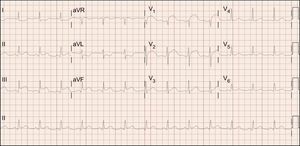
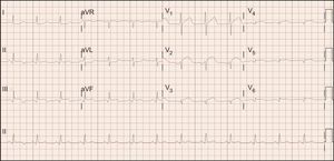
A 58-year-old female smoker with dyslipidemia presented to the emergency room with a 1-hour history of typical chest pain and hypotension (systolic blood pressure, 80mmHg). An electrocardiogram (ECG) was recorded (Figure 1). This showed simultaneous ST elevation in the inferior leads and in leads V1-V3. An ECG in the right leads was also recorded (Figure 2). This showed an ST elevation pattern in III > II, along with diffuse ST depression in AVL > I, with ST elevation greater than 1mm in leads V3 and V4.


What was the most probable coronary artery disease in this patient?
- 1.
Simultaneous occlusion of the proximal right coronary artery and proximal left anterior descending artery
- 2.
Occlusion of a distal segment of the left anterior descending artery on passing over the cardiac apex
- 3.
Occlusion of the left coronary artery
- 4.
Occlusion of the proximal right coronary artery and a large right ventricular branch
Suggest a diagnosis to this ECG Contest at http://www.revespcardiol.org/es/electroreto/70/09. The answer will be published in the next issue (October 2017). #EKGchallenge.