ISSN: 1885-5857 Impact factor 2024 4.9
Vol. 78. Num. 10.
Pages 876-885 (October 2025)

Original article
Fractional flow reserve or OCT to guide management of complex and noncomplex angiographically intermediate coronary stenosis

Reserva fraccional de flujo u OCT para guiar el tratamiento de estenosis coronarias complejas y no complejas angiográficamente intermedias

Andrea ZitoaFrancesco BurzottaabCristina AurigemmabEnrico RomagnolibFrancesco BianchinibEmiliano BianchiniaLazzaro ParaggiobMattia LunardibCarolina IerardibFilippo CreaacAntonio Maria LeoneacCarlo Traniab
https://doi.org/10.1016/j.rec.2025.02.012

Options

Supplementary data
Imagen extra
Rev Esp Cardiol. 2025;78:876-85
Abstract
Introduction and objectives

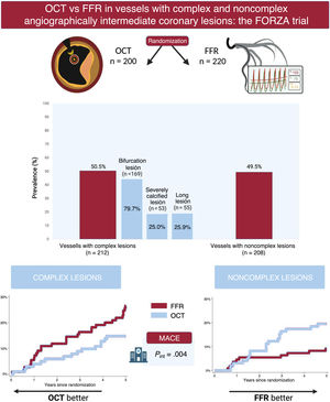
The management of patients with coronary artery disease can benefit from devices that improve functional or anatomical evaluation. This study aimed to compare the efficacy of optical coherence tomography (OCT) and fractional flow reserve (FFR) guidance for managing vessels with angiographically intermediate coronary lesions according to angiographic lesion complexity.
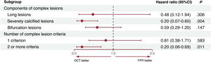
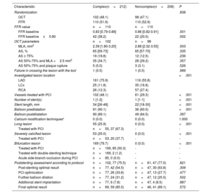
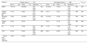
Methods

The FORZA trial (NCT01824030) was a randomized trial comparing the use of OCT or FFR for revascularization decisions and percutaneous coronary intervention optimization in patients with angiographically intermediate coronary lesions. Complex lesions were defined as long (length >38mm), severely calcified, or bifurcation lesions. The primary outcome was major adverse cardiac events (MACE), defined as a composite of all-cause death, myocardial infarction, or target vessel revascularization.
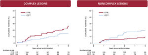
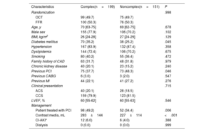
Results

A total of 420 vessels (200 OCT-guided and 220 FFR-guided) were enrolled, including 212 vessels with complex lesions. At the 5-year follow-up, the MACE rate was 20.8% in vessels with complex lesions and 13.9% in vessels with noncomplex lesions (HR, 1.52; 95%CI, 0.95-2.44; P=.078). Compared with FFR, OCT was associated with a lower risk of MACE in vessels with complex lesions (HR, 0.53; 95%CI, 0.28-0.98; P=.044), but with a higher risk of MACE in vessels with noncomplex lesions (HR, 2.23; 95%CI, 1.04-4.81; P=.040; Pinteraction=.004).

Conclusions

In vessels with angiographically intermediate coronary lesions, angiographic lesion complexity may modulate the long-term efficacy of the guidance modality, with a potential benefit of OCT in complex lesions and FFR in noncomplex lesions.

Keywords

Intermediate coronary lesions
Complex lesion
Optical coherence tomography
Fractional flow reserve
Percutaneous coronary intervention
INTRODUCTION

Coronary angiography is burdened by suboptimal assessment of lesion severity, limited identification of high-risk or vulnerable plaques, and raw evaluation of stent implantation during percutaneous coronary intervention (PCI). Thus, adjunctive tools that better characterize coronary stenoses are currently being extensively investigated in various clinical settings.

Fractional flow reserve (FFR) is an invasive index used to assess the potential of coronary stenosis to cause myocardial ischemia and is highly effective in determining lesion severity. Supported by findings from large-scale randomized controlled trials, FFR represents the standard of care for guiding decisions on revascularization in angiographically intermediate coronary artery lesions (AICLs).1,2

Intravascular imaging devices, such as optical coherence tomography (OCT) and intravascular ultrasound, provide detailed anatomical assessments of the vessel lumen, wall, and plaque, enabling the identification of high-risk or vulnerable plaques. Initially developed as tools to improve PCI planning and optimization, these imaging devices were subsequently tested as alternatives to FFR to guide revascularization decisions in patients with AICLs.3

Randomized clinical trials comparing FFR and intravascular imaging in patients with AICLs have shown similar long-term clinical efficacy between these 2 guidance modalities.4–6 However, it remains unknown whether these tools have different prognostic potential for various lesion subsets. Notably, complex coronary lesions represent a high-risk angiographic subset that often results in inaccurate severity and morphological assessments and, when treated, frequently leads to complex procedures, suboptimal PCI results, and less favorable clinical outcomes.7

Therefore, it is clinically relevant to determine whether FFR and intravascular imaging guidance exhibit different efficacy based on angiographic lesion complexity. To address this knowledge gap, we performed this post-hoc analysis of the FORZA (FFR or OCT Guidance to RevasculariZe Intermediate Coronary Stenosis Using Angioplasty) trial,8 investigating the clinical efficacy of OCT and FFR guidance for managing AICLs in vessels with complex and noncomplex lesions.

METHODSStudy design

The FORZA (FFR or OCT Guidance to RevasculariZe Intermediate Coronary Stenosis Using Angioplasty; NCT01824030) trial is an open-label, single-center, prospective, randomized trial comparing clinical outcomes in patients with at least 1 AICL managed with OCT or FFR guidance. The study design and main results at 1-month, 13-month, 3-year, and 5-year follow-up have been previously published.6,8–13 The study was approved by the ethics committee of our institution (code 6261/13), and all patients signed a dedicated informed consent form.

Study participants

Patients with acute or chronic coronary syndrome and at least 1 AICL were screened for enrollment. An AICL was defined as a coronary lesion with a visually estimated percentage diameter stenosis ranging between 30% and 80% in the nondistal segment of a major epicardial vessel. Key exclusion criteria included age <18 years, left ventricular ejection fraction ≤ 30%, recent (< 7 days) ST-segment elevation myocardial infarction (STEMI), recent (< 48hours) non-STEMI, prior STEMI in the territory supplied by the vessel with the AICL under investigation, severe valvular heart disease, multivessel disease with 1 or more untreated angiographically critical stenoses, and multivessel disease requiring coronary artery bypass graft intervention. The full list of inclusion and exclusion criteria is reported in .

Eligible patients were randomly assigned in a 1:1 ratio to OCT or FFR guidance for both PCI performance and, in the case of revascularization, optimization of PCI outcomes. In patients with multiple AICLs in different vessels, all AICLs were managed according to the randomized arm. Operators were asked to strictly adhere to the guidance provided by the assigned tool, without utilizing the alternative modality.

Complex lesions were defined as those with at least 1 of the following angiographic features: lesion length >38mm, severe calcification, or bifurcation. Lesion length was assessed according to angiography. Severe calcification was defined according to angiography as fluoroscopic radiopacities involving both sides of the arterial wall in more than 1 location and with a total length of at least 15mm.14 Bifurcation lesions were defined as lesions adjacent to, and/or involving, the origin of a side branch of at least 2.5mm.15 “Complex patients” were defined as those with at least 1 vessel with a complex lesion.

OCT guidance protocol

OCT images were acquired at the site of the AICL using commercially available systems. The following measurements were collected: minimal luminal area (MLA), proximal reference luminal area (RLA), distal RLA, and mean RLA. Based on these parameters, the percentage of area stenosis (AS) was calculated using the following formula: (mean RLAMLA)/mean RLA×100. Plaque rupture was defined as a recess in the plaque beginning at the luminal-intimal border.16

PCI was performed when at least 1 of the following criteria was met: a) AS ≥75%; b) AS between 50% and 75% and MLA <2.5 mm2; and c) AS between 50% and 75% and plaque rupture.

In patients who underwent revascularization, OCT was also used to guide PCI and optimize results. Additional interventions were performed in the presence of major stent malapposition (defined as a distance between the strut and vessel wall >350μm or <350 and >200μm for a length >600μm), major underexpansion (defined as an in-stent minimal cross-sectional area <75% of the RLA), or major edge dissection (defined as a dissection starting at the level of the stent edge and propagating for a length >600μm). The absence of any of these abnormalities was deemed an “optimal OCT result”. No specific PCI optimization algorithm for complex lesions was set. The details of the OCT study protocol have been previously reported in the study design.8

FFR guidance protocol

FFR was obtained using a 0.014-inch pressure monitoring guidewire and hyperemia induction through intravenous administration of 140mg/kg/min adenosine. PCI was not performed when FFR was >0.80, while it was performed when FFR was ≤0.80. Poststenting FFR was assessed, and when it was <0.90, additional stent postdilation could be performed. If FFR remained <0.90, a guidewire pullback was performed to identify potential pressure drops at least 5mm away from the initial stent, potentially requiring further stent implantation according to physician preference.17 The final achievement of an FFR ≥0.90 was defined as an “optimal FFR result”. The details of the FFR study protocol have been previously reported in the study design.8

Study outcomes

The primary outcome was major adverse cardiac events (MACE), defined as a composite of all-cause death, myocardial infarction (MI), or target vessel revascularization. Key secondary outcomes included individual components of the primary outcome, the composite of cardiac death or MI, and cardiac death.

Statistical analysis

All analyses were performed according to the intention-to-treat principle and on a per-vessel basis. Categorical variables are reported as total numbers and percentages, while continuous variables are reported as mean±standard deviation or median [interquartile range (IQR)], according to their distribution assessed by the Shapiro-Wilk test. Comparisons among groups for categorical variables were performed with the chi-square or Fisher exact test, while those for continuous variables were performed with the Student t test or Mann-Whitney U test, as appropriate. The Kaplan–Meier method was used to characterize the time until the first event for the primary outcome. Hazard ratios (HR) and 95% confidence intervals (95%CI) were computed for primary and secondary outcomes with Cox proportional hazards time-to-event analyses. Subgroup analyses for the primary outcome were performed in complex and noncomplex cohorts according to deferred or performed PCI and in the complex lesion cohort according to the components of complex lesions and the number of complex lesion criteria. Statistical significance was defined as a P value <.05. Statistical analyses were performed using Stata version 18 (StataCorp).

RESULTSBaseline clinical and vessel characteristics

A total of 350 patients (with 420 vessels with AICLs) were randomly assigned to OCT (174 patients, 200 vessels) or FFR (176 patients, 220 vessels). When stratifying the study population based on lesion complexity, 199 patients (212 vessels) had at least 1 complex lesion, while 151 patients (208 vessels) had only noncomplex lesions.

The baseline clinical characteristics of the study population are reported in table 1. All baseline clinical features were well-balanced between patients with complex and noncomplex lesions, except for a higher rate of diabetes mellitus (35.2% vs 25.2%; P=.045) and a lower rate of previous PCI (37.7% vs 48.3%; P=.046) in patients with complex lesions (table 1). Patient characteristics were well-balanced between the treatment arms for both complex and noncomplex lesions ().

Table 1.

Patient characteristics according to lesion complexity

Characteristics  Complex(n=199)  Noncomplex(n=151)  P 
Randomization      .998 
OCT  99 (49.7)  75 (49.7)   
FFR  100 (50.3)  76 (50.3)   
Age, y  70 [63-75]  69 [62-75]  .678 
Male sex  155 (77.9)  106 (70.2)  .102 
BMI, kg/m2  26 [24-28]  27 [24-29]  .129 
Diabetes mellitus  70 (35.2)  38 (25.2)  .045 
Hypertension  167 (83.9)  132 (87.4)  .358 
Dyslipidemia  144 (72.4)  106 (70.2)  .675 
Smoking  80 (40.2)  55 (36.4)  .472 
Family history of CAD  63 (31.7)  48 (31.8)  .979 
Chronic kidney disease  40 (20.1)  23 (15.2)  .240 
Previous PCI  75 (37.7)  73 (48.3)  .046 
Previous CABG  6 (3.0)  3 (2.0)  .547 
Previous MI  44 (22.1)  41 (27.2)  .276 
Clinical presentation      .715 
ACS  40 (20.1)  28 (18.5)   
CCS  159 (79.9)  123 (81.5)   
LVEF, %  60 [55-62]  60 [55-63]  .546 
Management       
Patient treated with PCI  98 (49.2)  52 (34.4)  .006 
Contrast media, mL  283±144  227±114  <.001 
CI-AKI*  12 (6.0)  6 (4.0)  .388 
Dialysis  0 (0.0)  0 (0.0)  .999 

ACS, acute coronary syndrome; BMI, body mass index; CAD, coronary artery disease; CABG, coronary artery bypass grafting; CI-AKI, contrast-induced acute kidney injury; CCS, chronic coronary syndrome; FFR, fractional flow reserve; LVEF, left ventricular ejection fraction; MI, myocardial infarction; OCT, optical coherence tomography; PCI, percutaneous coronary intervention.

The data are expressed as No. (%), mean±standard deviation, or median [25th-75th percentile].

*

Defined according to the Acute Kidney Injury Network definition.

The baseline vessel characteristics are reported in table 2. Vessels with complex lesions were more frequently located in the left anterior descending artery (75.9% vs 55.8%; P<.001) and were more likely to undergo PCI (48.1% vs 29.3%; P<.001) than vessels with noncomplex lesions. Among vessels with complex lesions, 55 (25.9%) had a long lesion (of which 37 [67.3%] were treated with PCI), 53 (25.0%) had a severely calcified lesion (of which 20 [37.7%] were treated with PCI), and 169 (79.7%) had a bifurcation lesion (of which 85 [50.3%] were treated with PCI) (). The OCT graphical representation of the most common complexity criterion, along with the most frequent OCT criterion for performing PCI, is shown in figure 1.

Table 2.

Vessel characteristics according to lesion complexity

Characteristic  Complex(n=212)  Noncomplex(n=208)  P 
Randomization      .838 
OCT  102 (48.1)  98 (47.1)   
FFR  110 (51.9)  110 (52.9)   
FFR value  n=110  n=110   
FFR baseline  0.83 [0.79-0.88]  0.86 [0.82-0.91]  .001 
FFR baseline0.80  42 (38.2)  22 (20.0)  .002 
OCT parameters  n=102  n=98   
MLA, mm2  2.39 [1.80-3.20]  2.88 [2.32-3.55]  .003 
AS, %  65 [59-73]  65 [57-70]  .335 
AS ≥ 75%  19 (18.8)  12 (12.5)  .236 
AS 50%-75% and MLA <2.5 mm2  35 (34.7)  28 (29.2)  .357 
AS 50%-75% and plaque rupture  5 (5.0)  3 (3.1)  .526 
Failure in crossing the lesion with the tool  1 (0.5)  1 (0.5)  .989 
Investigated lesion location      <.001 
LAD  161 (75.9)  116 (55.8)   
LCx  25 (11.8)  35 (16.8)   
RCA  26 (12.3)  57 (27.4)   
Vessels treated with PCI  102 (48.1)  61 (29.3)  <.001 
Number of stent(s)  1 [1-2]  1 [1-1]  <.001 
Stent length, mm  34 [26-48]  22 [18-30]  <.001 
Balloon predilatation  91 (90.1)  36 (60.0)  <.001 
Balloon postdilatation  90 (89.1)  49 (84.5)  .397 
Calcium modification techniques*  0 (0.0)  0 (0.0)  1.000 
Long lesion  55 (25.9)  0 (0.0)  <.001 
Treated with PCI  n=55, 37 (67.3) 
Severely calcified lesion  53 (25.0)  0 (0.0)  <.001 
Treated with PCI  n=53, 20 (37.7) 
Bifurcation lesion  169 (79.7)  0 (0.0)  <.001 
Treated with PCI  n=169, 85 (50.3) 
Treated with double-stenting technique  n=169, 2 (1.2) 
Acute side-branch occlusion during PCI  n=85, 0 (0.0) 
Poststenting assessment according to protocol  n=102, 77 (75.5)  n=61, 47 (77.0)  .821 
Post-stenting optimal result  n=77, 42 (54.5)  n=47, 30 (63.8)  .309 
PCI optimization  n=77, 26 (33.8)  n=47, 13 (27.7)  .477 
Further balloon dilation  n=77, 24 (31.2)  n=47, 12 (25.5)  .502 
Additional stent implantation  n=77, 6 (7.8)  n=47, 4 (8.5)  .887 
Final optimal result  n=69, 59 (85.5)  n=46, 41 (89.1)  .572 

AS, aortic stenosis, FFR, fractional flow reserve; LAD, left anterior descending artery; LCx, left circumflex artery; MI, myocardial infarction; MLA, minimal luminal area; OCT, optical coherence tomography; PCI, percutaneous coronary intervention; RCA, right coronary artery.

The data are expressed as No. (%) or median [25th-75th percentile].

*

Rota-ablation (Boston Scientific, USA), ShockWave (Shockwave Medical, USA), or cutting balloon.

Figure 1.

OCT representation of the most common angiographic complexity and PCI criteria. Bifurcation was the most common angiographic complexity criterion, while an AS of 50%-75% with an MLA <2.5 mm2 was the most frequent criterion for PCI in the OCT arm. The illustrated case shows a bifurcation of the LAD-D1, with a lesion mainly affecting the proximal main vessel, an AS of 62%, and an MLA of 2.3 mm2. AS, area stenosis; D1, first diagonal branch; LAD, left anterior descending artery; MLA, minimal lumen area; MV, main vessel; OCT, optical coherence tomography; PCI, percutaneous coronary intervention.

(0.57MB).

Vessels with complex and noncomplex lesions had similar rates of poststenting optimal results (54.5% vs 63.8%; P=.309), PCI optimization (33.8% vs 27.7%; P=.477), optimization by further balloon dilation (31.2% vs 25.5%; P=.502), optimization by additional stent implantation (7.8% vs 8.5%; P=.887), and final optimal results (85.5% vs 89.1%; P=.572).

Vessels in the OCT group were more likely to undergo PCI than those in the FFR group, both in complex (58.8% vs 38.2%; P=.003) and noncomplex lesions (39.8% vs 20.0%; P=.002) (). The rates of OCT criteria for PCI were similar between vessels with complex or noncomplex lesions, including AS ≥ 75% (18.8% vs 29.2%; P=.236), AS 50% to 75% and MLA <2.5 mm2 (34.7% vs 29.2%; P=.357), and AS 50% to 75% with plaque ulceration (5% vs 3.1%; P=.526).

Finally, optimal results were more frequent in the OCT group than in the FFR group, both at the poststenting assessment (complex: 41.2% vs 19.2%, P=.054; noncomplex: 36.4% vs 7.1%, P=.041) and the final assessment (complex vessels: 100.0% vs 58.3%, P<.001; noncomplex vessels: 100.0% vs 64.3%, P<.001).

5-year clinical outcomes according to angiographic lesion complexity

Clinical outcomes according to angiographic lesion complexity and randomization are presented in . Compared with vessels with noncomplex lesions, those with complex lesions had numerically higher rates of MACE (20.8% vs 13.9%; HR, 1.52; 95%CI, 0.95-2.44; P=.078), cardiac death or MI (4.7% vs 3.4%; HR, 1.42; 95%CI, 0.54-3.73; P=.476), all-cause death (12.7% vs 8.2%; HR, 1.57; 95%CI, 0.86-2.88; P=.145), cardiac death (2.8% vs 2.4%; HR, 1.19; 95%CI, 0.36-3.90; P=.775), MI (2.4% vs 1.0%; HR, 2.48; 95%CI, 0.48-12.76; P=.279), and target vessel revascularization (8.0% vs 5.8%; HR, 1.40; 95%CI, 0.67-2.94; P=.368).

5-year clinical outcomes according to angiographic lesion complexity and randomization

Clinical outcomes according to angiographic lesion complexity and randomization are presented in table 3.

Table 3.

Clinical outcomes according to lesion complexity and treatment arm

Outcome  Complex lesion (n=212)Noncomplex lesion (n=208)Pinteraction 
  OCT(n=102)  FFR(n=110)  HR(95%CI)  P  OCT(n=98)  FFR(n=110)  HR(95%CI)  P   
MACE  15 (14.7)  29 (26.4)  0.53 (0.28-0.98)  .044  19 (19.4)  10 (9.1)  2.23 (1.04-4.81)  .040  .004 
Cardiac death or MI  1 (1.0)  9 (8.2)  0.12 (0.01-0.91)  .040  4 (4.1)  3 (2.7)  1.59 (0.35-7.09)  .546  .044 
All-cause death  7 (6.9)  20 (18.2)  0.36 (0.15-0.85)  .020  12 (12.2)  5 (4.5)  2.86 (1.01-8.11)  .049  .003 
Cardiac death  0 (0.0)  6 (5.5)  3 (3.1)  2 (1.8)  1.83 (0.31-10.95)  .508  .999 
MI  1 (1.0)  4 (3.6)  0.27 (0.03-2.40)  .239  1 (1.0)  1 (0.9)  1.13 (0.07-18.07)  .931  .422 
TVR  8 (7.8)  9 (8.2)  0.93 (0.36-2.42)  .890  6 (6.1)  6 (5.5)  1.14 (0.37-3.54)  .820  .790 
Due to side-branch restenosis  1 (0.5)  0 (0.0) 

95%CI, 95% confidence interval; FFR, fractional flow reserve; HR, hazard ratio; MACE, major adverse cardiac events; MI, myocardial infarction; OCT, optical coherence tomography; TVR, target vessel revascularization.

Unless otherwise indicated, the data are expressed as No. (%).

Compared with FFR, OCT was associated with a lower risk of MACE in vessels with complex lesions (14.7% vs 26.4%; HR, 0.53; 95%CI, 0.28-0.98; P=.044) but a higher risk of MACE in vessels with noncomplex lesions (19.4% vs 9.1%; HR, 2.23; 95%CI, 1.04-4.81; P=.040), with a significant treatment-by-subgroup interaction for lesion complexity (Pinteraction=.004) (figure 2). Similar findings were found for the outcomes of cardiac death or MI (vessels with complex lesions: HR, 0.12; 0.01-0.91; P=.040; vessels with noncomplex lesions: HR, 1.59; 95%CI, 0.35-7.09; P=.546; Pinteraction=.044) and all-cause death (vessels with complex lesions: HR, 0.36; 95%CI, 0.15-0.85; P=.020; vessels with noncomplex lesions: HR, 2.86; 95%CI, 1.01-8.11; P=.049; Pinteraction=.003). Similar trends, although not statistically significant, were found for the outcomes of cardiac death (Pinteraction=1.000), MI (Pinteraction=.422), and target vessel revascularization (Pinteraction=.790) (table 3).

Figure 2.

Cumulative incidence of major adverse cardiac events at 5 years in complex and noncomplex groups. FFR, fractional flow reserve; OCT, optical coherence tomography.

(0.18MB).
Subgroup analyses

Results for the primary outcome were consistent among the subgroups of deferred and performed PCI in both complex (Pinteraction=.451) and noncomplex (Pinteraction=.614) cohorts ().

Results for the primary outcome in the complex lesion cohort revealed a consistent trend among the individual components of the definition of complex lesions (figure 3) with a significant risk reduction with OCT in the subgroup of severely calcified lesions (OCT vs FFR: HR, 0.20; 95%CI, 0.07-0.60; P=.004) and a nonsignificant reduction in the subgroups of lesions longer than 38mm (OCT vs FFR: HR, 0.48; 95%CI, 0.12-1.94; P=.306) and bifurcation lesions (OCT vs FFR: HR, 0.59; 95%CI, 0.29-1.20; P=.147).

Figure 3.

Subgroup analyses for major adverse cardiac events in the complex lesion cohort. 95%CI, 95% confidence interval; FFR, fractional flow reserve; OCT, optical coherence tomography.

(0.17MB).

Finally, when assessing the impact of the number of criteria for complex lesions (figure 3), OCT was associated with a significant reduction in the subgroup with 2 or more criteria for complex lesions (OCT vs FFR: HR, 0.20; 95%CI, 0.06-0.69; P=.011) and a nonsignificant reduction in the subgroup with a single criterion for complex lesions (OCT vs FFR: HR, 0.81; 95%CI, 0.38-1.71; P=.583).

DISCUSSION

The search for improved clinical management of patients with AICLs is an evolving field, and the rational selection of invasive devices is highly desirable. To date, FFR remains the gold standard for managing AICLs, while OCT represents a cutting-edge imaging modality for assessing stent-vessel interactions during PCI. Angiographic lesion complexity is an easily evaluable parameter that reflects different plaque morphologies and the risk of PCI-related complications. In this context, the post-hoc analysis of the FORZA trial suggests that the long-term clinical efficacy of OCT and FFR in guiding the management of vessels with AICLs may be influenced by angiographic lesion complexity, with a potential benefit of OCT in complex lesions and FFR in noncomplex lesions (figure 4). These findings are hypothesis-generating and pave the way for a more personalized selection of adjunctive devices in patients with AICLs.

Figure 4.

Central illustration. Main study findings. FFR, fractional flow reserve; HR, hazard ratio; MACE, major adverse cardiac events; OCT, optical coherence tomography.

(0.64MB).

Adjunctive tools to angiography have significantly enhanced the assessment of coronary lesions, improving procedural and long-term outcomes in patients undergoing PCI.2 Both FFR and intravascular imaging devices have been evaluated as guidance tools for managing AICLs, which are difficult to assess solely by angiography due to significant interobserver variability.4–6The FORZA trial was the first to compare OCT and FFR to guide the management of patients with AICLs.8 Since the anatomical criteria for revascularization had not been tested prior to the study design, they were selected based on prognostic factors related to the degree of luminal narrowing (ie, AS with or without MLA) and the potential presence of an underlying complicated morphology (ie, plaque rupture).8 The results of the FORZA trial showed a higher rate of invasive management with OCT and a similar rate of MACE between the 2 guidance modalities at 5 years of follow-up.6,9 These findings were consistent with those of 2 other randomized controlled trials showing comparable clinical outcomes between FFR- and intravascular ultrasound-guided management, thus supporting the long-term safety of intravascular imaging as a guidance tool for the management of AICLs.4,5

The distinct roles of physiology and imaging warranted further investigation to determine the angiographic or clinical settings in which each modality is most effective. This study presents an intriguing hypothesis and helps bridge this gap, potentially reflecting the fundamental differences between FFR and OCT. Specifically, FFR has traditionally been developed to support revascularization decision-making, whereas OCT and other intravascular imaging devices have predominantly been used for PCI planning and optimization.

Given this background, an anatomical assessment may be more effective in evaluating and guiding the revascularization of complex lesions, which often involve vulnerable morphologies that are not visible angiographically and can lead to technically challenging procedures and suboptimal PCI outcomes.7 Intravascular imaging devices can precisely assess plaque composition, lesion characteristics, reference segments, and landing zones, facilitating the selection of appropriate tools for lesion preparation, optimal stent deployment, and the identification of postintervention stent-related complications, such as malapposition, underexpansion, deformation, or edge dissection.3,18,19

Supporting this assumption, subgroup analysis revealed that lesions with 2 or more complex criteria—presumably posing greater challenges than those with a single complex criterion—showed the most significant benefit from OCT guidance compared with FFR guidance. Conversely, noncomplex lesions may be better suited for physiological assessment, allowing for a more selective approach to PCI. This strategy ensures that PCI is reserved for AICLs capable of inducing myocardial ischemia, thereby reducing the risk of procedure-related complications, stent restenosis, and the prolonged need for dual antiplatelet therapy.

A notable strength of this study is its per-vessel analysis, which enables a precise evaluation of the efficacy of each guidance modality based on angiographic lesion complexity. This is particularly important in patients with multivessel disease, who may have both complex and noncomplex lesions in different vessels, often necessitating distinct management strategies.

Previous studies have used varying definitions of complex lesions, often including unprotected left main coronary artery lesions, chronic total occlusions, and in-stent restenosis.20–22 However, these conditions were exclusion criteria in the FORZA trial, which included only patients with de novo AICLs located outside the left main artery. Therefore, our findings should be interpreted within this specific context and applied only to the lesions classified as complex in this study.

Currently, there is growing interest in determining whether performing PCI in patients with vulnerable nonflow-limiting plaques might reduce the risk of future cardiovascular events.23,24 Interestingly, in the COMBINE OCT-FFR trial, thin-cap fibroatheroma was detected in one-fourth of FFR-negative lesions in patients with diabetes mellitus and was associated with a 5-fold higher rate of MACE at 18-month follow-up.25 Although underpowered for clinical outcomes, the PREVENT trial recently showed that preventive PCI in patients with nonflow-limiting vulnerable coronary plaques, identified by intravascular imaging, reduced the risk of MACE at 2-year follow-up compared with optimal medical therapy alone.26

Physiology and imaging currently represent parallel entities due to concerns about procedural time and costs. The FORZA trial compared these modalities as standalone strategies for guiding revascularization decisions and optimizing PCI outcomes. Therefore, our findings should be interpreted within this context, as they do not explore the potential complementary role of physiology and imaging. However, their synergistic use may improve lesion assessment, procedural strategies, and clinical outcomes. By combining both approaches, clinicians could identify lesions warranting revascularization based on concordant prognostic parameters while ensuring precise PCI planning, execution, and optimization. In the near future, the development of intravascular devices capable of simultaneously acquiring both functional and anatomical information may herald a paradigm shift, enabling more precise and effective PCI.

Study limitations

Our study has some limitations. First, it is a post-hoc subgroup analysis of a small, single-center trial, which limits the generalizability of the results and may introduce selection bias due to the lack of randomization based on angiographic lesion complexity. Additionally, the study lacked sufficient statistical power to detect significant differences in individual outcomes, with few events such as MI or vessel revascularization. Second, our findings should be applied specifically to the target population included in the study, which mainly consisted of patients with chronic coronary syndrome and preserved left ventricular function. Third, the FORZA trial was an open-label study, with both physicians and patients being aware of the trial-group assignments. Fourth, no correction for multiple comparisons was applied. Fifth, some vessels lacked post-stenting and/or final OCT or FFR assessment, limiting outcome comparisons based on optimal results. Sixth, noncardiac deaths accounted for a large proportion of overall mortality. However, their distribution was balanced between the randomized groups, suggesting they did not influence the overall findings. Seventh, the inclusion of angiographic intermediate lesions inherently involved plaques with a limited burden, and consequently, the definition of “complex lesion” should be interpreted within this specific context. Eighth, in the OCT arm, there was no predefined protocol for PCI optimization. Ninth, the absence of a trial screening log limited our ability to thoroughly analyze and characterize the clinical or procedural criteria for patient exclusion. Finally, the potential impact of sex and gender was not evaluated in this analysis. Given these limitations, our findings should be interpreted with caution and considered hypothesis-generating.

CONCLUSIONS

In vessels with AICLs, angiographic lesion complexity may modulate the long-term efficacy of the type of adjunctive device used to guide revascularization and optimize PCI. OCT may provide a potential benefit in complex lesions, while FFR may be more effective in noncomplex lesions.

FUNDING

The FORZA trial was funded by academic grants (Bando Linea D. 1, Università Cattolica del Sacro Cuore, Rome, Italy).

ETHICAL CONSIDERATIONS

The study was approved by the ethics committee of our Institution (code 6261/13) and all patients signed a dedicated informed consent form. Potential impact of sex and gender was not evaluated in this analysis. Data are available upon reasonable request to the corresponding author (F. Burzotta).

STATEMENT ON THE USE OF ARTIFICIAL INTELLIGENCE

No artificial intelligence techniques were used in this study.

AUTHORS’ CONTRIBUTIONS

A. Zito: conception and design, data analysis, drafting of the article. F. Burzotta: conception and design, data analysis, patient enrollment, administrative support, drafting of the article. C. Aurigemma, E. Romagnoli: critical revision of the article, patient enrollment. F. Bianchini, E. Bianchini, L. Paraggio, M. Lunardi, C. Ierardi: critical revision of the article. F. Crea: critical revision of the article, administrative support. A. Maria Leone: conception and design, critical revision of the manuscript, patient enrollment, administrative support. C. Trani: critical revision of the manuscript, patient enrollment, administrative support.

CONFLICTS OF INTEREST

F. Burzotta has participated in advisory board meetings or received speaker's fees from Medtronic, Abbott, Terumo, Daiichi-Sankyo, and Abiomed. A.M. Leone has participated in advisory board meetings or received speaker's fees from Abbott, Medtronic, Daiichi-Sankyo, Bayer, Bruno Farmaceutici, and Menarini.C. Trani has participated in advisory board meetings or received speaker's fees from Medtronic, Abbott, Terumo, Daiichi-Sankyo, and Abiomed. All other authors declare no competing interests.

ACKNOWLEDGMENTS

BioRender platform and templates were used to create the central illustration.

WHAT IS KNOWN ABOUT THE TOPIC?

  • -

    For the management of patients with angiographically intermediate coronary stenosis, fractional flow reserve (FFR) and optical coherence tomography (OCT) guidance is associated with similar outcomes at 5-year follow-up. However, it remains unknown whether the efficacy of FFR and OCT differs based on angiographic lesion complexity.

WHAT DOES THIS STUDY ADD?

  • -

    This study suggests that angiographic lesion complexity may influence the long-term efficacy of the guidance modality, with a potential benefit of OCT in complex lesions and FFR in noncomplex lesions.

References
[1]
P.A.L. Tonino, B. De Bruyne, N.H.J. Pijls, et al.
Fractional Flow Reserve versus Angiography for Guiding Percutaneous Coronary Intervention.
N Engl J Med., (2009), 360 pp. 213-224
[2]
F.J. Neumann, M. Sousa-Uva, A. Ahlsson, et al.
2018 ESC/EACTS Guidelines on myocardial revascularization.
Eur Heart J., (2019), 40 pp. 3096
[3]
A.G. Truesdell, M.A. Alasnag, P. Kaul, et al.
Intravascular Imaging During Percutaneous Coronary Intervention: JACC State-of-the-Art Review.
J Am Coll Cardiol., (2023), 81 pp. 590-605
[4]
H. Zuo, Q. Liu, Z. Zhang, et al.
[Outcomes of percutaneous coronary intervention for intermediate coronary artery disease guided by intravascular ultrasound or fractional flow reserve].
Nan Fang Yi Ke Da Xue Xue Bao., (2014), 34 pp. 704-708
[5]
B.K. Koo, X. Hu, J. Kang, et al.
Fractional Flow Reserve or Intravascular Ultrasonography to Guide PCI.
N Engl J Med., (2022), 387 pp. 779-789
[6]
F. Burzotta, A. Zito, C. Aurigemma, et al.
Fractional flow reserve or optical coherence tomography for angiographically intermediate coronary stenoses: 5-year outcomes in the FORZA trial.
Eur Heart J., (2024), 45 pp. 2785-2788
[7]
N. Werner, G. Nickenig, J.M. Sinning.
Complex PCI procedures: challenges for the interventional cardiologist.
Clin Res Cardiol., (2018), 107 pp. 64-73
[8]
F. Burzotta, A.M. Leone, G.L. De Maria, et al.
Fractional flow reserve or optical coherence tomography guidance to revascularize intermediate coronary stenosis using angioplasty (FORZA) trial: Study protocol for a randomized controlled trial.
[9]
A.M. Leone, F. Burzotta, C. Aurigemma, et al.
Prospective Randomized Comparison of Fractional Flow Reserve Versus Optical Coherence Tomography to Guide Revascularization of Intermediate Coronary Stenoses: One-Month Results.
J Am Heart Assoc., (2019), 8 pp. e012772
[10]
F. Burzotta, A.M. Leone, C. Aurigemma, et al.
Fractional Flow Reserve or Optical Coherence Tomography to Guide Management of Angiographically Intermediate Coronary Stenosis: A Single-Center Trial.
ACC Cardiovasc Interv., (2020), 13 pp. 49-58
[11]
A.M. Leone, F. Burzotta, C. Aurigemma, et al.
PCI Deferral Based on Fractional Flow Reserve or Optical Coherence Tomography: Two-Year Results of the Forza Trial.
Rev Cardiovasc Med., (2022), 23 pp. 361
[12]
D. Ding, S. Tu, Y. Li, et al.
Quantitative flow ratio modulated by intracoronary optical coherence tomography for predicting physiological efficacy of percutaneous coronary intervention.
Catheter Cardiovasc Interv., (2023), 102 pp. 36-45
[13]
C. Aurigemma, D. Ding, S. Tu, et al.
Three-Year Clinical Impact of Murray Law-Based Quantitative Flow Ratio and OCT- or FFR-Guidance in Angiographically Intermediate Coronary Lesions.
Circ Cardiovasc Interv., (2024), 17 pp. e013191
[14]
R.F. Riley, M.P. Patel, J.D. Abbott, et al.
SCAI Expert Consensus Statement on the Management of Calcified Coronary Lesions.
J Soc Cardiovasc Angiogr Interv., (2024), 3 pp. 101259
[15]
F. Burzotta, Y. Louvard, J.F. Lassen, et al.
Percutaneous coronary intervention for bifurcation coronary lesions using optimised angiographic guidance: the 18th consensus document from the European Bifurcation Club.
EuroIntervention., (2024), 20 pp. e915-e926
[16]
G.J. Tearney, E. Regar, T. Akasaka, et al.
Consensus standards for acquisition, measurement, and reporting of intravascular optical coherence tomography studies: a report from the International Working Group for Intravascular Optical Coherence Tomography Standardization and Validation.
J Am Coll Cardiol., (2012), 59 pp. 1058-1072
[17]
P.A.L. Tonino, N.P. Johnson.
Why Is Fractional Flow Reserve After Percutaneous Coronary Intervention Not Always 1.0?.
JACC Cardiovasc Interv., (2016), 9 pp. 1032-1035
[18]
E. Romagnoli, F. Burzotta, R. Vergallo, et al.
Clinical impact of OCT-derived suboptimal stent implantation parameters and definitions.
Eur Heart J Cardiovasc Imaging., (2023), 25 pp. 48-57
[19]
Z.A. Ali, U. Landmesser, A. Maehara, et al.
OCT-Guided versus Angiography-Guided Coronary Stent Implantation in Complex Lesions: An ILUMIEN IV Substudy.
J Am Coll Cardiol., (2024), 84 pp. 368-378
[20]
J.M. Lee, K.H. Choi, Y. Song, Bin, et al.
Intravascular Imaging-Guided or Angiography-Guided Complex PCI.
N Engl J Med., (2023), 388 pp. 1668-1679
[21]
Z.A. Ali, U. Landmesser, A. Maehara, et al.
Optical Coherence Tomography-Guided versus Angiography-Guided PCI.
N Engl J Med., (2023), 389 pp. 1466-1476
[22]
J.J. Wykrzykowska, L.S.M. Kerkmeijer.
Complex PCI: time for a universal definition.
EuroIntervention., (2020), 16 pp. 536-537
[23]
P. Gaba, B.J. Gersh, J. Muller, J. Narula, G.W. Stone.
Evolving concepts of the vulnerable atherosclerotic plaque and the vulnerable patient: implications for patient care and future research.
Nat Rev Cardiol., (2023), 20 pp. 181-196
[24]
F. Prati, E. Romagnoli, L. Gatto, et al.
Relationship between coronary plaque morphology of the left anterior descending artery and 12 months clinical outcome: the CLIMA study.
Eur Heart J., (2020), 41 pp. 383-391
[25]
E. Kedhi, B. Berta, T. Roleder, et al.
Thin-cap fibroatheroma predicts clinical events in diabetic patients with normal fractional flow reserve: the COMBINE OCT-FFR trial.
Eur Heart J., (2021), 42 pp. 4671-4679
[26]
S.J. Park, J.M. Ahn, D.Y. Kang, et al.
Preventive percutaneous coronary intervention versus optimal medical therapy alone for the treatment of vulnerable atherosclerotic coronary plaques (PREVENT): a multicentre, open-label, randomised controlled trial.
Lancet., (2024), 403 pp. 1753-1765

These 2 senior authors equally contributed and should be considered co-last authors.

Copyright © 2025. Sociedad Española de Cardiología