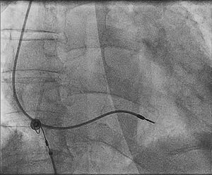
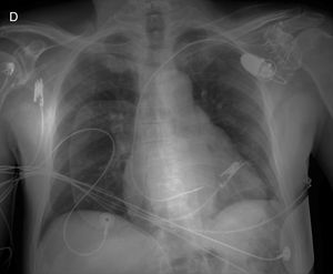


Intermittent capture of the pacemaker lead can be observed. The lead has become loose in the right ventricular outflow tract, and is manifest as QRS axis deviation during captured beats. Ectopic beats are produced with mechanical and direct myocardial pacing on impact with this structure (response 2, correct). Figure 1 shows a fluoroscopic image in the anteroposterior projection after completion of pacemaker implantation. Figure 2 shows the chest X-ray taken at the onset of capture failures and ventricular tachycardia. A clear displacement of the electrode is observed. Response 1 is incorrect, as a lower axis is observed, which rules out apical pacing. Response 3 is incorrect because supraventricular tachycardia with aberrant conduction would not explain the capture failures observed. Response 4 is incorrect because the ventricular ectopic beats are monomorphic and have a morphology similar to paced beats, and so the there is some relationship with the electrode.
ISSN: 1885-5857
Impact factor 2024
4.9