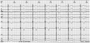
The PR intervals shorten from beat to beat, until they are superimposed on the QRS complex. Negative T waves are normal until V4 in children and the QTc interval is normal. Another blocked P wave can be seen in the T wave, thus ruling out isorhythmic atrioventricular dissociation (responses 1, 2, and 4 incorrect). An atrial frequency of 100 bpm is observed along with nodal escape rhythm or infra-Hisian rhythm of 48 bpm (response 3, correct). The rhythm strip confirms diagnosis of complete atrioventricular block (figure 1). Cardiac malformation and maternal anti-Ro/SSA and anti-La/SSB antibodies should be ruled out.1 Studies (echocardiography, exercise testing, Holter study) are recommended to stratify the risk of sudden cardiac death and inform the need for permanent pacemaker placement.2
ISSN: 1885-5857
