
ISSN: 1885-5857 Impact factor 2024 4.9
Vol. 78. Num. 2.
Pages 156-158 (February 2025)

Scientific letter
A minimalist approach for left atrial appendage occlusion with the use of a mini-transesophageal 4D probe

Abordaje minimalista para el cierre percutáneo de la orejuela izquierda mediante minisonda transesofágica 4D

Laura SanchisabPedro Cepas-GuillénabEduardo FloresabAnder RegueiroabMarta SitgesabXavier Freixaab
https://doi.org/10.1016/j.rec.2024.08.005

Options

Imagen extra
Rev Esp Cardiol. 2025;78:156-8
To the Editor,

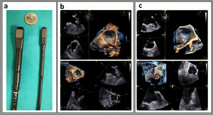
Left atrial appendage occlusion (LAAO) is typically performed under 3-dimensional (3D) transesophageal echocardiographic (TEE) guidance, which usually requires general anesthesia.1 A minimalist approach has been described,2 involving preprocedural 3D planning (using TEE or computed tomography [CT]) and intervention guidance with a micro- or mini-2-dimensional (2D) TEE probe under conscious sedation, thus avoiding the need for anesthesiology. This approach is safe and shows similar performance in experienced centers, but its use in complex cases is limited by the absence of 3D capabilities. A novel mini-TEE with 4D capabilities, initially intended for pediatric use,3 is currently available (figure 1A). Reports of small series have suggested the potential usefulness of this approach for LAAO with conscious sedation.4 Our aim was to evaluate its performance in a larger series of patients and to compare 3D measurements taken during planning and intervention. The study was approved by our ethics committee.

Figure 1.

A: standard 4-dimensional (4D) probe 6VT-D (left), mini-4D-probe 9VT-D (right); B: 3-dimensional imaging of the same patient with standard 4D-probe 6VT-D and 4D-mini-probe 9VT-D.

(0.51MB).

This observational, single-center study included all consecutive patients undergoing LAAO since the mini-4D-TEE became available. All patients provided written informed consent for the intervention, and all analyzed data were anonymized. Sixty-three patients were scheduled for LAAO guided by the mini-4D-TEE probe within 23 weeks. One patient was rescheduled for general anesthesia due to disorientation during probe insertion. Thus, LAAO with a minimalist approach using mild sedation was successfully performed in 62 (98.4%) patients (midazolam 1.1±0.5mg [0-2] and fentanyl 37.4±14.7μg [0-50], plus topical pharyngeal lidocaine): 77.1±7.1 years; 59.7% (n=37) male; weight 72.8±15.8kg. Noninvasive blood pressure and O2 saturation were monitored during the intervention. Vascular echography was used to guide venous femoral access and biplane imaging was used to guide transeptal puncture. Left atrial pressure was routinely measured to ensure a mean pressure ≥ 12mmHg (intravenous serum was administered if needed).

The 3D volume of the left atrial appendage (LAA) was obtained, and the landing zone (LZ) was measured using multiplanar reconstruction (maximal, minimal, and mean). Based on LAA morphology and measurements, the type and size of the device were selected: Amulet (Abbott, United States) (72.6%; n=45), Lambre (Lifetech, China) (19.4%; n=12), and Watchman FLX (Boston Scientific, United States) (8.1%; n=5). A total of 64.5% of the patients (n=40) had previously undergone 3D screening (93.5% TEE) (figure 1B). Among these patients, those with chicken-wing anatomies were excluded, as the landing zone (LZ) measurement is not standardized in these cases. For the remaining patients, there were no significant differences between the LZ mean diameter and the intraprocedural imaging measurements (figure 1C): standard 4D probe 17.8±5.3mm vs mini-4D-TEE 18.3±4.5mm; P=.308. In all cases, the device selected after LAA measurements was successfully implanted (pulmonary ridge coverage 74.2%; 1 significant leak [> 3mm]).

Contrast was used in some patients with complex anatomy (21%; n=13), the intervention time was 47.3±18.8minutes, and X-ray exposure time was 8.1±5.3minutes. No intraprocedural complications were observed. Same-day discharge was decided for 51 patients (82.3%) after we performed a transthoracic echocardiogram to exclude pericardial effusion or embolization. Regarding the remaining 11 patients: 5 were already hospitalized due to bleeding, 1 due to heart failure, 2 were combined with transcatheter aortic valve implantation, and 4 lived too far from the hospital to be included in the same-day discharge program.

As shown in this series, tolerance to the 4D mini-TEE with conscious sedation was excellent. The short intervention times and the absence of anesthesia recovery allowed for fast patient turnover, with most procedures performed on an outpatient basis. In our center, patients previously always underwent preprocedural 3D planning. However, in the present cohort, LAAO was performed without preprocedural 3D imaging in some patients, given the high imaging quality of the 4D mini-TEE and the need to accommodate some in-hospital patients without available slots for the preferred standard 3D-TEE screening.

As shown, the concordance between screening and interventional measurements was good. Therefore, in high-volume centers with various device types readily available, direct LAAO without prior planning may be considered. No contraindications for closure were found in our series (such as protruding thrombus or extreme dimensions of the LAA).

The main limitation of the study is its observational nature. In addition, the study was conducted in a high-volume center with experienced operators (both interventional cardiologists and imaging specialists), and consequently the results may not be extrapolated to centers with lower volumes or less experience.

According to our initial observations, LAAO is feasible and safe using a minimalist approach with 4D mini-TEE guidance and mild sedation in most patients. This strategy allowed for rapid patient turnover and same-day discharge.

FUNDING

None.

ETHICAL CONSIDERATIONS

The study was approved by the ethics committee of Hospital Clínic, Barcelona. As this was a retrospective study, consecutive patients were included without gender discrimination.

STATEMENT ON THE USE OF ARTIFICIAL INTELLIGENCE

No artificial intelligence was used in the present study.

AUTHORS’ CONTRIBUTIONS

L. Sanchis is an associate editor of Rev Esp Cardiol. The journal's editorial procedure to ensure impartial processing of the manuscript was followed; she was in charge of the conception of the work, analysis, writing and final approval. P. Cepas-Guillén: drafting, data acquisition, and final approval. E. Flores: data acquisition, drafting, and final approval. A. Regueiro: interpretation of the data, critical review, and final approval. M. Sitges: critical review, final approval. X. Freixa: interpretation of the data, critical review, and final approval.

CONFLICTS OF INTEREST

M. Sitges and L. Sanchis have received speakers’ honoraria from GE HealthCare. The other authors have no conflicts to declare.

REFERENCES
[1]
M. Glikson, R. Wolff, G. Hindricks, et al.
EHRA/EAPCI expert consensus statement on catheter-based left atrial appendage occlusion - An update.
EuroIntervention., (2020), 15 pp. 1133-1180
[2]
A. Aminian, N. Leduc, X. Freixa, et al.
Left Atrial Appendage Occlusion Under Miniaturized Transesophageal Echocardiographic Guidance and Conscious Sedation: Multicenter European Experience.
JACC Cardiovasc Interv., (2023), 16 pp. 1889-1898
[3]
C. Karsenty, K. Hadeed, P. Acar.
First experience with 3-dimensional pediatric transesophageal echocardiography.
Rev Esp Cardiol., (2023), 76 pp. 487
[4]
L. Sanchis, A. Regueiro, P. Cepas-Guillen, et al.
First experience of left atrial appendage occlusion using a 3D mini transoesophageal echocardiographic probe with conscious sedation.
EuroIntervention., (2023), 18 pp. 1460-1461
Copyright © 2024. Sociedad Española de Cardiología