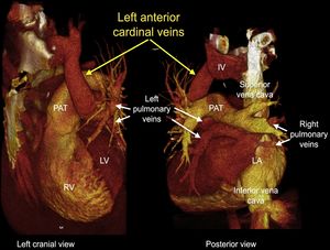
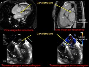
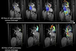
A 27-year-old woman was seen for progressive dyspnea with onset 1 month earlier. Initially, computed tomography angiography of the pulmonary arteries ruled out pulmonary thromboembolism, although marked dilatation of the right ventricle (RV) and the pulmonary artery trunk (PAT) was detected. Likewise, persistence of the left anterior cardinal vein (LACV) was observed, with outflow in the roof of the left atrium (LA) and cranially in the innominate vein (IV). The LACV collected venous drainage from the left lung, while the right pulmonary veins drained with independent ostia into the LA (Figure 1). Echocardiography and cardiac magnetic resonance revealed a left atrial membrane, with limited fenestration, consistent with cor triatriatum (Figure 2). Cardiac magnetic resonance showed LV dilatation (end-diastolic volume index of 188mL/m2), RV ejection fraction of 55%, and a left-right shunt with QP/QS of 4.3:1. The 4D flow sequence showed complete anomalous venous drainage of the left lung and anatomically normal but functionally mixed right venous drainage: cardiac and towards the LACV (Figure 3 and ).


Cor triatriatum and LACV rarely co-occur, as the usual presentation is with an atrial shunt. In this case, the use of 4D flow helped to elucidate how the presence of cor triatriatum transformed normal right pulmonary drainage into partially anomalous drainage and confirmed the occurrence of complete left pulmonary drainage toward the LCAV, thus explaining the substantial left-right shunt.