To the Editor:
We describe the case of a 68-year-old man seen in the emergency room for sudden piercing chest pain radiating toward the back. The electrocardiogram and analytical parameters were normal; however, the chest x-ray showed mediastinal widening. The chest and abdominal computed tomography (CT) revealed DeBakey type 1 aortic dissection and TTE showed severe aortic regurgitation. The patient underwent emergency surgery. The entry site was at the origin of the left subclavian artery, with retrograde progression to the aortic valve and anterograde progression toward the thoracic aorta. Under deep hypothermia and antegrade cerebral perfusion, the ascending aorta and aortic arch were replaced, with reimplantation of the supraaortic branch and aortic valve resuspension.
There were no complications in the postoperative care unit and the patient was discharged at 48 h.
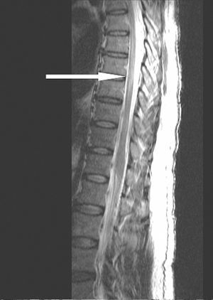
On postoperative day 3, the patient began to experience symptoms of dysesthesia/paresthesia in the left leg that progressed within 24 h to paraplegia of the legs with loss of overall sensitivity (D10). Cranial CT study showed no signs of ischemia and/or hemorrhage, and the chest and abdominal CT indicated no progression of dissection. Magnetic resonance imaging (MRI) of the thoracolumbar spine was consistent with spinal cord ischemia (Figure 1).

Figure 1. Magnetic resonance of the thoracolumbar spine showing a hyperintense image in the T2 sequence (that was hypointense on T1) from D7 to L2, consistent with spinal cord ischemia.
On postoperative day 4, a lumbar catheter was inserted for cerebrospinal fluid (CSF) drainage, with a CSF opening pressure of 14 mm Hg and maintaining the intradural pressure at 5 mm Hg. The motor deficit resolved in both legs 8 h after catheter implantation; all other neurological symptoms improved progressively.
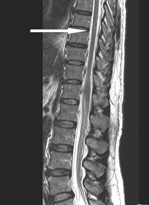
The CSF drain was withdrawn after 4 days. Follow-up MRI of the thoracolumbar spine showed no evidence of spinal cord ischemia (Figure 2).

Figure 2. Magnetic resonance of the thoracolumbar spine shows no signal intensity changes in the spinal cord indicative of ischemia.
The patient was discharged 24 days after the surgery. At the 3-month visit following discharge, he presented complete remission of the neurological symptoms and normal functional capacity.
Perioperative spinal cord ischemia is one of the most serious complications of thoracic and/or abdominal aorta surgery, with a prevalence of 4% to 25%.1-3 Nevertheless, spinal cord ischemia during the postoperative period following acute aortic dissection is rare and only a few cases have been reported.4,5 The etiology of postoperative spinal cord ischemia is multifactorial and includes intraoperative factors (eg, insufficient arterial flow to the spinal cord during aortic clamping and circulatory arrest, proximal hypertension with elevated CSF pressure),1,2 acute episodes of postoperative hypotension and hypovolemia,5 embolic/ thrombotic occlusion of the spinal arteries originating from the false lumen,4,5 or mechanical compression due to the dissection flap.
When paraplegia is established, there are few therapeutic options and the efficacy of CSF drainage, corticosteroid therapy, or hyperbaric oxygenation is uncertain.2,6 In the case described, even though the patient's paraplegia had lasted 24 h, he experienced rapid clinical improvement after CSF drainage, with complete resolution of the motor deficit only a few hours later.
Although the efficacy of these therapeutic measures is described in the literature as uncertain, CSF drainage is a simple, rapid technique that, in our case, was effective for treating an established motor deficit.