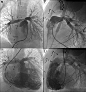
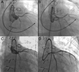
A 9-month-old male infant (6.9kg body weight) with aortic valve stenosis had been treated surgically with the Ross procedure. He underwent cardiac catheterization, which revealed severe obstruction at the origin of both pulmonary arteries (Figure 1A and B) that resolved with implantation of Formula™ 414 in both pulmonary arteries (Figure 1C and D); 12hours later, the patient developed signs of ischemia in left coronary artery territory, with left ventricular dysfunction, and was placed on extracorporeal membrane oxygenation. Once he had been stabilized, coronary angiography was performed; this test revealed a critical obstruction in left main coronary artery (Figure 2A) that resolved after implantation of a 2.25mm×12mm Taxus Liberte™ stent into coronary artery (Figure 2B). Ventricular function began to improve, and extracorporeal membrane oxygenation was discontinued 5 days later. When the child was 3 years old, follow-up coronary angiography revealed endothelial proliferation within the stent, with a considerable reduction in the lumen (Figure 2C), that resolved after implantation of a 2.5mm×11mm BioMatrix Flex™ stent (Figure 2D).


Coronary stenosis following stent implantation into the pulmonary arteries is a very uncommon complication that can be avoided by performing imaging studies or coronary angiography simultaneously with intra-arterial balloon inflation prior to the intervention.
A high index of suspicion, together with stabilization under extracorporeal membrane oxygenation and intracoronary stent implantation, can save the patient's life.