Echocardiography imaging guidance plays an essential role in the safe and accurate execution of transcatheter structural interventions. Transesophageal echocardiography (TEE) is the preferred option in the guidance of left atrial appendage occlusion (LAAO) and transcatheter valve interventions.1 Procedures with general anesthesia increase the risk of hypotensive periods, respiratory disorders, and the time and cost of interventional procedures. In elderly patients, general anesthesia is associated with perioperative pulmonary complications, neuromuscular block, and a higher risk of cognitive decline.2 In addition, the use of a standard TEE probes for more than 60minutes has been associated with a higher risk of oropharyngeal lesions.3
Currently, a large number of structural procedures are performed under local rather than general anesthesia. In prolonged procedures, however, TEE imaging is generally performed under general anesthesia, as standard TEE probes may not be very well tolerated with just light sedation. Intracardiac echocardiography (ICE) has been proposed as a valid alternative to TEE for some structural procedures such as LAAO to avoid general anesthesia. Despite the lower imaging quality, the higher cost and the need for additional vascular and transseptal access, some operators have moved to this less invasive approach. The recent arrival of novel micro-TEE probes might represent a very valid alternative to allow a minimally-invasive approach while maintaining proper imaging quality. Although some other experiences with micro-TEE probes have already been published,4,5 the current report presents the initial experience with a latest-generation micro-TEE probe (10T-D General electrics), which allows multiplane 2-dimensional (2D) imaging with a very small distal tip diameter of 5.6 ·× 7.7 mm.
We present our initial experience with 2 cases of LAAO in patients with atrial fibrillation with a high risk of stroke and with contraindications to long-term anticoagulation. The procedures were performed under light sedation and local anesthesia guided with a new-generation micro-TEE probe together with angiography. The micro-TEE probe provided excellent imaging quality for transseptal puncture and LAA assessment in both cases compared with the standard 2D-TEE, which was performed 1 week before the procedure (Figure). A 22-mm and 28-mm Amulet (St Jude Medical) were successfully implanted without complications. A final angiogram confirmed the correct placement of the device. The procedures lasted 52 and 45minutes, respectively, and the patients were discharged the day after the procedure.
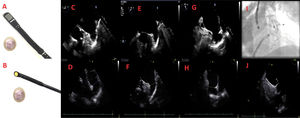
 and micro (B) probes. Multiple 2D-TEE views with the adult (C,E,G) and micro probe (D,F,H). Final result after LAAO with fluoroscopy (I) and micro-TEE probe (J). LAAO, left atrial appendage occlusion; TEE, transesophageal echocardiographic.")
Comparison between adult and micro 2D-TEE probe. 2D-TEE adult (A) and micro (B) probes. Multiple 2D-TEE views with the adult (C,E,G) and micro probe (D,F,H). Final result after LAAO with fluoroscopy (I) and micro-TEE probe (J). LAAO, left atrial appendage occlusion; TEE, transesophageal echocardiographic.
LAAO requires transseptal puncture, accurate LAA assessment and continuous imaging guidance for device deployment. The patient must be immobile during all these phases, as a sudden movement can increase the risk of significant complications, including atrial perforation and pericardial tamponade. For these reasons, most operators using TEE guidance tend to use general anesthesia.6 In our series, the micro-TEE probe was well tolerated by patients, and provided sufficient image quality for procedure guidance.
A previous study comparing micro-TEE with ICE and standard TEE showed that micro-TEE offers comparable image quality with a much wider field of vision than ICE and a slightly reduced but sufficient image quality and comparable accuracy to standard TEE.5 One of the main limitations of current micro-TEE probes and ICE is the absence of 3D imaging. For this reason, before LAAO, previous imaging assessment with 3-dimensional (3D) techniques (3D-TEE or computed tomography scan) is generally recommended. It is well established that 2D imaging might be sufficient to guide some structural procedures but definitely not enough to provide an accurate morphological assessment of cardiac structures like LAA. Structural procedures are the most demanding and 3D-TEE and simultaneous orthogonal views are very important during the procedure and if there are complications.
Although this initial experience focused on LAAO, the high patient-comfort and the excellent imaging quality suggested that latest-generation micro-TEE probes might represent a very valid alternative not only for LAAO but also for other percutaneous structural heart interventions and could be an option in patients without intubation or general anesthesia, in particular in elderly patients or those with severe comorbidities. Additional experience will be necessary to confirm that high quality imaging is also observed in a larger sample of patients.