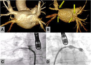

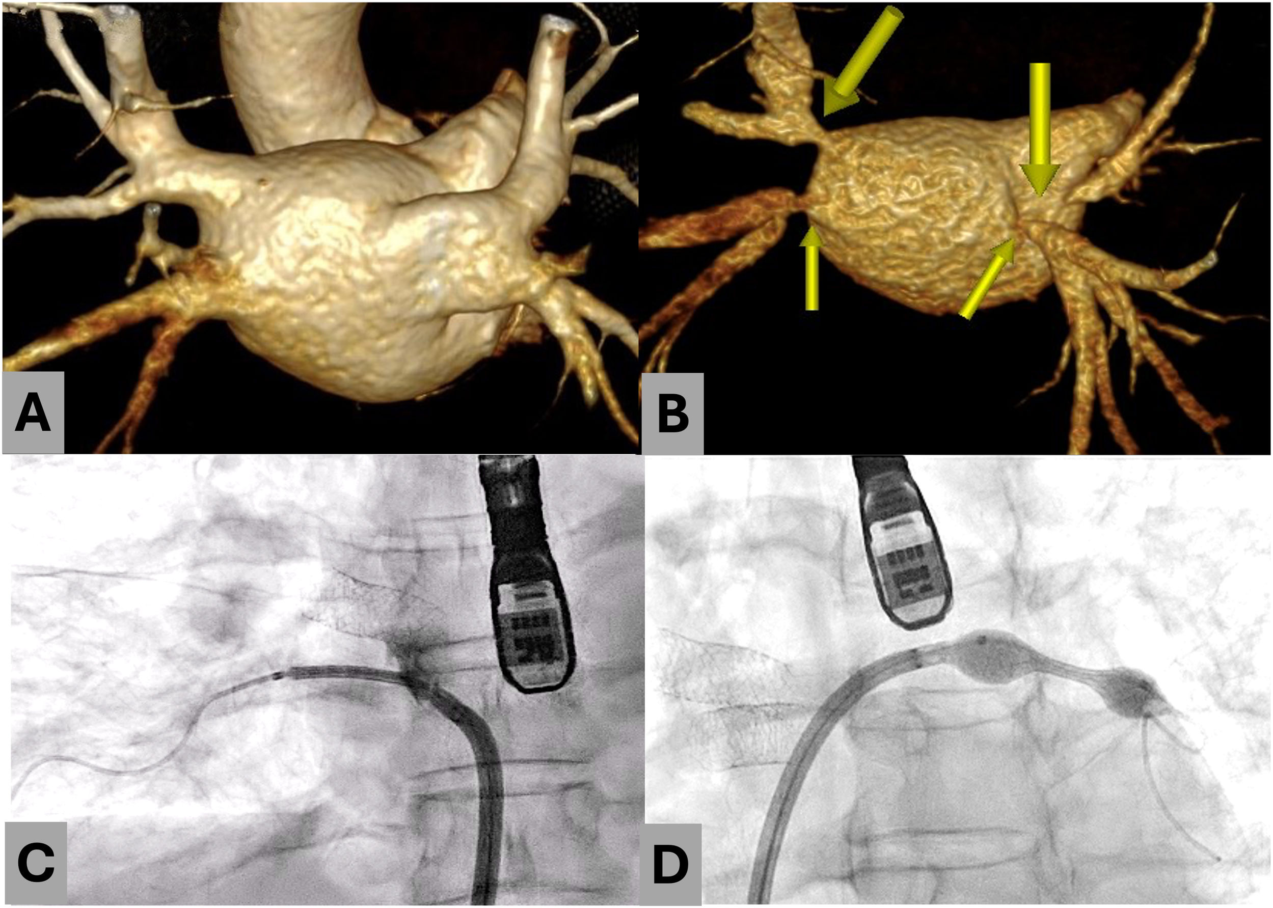
We present the case of a 46-year-old woman who presented with dyspnea and hemoptysis. She had paroxysmal atrial fibrillation that had been treated with pulmonary vein ablation via radiofrequency 6 months earlier. The patient was a smoker and a carrier of the SCN5A gene. We performed computed tomography angiography (CTA), which revealed stenosis of both right pulmonary veins and of a common trunk (figure 1A, 3-dimensional reconstruction of the pulmonary veins before ablation; figure 1B, stenosis after ablation). The patient underwent percutaneous angioplasty with placement of 3 stents. Postsurgical CTA confirmed the initial success of the procedure (figure 1C,D).

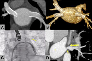
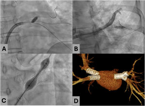
In the following weeks, the patient developed hemoptysis, which became life-threatening. A subsequent CTA scan revealed permeable stents (figure 2A,B) with left superior pulmonary vein (LSPV) occlusion, indicating critical ostial stenosis before stent placement (figure 2C,D; LA, left atrium). We performed a new percutaneous procedure by advancing a coronary angioplasty guidewire through the struts of the previous stent into the presumed LSPV. The struts were dilated with coronary angioplasty balloons (figure 3A). Using a balloon as an anchor, we then advanced a hydrophilic catheter to assess branch patency (figure 3B). A new stent was placed inside the previous stent (figure 3C and video 1 of the supplementary data), with procedural success confirmed via CTA (figure 3D). Follow-up CTA showed the stent positioned toward the superior branch of the left inferior pulmonary vein (figure 3D). As the patient's condition was stable, we opted for a conservative approach.


Pulmonary vein stenosis is a rare but potentially severe complication of atrial fibrillation ablation. Due to the anatomical variability and complexity of these lesions, percutaneous repair may prove challenging and necessitate the use of imaging techniques.
FUNDINGNone.
ETHICAL CONSIDERATIONSThe patient gave her consent for publication of this case report. Potential sex and gender biases according to the SAGER guidelines were considered during the drafting of the present article.
STATEMENT ON THE USE OF ARTIFICIAL INTELLIGENCENo artificial intelligence was used during the preparation of this work.
AUTHORS’ CONTRIBUTIONSAll the authors participated in the drafting and editing of the manuscript.
CONFLICTS OF INTERESTThe authors declare that they have no conflicts of interest.