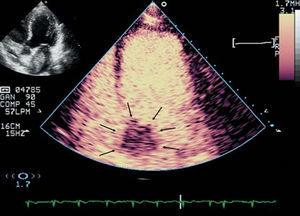
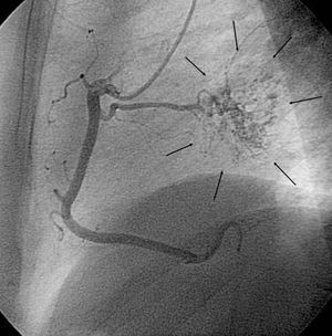
A 59-year-old woman consulted for palpitations of one month's evolution. The electrocardiogram documented frequent premature atrial beats, occurring shortly after episodes of paroxysmal atrial fibrillation. Echocardiography performed to investigate these findings showed a left atrial mass, 50×35 mm in diameter, adhering to the interatrial septum. On the contrast myocardial perfusion study (Figure 1, apical view), the mass showed mild enhancement with respect to the adjacent myocardium. Preoperative coronary angiography (Figure 2, left lateral view) revealed an atrial branch arising from the right coronary artery that provided vascular support to the mass and produced an image of "tumor blush" at the level of the left atrium (arrows).

Figure 1.

Figure 2.
The patient underwent surgery through a biatrial approach to remove the mass, with resection of the portion of the interatrial septum where the pedicle inserted. Pathological study confirmed the diagnosis of atrial myxoma.