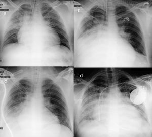
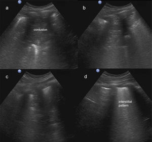
A 26-year-old man with recently diagnosed hypertrophic cardiomyopathy experienced a witnessed cardiorespiratory arrest while exercising and underwent immediate basic cardiopulmonary resuscitation with cardiac massage. The emergency services noted an initial rhythm in ventricular fibrillation; after 20minutes of cardiopulmonary resuscitation, sinus rhythm was regained. The patient was admitted to the ICU, and after completion of the hypothermia protocol, respiratory progress was attempted. He was neurologically conscious and obeyed orders, but did not tolerate weaning from respiratory support. The chest radiograph at 48hours following admission depicted an infiltrate in the right middle lung field (Figure 1A). Chest ultrasonography showed a contusion in the right anteroinferior region of the lung (Figure 2A and ). At 72hours, the pulmonary infiltrate and right basal atelectasis had progressed, being more evident in the chest radiograph (Figure 1B), in the context of contusion (Figure 2B). At 96hours, the atelectasis and contusion showed some improvements (Figure 1C and Figure 2C). At 5 days, the pulmonary infiltrates persisted on radiography, with improvements in blood gas measures that enabled extubation without incidents. The patient underwent automatic implantable defibrillator implantation (Figure 1D). On ultrasound study, an interstitial pattern was detected in the area of the contusion (Figure 2D and ). Culture of bronchial aspirate was negative. At 9 days, the patient was discharged from hospital, asymptomatic. Ultrasound, a simple, inexpensive technique, is useful for the diagnosis of lung lesions caused by chest trauma. It can be performed at the bedside and is a radiation-free technique, unlike chest computed tomography or radiography.
ISSN: 1885-5857
Impact factor 2024
4.9

