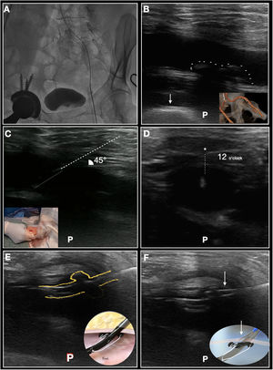

Ultrasound (US)-guided access of the common femoral artery (CFA) to obtain large bore vascular access for procedures such as transcatheter aortic valve replacement (TAVR) has become the standard of care. Use of Perclose ProGlide (Abbott Vascular, United States) sutures is routine for preclosure of the access site. However, in patients with a long tissue tract or posterior wall calcification, the positioning of the ProGlide in the artery to obtain optimal suture deployment can be difficult. Here, we show a stepwise approach to this process in a patient with prior right total hip replacement (figure 1A), with the long-axis view at the level of the CFA showing calcification in the posterior wall (demarcated with dashed points) (figure 1B). The anterior wall access at a location free of calcification was obtained in both CFA using the standard US-guided technique. Figure 1C shows the needle penetrating the dermal layers and approaching the CFA at a 45° angle (the left lower panel shows the positioning of the ultrasound [left hand] and needle [right hand] by the operator). Figure 1D shows the wire approximating the anterior wall of the CFA at the 12 o’clock position. After foot deployment, the device was pulled back under direct US visualization to confirm the position of the foot against the anterior wall (figure 1E) and avoidance of engagement with the posterior wall calcification during needle deployment (Figure 1F and ). After providing informed consent, the patient underwent a successful TAVR procedure with SAPIEN Ultra 26mm and the vascular access was closed uneventfully.
FUNDING
No funding.
AUTHORS’ CONTRIBUTIONSAll authors have read and approved the manuscript.
CONFLICTS OF INTERESTThe authors have no conflicts of interest to declare.
Supplementary data associated with this article can be found in the online version available at https://doi.org/10.1016/j.rec.2021.11.014