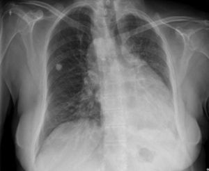
A 64-year-old female with no family history of heart disease came to our hospital due to an episode of respiratory superinfection. An x-ray (Fig. 1) showed loss of volume in the left hemithorax and an abnormal cardiac silhouette, so we decided to perform a magnetic resonance (MR) imaging study.

Using a 1.5T MR imaging, black blood sequences were acquired in all three imaging planes and a multiphase angiography was performed after administering intravenous contrast (gadolinium).
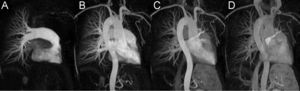
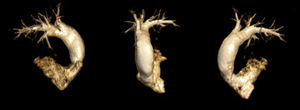
The multiphase MR angiography revealed agenesis of the left pulmonary artery (Fig. 2A) in the pulmonary artery phase. The systemic arterial phase (Fig. 2B) revealed a right aortic arch and left pulmonary arterial vascularization provided by systemic collateral vessels dependent on the left subclavian artery. The supra-aortic trunks originated from a left brachiocephalic trunk that gave rise to the common left carotid artery and the left subclavian artery, and from a right common carotid artery and a right subclavian artery originating independently from the aortic arch (Fig. 2C). The systemic venous phase (Fig. 2D) showed a delay in left pulmonary venous return. Volumetric reconstructions from MR data show, in different obliquities, the morphology of the right ventricular outflow tract, the trunk of the pulmonary artery and the right pulmonary artery (Fig. 3).


Multiphase MR angiography, with its high temporal resolution, enables us to obtain anatomical and functional information about thoracic vascular structures with a single injection of intravenous contrast and no ionizing radiation.