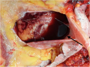
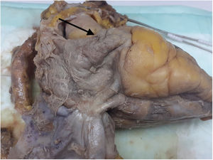
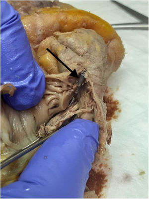

The perforation and resulting cardiac tamponade associated with the implantation of permanent pacemaker leads is a highly infrequent complication, with a rate of about 0.8% in Spain and an apparently very low associated mortality. Accordingly, pacemakers are generally safe devices. We present the case of a male patient who underwent implantation of a dual-chamber (right atrium and ventricle) permanent pacemaker that was complicated by perforation of the right atrium by an active fixation lead. There were no complications or events during the implantation and the patient was discharged to home at 24hours. The patient attended the emergency department 18hours after discharge due to respiratory failure and with mild radiological infiltrates and was admitted to the hospitalization ward with suspected pneumonia, without hemodynamic instability in the emergency department. In the ward, the patient developed electromechanical dissociation, and cardiopulmonary resuscitation was unsuccessful. Consent was obtained from the family and a clinical autopsy was performed; opening of the pericardial cavity, which was noted to be convex, revealed abundant hemopericardium (figure 1). In the right atrial appendage, the helical anchor of the lead (figure 2) reached the exterior of the atrial cavity. It was clearly evident that the anchoring system (figure 3) had pierced the entire thickness of the atrial wall. According to the pathologist who performed the autopsy, the wall was abnormally thin (1.1- to 1.5-mm-thick), which was possibly associated with the patient's underlying disease (multiple myeloma) and the chemotherapy and radiotherapy received.
FUNDING


No funding was received.
AUTHORS’ CONTRIBUTIONSThe authors contributed equally.
CONFLICTS OF INTERESTThere are no conflicts of interest.