To the Editor:
Persistent left superior vena cava is the most common congenital venous abnormality of the thorax and has a prevalence of 0.5% among the general population.1 Other related malformations include absent right superior vena cava and presence of a left azygos vein. In these situations, the right side of the head and the right arm drain mainly through the innominate vein in the left superior vena cava which itself drains through an extremely dilated coronary sinus.
We describe a 59-year-old man, with a history of smoking, tuberculosis during childhood, and a hiatal hernia, who had dilated cardiomyopathy in functional class III/IV and was waiting for a heart transplant. No evidence of congenital anomaly was observed in the preoperative assessment.
During the surgical procedure, the right superior vena cava was found to be absent and the innominate vein was seen to drain into the left superior vena cava, with the vena cava draining into the coronary sinus. Conventional aortic cannulation of the inferior vena cava into the right atrium and selective cannulation of the left superior vena cava were performed.
The only surgical variations to the conventional implant technique were as follows: a) the left superior vena cava was selectively cannulated; b) the coronary sinus and its junction in the remaining right atrium were left intact during cardiotomy; and c) the atrioventricular groove of the recipient was left intact. The extracorporeal circulation and ischemia times were 135 and 145 min, respectively.
Postoperative cardiologic progress was satisfactory and postoperative angiography follow-up showed good drainage through the left superior vena cava with no morphological abnormalities.
Persistent left superior vena cava is a relatively rare abnormality that may require more complex surgical management if not diagnosed before the operation, a situation reported rather often in the literature. This malformation may be suspected before surgery when there is evidence of mediastinal widening, abnormal positioning of the central venous catheter on chest x-ray, and a large, echographic signal-free space in the retrocardial area corresponding to a dilated coronary sinus.
Several surgical alternatives to solve this problem have been described. Placement of a prosthesis between the right atrium and the innominate vein plus ligation of the left superior vena cava would allow conventional heart transplantation.2 However, a disadvantage of this technique is a higher risk of prosthesis-related infection in immunocompromised patients.
Other techniques have been developed, but require prior knowledge of the malformation to retain a sufficiently long segment of superior vena cava from the donor for the purpose of combining various anastomosis possibilities and thus expand the range of surgical options.3 The first method consists of reconstructing the right superior vena cava from the division of the innominate vein where it enters the left superior vena cava and anastomosing it to the right superior vena cava of the donor. The venous drainage of the upper body would be left with 2 superior venae cavae. Another option is end-to-side anastomosis of the recipient innominate vein to the donor superior vena cava, with ligation to the left superior vena cava. The third method described is to mobilize the left superior vena cava and to anastomose the donor superior vena cava after tunneling it by the transverse sinus.4
Our proposal appears to be technically simpler, does not lengthen the time of extracorporeal circulation excessively, and does not assume preoperative knowledge of this malformation. Moreover, it can be used without modifying the resection technique employed with the donor heart.
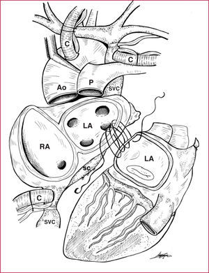
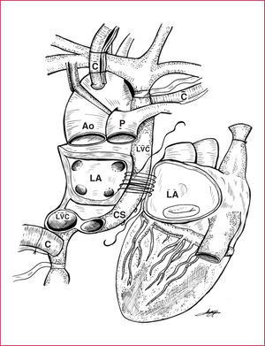
The standard orthotopic transplantation and the bicaval anastomosis technique both require selective cannulation of the left superior vena cava and isolation of the coronary sinus in the atrioventricular groove of the heart recipient, with an incision made between the upper edge of the left pulmonary veins and the lower edge of the coronary sinus, plus ligation of the small coronary veins that drain into the coronary sinus, thus isolating the coronary sinus and the inferior vena cava in the remaining right atrium.5,6 (Figures 1 and 2).

Figure 1. Standard heart transplantation. Note the cannulation of the left superior vena cava and the isolation of the coronary sinus. Ao indicates aorta; C, extracorporeal circulation cannulae; CS, coronary sinus; IVC, inferior vena cava; LA, left atrium; P, pulmonary artery; RA, right atrium; SVC, left superior vena cava.

Figure 2. Heart transplantation using the bicaval technique. Start of anastomoses in the left atrium. Ao indicates aorta; C, extracorporeal circulation cannulae; CS, coronary sinus; IVC, inferior vena cava; LA, left atrium; P, pulmonary artery; RA, right atrium; SVC, left superior vena cava.
In the end, 2 coronary sinuses would be left in both scenarios: that of the heart donor and that of the recipient which drains the left vena cava.