ISSN: 1885-5857 Impact factor 2024 4.9
Vol. 78. Num. 3.
Pages 176-184 (March 2025)

Original article
Early unloading and clinical outcomes in patients with fulminant myocarditis undergoing VA-ECMO: results of a multicenter retrospective study

Descarga precoz y evolución clínica en pacientes con miocarditis fulminante sometidos a ECMO-VA: resultados de un estudio retrospectivo multicéntrico

Minjung BakaJunho HyunbHyukjin ParkcHyung Yoon KimdSeonhwa LeeeIn-Cheol KimeSo Ree KimfMi-Na KimfKyung-Hee KimgJeong Hoon Yanga
https://doi.org/10.1016/j.rec.2024.06.004

Options

Supplementary data
Imagen extra
Rev Esp Cardiol. 2025;78:176-84
Abstract
Introduction and objectives

Although venoarterial extracorporeal membrane oxygenation (VA-ECMO) provides effective cardiocirculatory support in patients with fulminant myocarditis, the most effective timing of venting is uncertain. We aimed to investigate the benefit of early venting among patients who underwent VA-ECMO for fulminant myocarditis.
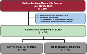
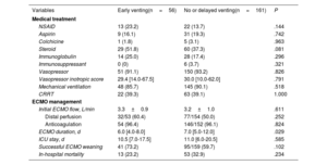
Methods

Among 841 patients with acute myocarditis from 7 hospitals in the Republic of Korea, 217 patients with fulminant myocarditis who underwent VA-ECMO were included in this analysis. The patients were categorized into 2 groups: an early unloading group that underwent venting within 24hours of ECMO insertion, and the no or delayed unloading group. The primary outcome was a composite of death, cardiac replacement, or cardiovascular rehospitalization.
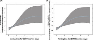
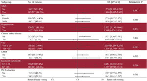
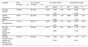
Results

Among 217 patients, 56 underwent early venting, 54 underwent delayed venting, and 107 did not undergo venting. On spline curves in 110 patients who underwent venting, rapid deterioration was observed as the timing of venting was delayed. The incidence of the primary outcome was lower in the early venting group than in the no or delayed unloading group (37.5% vs 58.4%; HR, 0.491; 95%CI, 0.279-0.863; P=.014). Among patients not experiencing the primary outcome within 6 months, clinical outcomes were similar after 6 months (P=.375).

Conclusions

Early left heart unloading within 24hours of ECMO insertion is associated with a lower risk of a composite of death, cardiac replacement therapy, and cardiovascular rehospitalization in patients with fulminant myocarditis undergoing VA-ECMO. Registered at ClinicalTrials.gov (NCT05933902).

Keywords

Fulminant myocarditis
Unloading
Extracorporeal membrane oxygenation
INTRODUCTION

Fulminant myocarditis poses significant challenges to clinicians due to its unpredictable clinical course and the absence of well-established treatment strategies. The introduction of extracorporeal membrane oxygenation (ECMO) has allowed additional time for myocardial recovery in patients with fulminant myocarditis.1,2 Nevertheless, fulminant myocarditis continues to have a poor prognosis, with mortality rates ranging from 30% to 40%.3,4

Some ECMO registries have introduced early unloading or planned unloading as methods to improve prognosis.5–7 In circumstances of left ventricle (LV) distension due to increased afterload during venoarterial ECMO (VA-ECMO), the oxygen demand is increased, and myocardial recovery may be delayed.8,9 Theoretically, left heart unloading could alleviate this LV distension, suppress arrhythmias, increase pulsatility, improve coronary perfusion, and promote myocardial recovery.10 However, there is debate regarding the need for early or planned unloading for all cardiogenic shock patients requiring VA-ECMO.11–13 Furthermore, inconsistent results on unloading in the context of VA-ECMO can largely be attributed to heterogeneity in baseline characteristics and etiology in study populations. Due to the limited proportion of acute myocarditis as an etiology and inherent limitations of code extraction data in previous studies, the role of LV unloading in fulminant myocarditis has not been elucidated.14

Therefore, we investigated whether early LV unloading in patients with acute myocarditis who underwent VA-ECMO could improve clinical outcomes based on data from a multicenter myocarditis dedicated registry.

Methods

The datasets used and analyzed during the current study are available from the corresponding author upon reasonable request.

Study design and population

The study population of this multicenter observational study was retrospectively gathered from 7 hospitals (Samsung Medical Center, Chonnam National University Hospital, Asan Medical Center, Keimyung University Dongsan Medical Center, Bucheon Sejong Hospital, Korea University Anam Hospital, and Incheon Sejong Hospital) in the Republic of Korea from January 2004 to December 2022. Patients who met the diagnostic criteria of biopsy-proven or clinically suspected acute myocarditis were included. Biopsy-proven myocarditis was defined in accordance with the Dallas criteria.15 The decision to perform additional immunohistochemistry and viral genome analysis on biopsy specimens was made by the attending physician or the pathologist. Clinically suspected myocarditis was defined based on the European Society of Cardiology position statement.16 Patients with coronary artery disease with significant epicardial artery stenosis (diameter stenosis >70%), known pre-existing cardiovascular (CV) disease or extra-cardiac causes that could explain the syndrome, or chronic inflammatory cardiomyopathy were excluded. All patients admitted with myocarditis were included in the analysis, regardless of the location of admission. The definition of fulminant myocarditis was a case requiring hemodynamic support by inotrope, vasopressor, or mechanical circulatory support, according to previous studies.17,18

Based on these criteria, data were collected from 841 patients. Among these 841 patients with acute myocarditis enrolled in this registry, 217 with fulminant myocarditis requiring VA-ECMO insertion were included in the analysis. We excluded patients with nonfulminant myocarditis (n=420), patients with fulminant myocarditis who used only an intra-aortic balloon pump (IABP) without ECMO (n=19), and patients who used only vasopressor or inotrope without mechanical circulatory support (n=185) (figure 1).

Figure 1.

Study flow. MCS, mechanical circulatory support; VA-ECMO, venoarterial extracorporeal membrane oxygenation.

(0.26MB).
Data collection and curation

The following data were collected retrospectively by reviewing electronic medical records: demographic features, comorbidities, clinical presentations, laboratory findings, electrocardiogram (ECG) results, echocardiography, coronary angiography, coronary computerized tomography angiography, cardiac magnetic resonance imaging, cardiac pathological findings, in-hospital management (including medications), organ support devices, and clinical outcomes.

The definitions of underlying disease followed the criteria outlined in the International Classification of Diseases.19 Interpretation of data requiring medical judgment, such as ECG findings, echocardiography, coronary angiography, coronary computerized tomography angiography, cardiac magnetic resonance imaging, and cardiac pathological diagnosis was conducted by physicians with expertise in the respective fields. The outcomes were summarized by independent researchers, distinct from those who had collected the baseline information. In cases where mortality information was not recorded in the charts, data from Statistics Korea were used. The current study protocol was approved by the institutional review board at each of the participating sites and conducted according to the principles of the Declaration of Helsinki. All boards waived the requirement for informed consent as patients were retrospectively enrolled and data were collected after being anonymized.

Procedure and outcome

Venting techniques included trans-septal venting, pulmonary artery drainage, transaortic left ventricular unloading, surgical venting, and IABP insertion.20 Transseptal venting refers to the placement of a drainage cannula in the left atrium.21–23 Cases in which an IABP was inserted for bridging during ECMO removal were not counted as part of the venting group. LV unloading was performed based on the criteria proposed by the Extracorporeal Life Support Organization.24

In this study, the primary outcome was a composite of all-cause mortality, cardiac replacement therapy, or cardiovascular rehospitalization. Cardiac replacement therapy was defined as either heart transplantation or insertion of a durable left ventricular assist device. CV rehospitalization was defined as hospital admission with a cardiovascular cause, such as arrhythmia or heart failure, excluding routine heart biopsy.

Patient management of fulminant myocarditis

In hemodynamically unstable fulminant myocarditis patients, vasopressors, inotropic agents, mechanical ventilation, and renal replacement therapy were recommended according to the accompanying organ dysfunction. In cases of refractory cardiogenic shock or cardiac arrest, temporary mechanical circulatory support such as ECMO or IABP was applied. All decisions regarding ECMO insertion and unloading in patients with fulminant myocarditis were made by individual clinicians. If weaning from temporary mechanical circulatory support was not appropriate, an exit strategy with a durable left ventricular assist device or heart transplantation was considered after assessing the eligibility.

Statistics

Continuous variables are presented as mean±standard deviation or median using Student t test or Mann-Whitney U test. The Shapiro-Wilk test was used for normality assumptions of continuous variables. Categorical variables were compared between groups using chi-square test and were presented as numbers and relative frequencies. The relationship between unloading time after ECMO insertion and risk of the primary clinical outcome is graphically presented with restricted cubic spline (df=3) in patients who underwent unloading (n=110). The cumulative incidence of events is presented as Kaplan-Meier estimates, with the significance level assessed with log-rank test. Patients were censored when lost to follow-up or when events occurred. Clinical outcomes were compared between groups of patients with early unloading and no or delayed unloading using a Cox proportional hazards regression model to calculate hazard ratio (HR) and 95% confidence interval (95%CI). We included covariates that were significant in univariate analysis and those that were clinically relevant in multivariable models. All P values were 2-sided, and P values <.05 were considered statistically significant. Statistical analyses were performed using R statistical software version 4.1.0 (R Foundation for Statistical Computing, Austria).

ResultsBaseline characteristics of the study population

Among 217 patients, 110 underwent LV venting (56 underwent early venting within 24hours, while the remaining 54 underwent venting after 24hours). We classified the study population into groups with early venting and delayed or no venting (figure 1).

The median follow-up duration was 26 (9-54) months. The median age was 39 (27-54) years, and 99 patients (45.6%) were male. Of the 110 patients who had undergone venting, transseptal venting occurred in 77 (70.0%), IABP in 17 (15.5%), surgical venting in 13 (11.8%), pulmonary artery drainage in 2 (1.8%), and transaortic left ventricular unloading in 1 (0.9%). All cardiopulmonary resuscitations were was performed before or during ECMO implantation. The total number of cardiac arrest patients was 84, of which 62 were patients with extracorporeal cardiopulmonary resuscitation.

Baseline characteristics are presented in table 1. There were no significant differences between the groups in vital signs, symptoms or signs, ECG findings, echocardiographic parameters, and pathologic findings on biopsy except for the incidence of chronic kidney disease, and the levels of total bilirubin, creatine kinase-MB, and C-reactive protein. There were also no significant differences between the 2 groups in terms of medication usage, use of invasive life support devices, and ECMO management (table 2).

Table 1.

Baseline characteristics

Variables  Early venting(n=56)  No or delayed venting(n=161)  P 
Age, y  43.0 [29.5-56.0]  37.0 [26.0-53.0]  .256 
Sex, male  31 (55.4)  68 (42.2)  .123 
Body mass index, kg/m2  23.0±4.8  22.9±3.9  .902 
Current smoker  12 (21.4)  24 (14.9)  .357 
Comorbidities
Hypertension  16 (28.6)  25 (15.5)  .051 
Diabetes mellitus  6 (10.7)  15 (9.3)  .966 
Chronic kidney disease  4 (7.1)  1 (0.6)  .022 
Cerebrovascular disease  1 (1.8)  4 (2.5)  1.000 
Autoimmune disease  0 (0)  4 (2.5)  .539 
Malignancy  1 (1.8)  11 (6.8)  .278 
Vital signs
Mean blood pressure, mmHg  56.5 [39.0-69.0]  53.0 [43.0-62.0]  .278 
Heart rate  103.5 [88.0-119.0]  100.0 [74.0-127.0]  .761 
Respiratory rate  20.0 [16.0-22.0]  20.0 [16.0-26.5]  .331 
Body temperature  37.2±1.3  37.3±1.3  .738 
Symptoms or signs
Fever  20 (35.7)  56 (34.8)  1.000 
Chest discomfort  27 (48.2)  71 (44.1)  .706 
Dyspnea, NYHA III-IV  26/48 (54.2)  61/140 (43.6)  .270 
Nausea or vomiting  20 (35.7)  59 (36.6)  1.000 
Electrocardiogram at admission
QRS duration  122.0 [98.0-150.0]  126.0 [98.0-150.0]  .896 
Left bundle branch block  10/55 (18.2)  16/158 (10.1)  .183 
Ventricular arrhythmia  11/52 (21.2)  26/153 (17.0)  .642 
Atrioventricular block  6/52 (11.5)  10/153 (6.5)  .388 
Atrial fibrillation  2/52 (3.8)  5/153 (3.3)  1.000 
Left ventricular ejection fraction, %  22.0 [15.0-37.0]  23.0 [15.0-38.2]  .641 
RV dysfunction  28/49 (57.1)  66/136 (48.5)  .386 
Cardiac arrest  21 (37.5)  63 (39.1)  .955 
Respiratory PaO2/FiO2, mmHg <200 mmHg  8/53 (15.1)  34/151 (22.5)  .341 
Glasgow Coma Scale ≤ 9  18 (32.1)  94/157 (59.9)  .001 
Laboratory findings
White blood cell count, μL  11.3 [7.8-15.3]  12.2 [8.3-16.9]  .266 
Hemoglobin, g/dL  12.8±2.3  12.3±2.3  .144 
Platelets,×103/μL  202.0 [127.5-258.5]  174.0 [107.0-244.0]  .163 
Total bilirubin, mg/dL  0.6 [0.4-0.9]  0.8 [0.6-1.5]  .009 
Creatinine, mg/dL  1.1 [0.8-1.7]  1.1 [0.9-1.6]  .959 
CK-MB, ng/mL  68.0 [24.5-125.8]  45.6 [9.4-85.5]  .045 
NT-proBNP, pg/mL  14 791.0 [5033.0-31 553.0]  11 386.5 [4741.5-27 046.0]  .513 
C-reactive protein, mg/dL  7.8 [2.5-15.2]  4.0 [1.3-9.1]  .008 
Histologic findings      .210 
Nondiagnostic  4/36 (11.1)  7/69 (10.1) 
Lymphocytic  32/36 (88.9)  54/69 (78.3) 
Eosinophilic  0 (0)  5/69 (7.2) 
Giant cell  0 (0)  3/69 (4.3) 

CK-MB, creatine kinase-MB; NT-proBNP, N-terminal pro-B-type natriuretic peptide; NYHA, New York Heart Association; RV, right ventricle.

Data are expressed as No. (%), mean±standard deviation or median [interquartile range].

Table 2.

In-hospital management and clinical outcomes

Variables  Early venting(n=56)  No or delayed venting(n=161)  P 
Medical treatment
NSAID  13 (23.2)  22 (13.7)  .144 
Aspirin  9 (16.1)  31 (19.3)  .742 
Colchicine  1 (1.8)  5 (3.1)  .963 
Steroid  29 (51.8)  60 (37.3)  .081 
Immunoglobulin  14 (25.0)  28 (17.4)  .296 
Immunosuppressant  0 (0)  6 (3.7)  .321 
Vasopressor  51 (91.1)  150 (93.2)  .826 
Vasopressor inotropic score  29.4 [14.0-67.5]  30.0 [10.0-62.0]  .791 
Mechanical ventilation  48 (85.7)  145 (90.1)  .518 
CRRT  22 (39.3)  63 (39.1)  1.000 
ECMO management
Initial ECMO flow, L/min  3.3±0.9  3.2±1.0  .611 
Distal perfusion  32/53 (60.4)  77/154 (50.0)  .252 
Anticoagulation  54 (96.4)  146/152 (96.1)  .824 
ECMO duration, d  6.0 [4.0-8.0]  7.0 [5.0-12.0]  .029 
ICU stay, d  10.5 [7.0-17.5]  11.0 [6.0-20.5]  .585 
Successful ECMO weaning  41 (73.2)  95/159 (59.7)  .102 
In-hospital mortality  13 (23.2)  53 (32.9)  .234 

CRRT, continuous renal replacement therapy; ECMO, extracorporeal membrane oxygenation; ICU, intensive care unit; NSAID, nonsteroidal anti-inflammatory drug.

Data are expressed as No. (%), mean±standard deviation, or median [interquartile range].

Clinical outcomes

The ECMO duration was longer in the no or delayed venting group (6 [4-8] days in the early venting group vs 7 [5-12] days in the no or delayed venting group; P=.036). In-hospital mortality was higher in the no or delayed venting group (table 2).

During follow-up, the Kaplan-Meier estimated all-cause mortality rate was 33.9% at 6 months; the rate of all-cause mortality or cardiac replacement was 42.4%; and the rate of all-cause mortality, cardiac replacement therapy, or CV rehospitalization (the primary outcome) was 47.5% (figure 1 of the supplementary data). In patients who had undergone unloading (n=110), the probability of all-cause mortality and cardiac replacement therapy increased as venting was delayed after VA-ECMO insertion according to spline curve analysis (figure 2A). A trend of increased adverse event occurrence with delayed venting was observed in the composite outcome of all-cause mortality, cardiac replacement therapy, and CV rehospitalization (figure 2B). Under the assumption of a proportional relationship between event rate and venting time, the HR for all-cause mortality and cardiac replacement therapy was 1.098 (95%CI, 1.030-1.170; P=.004) for each additional day. Similarly, the HR for all-cause mortality, cardiac replacement therapy, and CV rehospitalization was 1.095 (95%CI, 1.029-1.165; P=.004) for each additional day. Unloading within 24hours resulted in an HR less than 1, indicating protective effects of early venting.

Figure 2.

Risk of change in clinical events according to timing of unloading. All-cause mortality and cardiac replacement therapy. All-cause mortality, cardiac replacement therapy, and CV rehospitalization. CV, cardiovascular; HR, hazard ratio.

(0.17MB).

Follow-up clinical outcomes are presented in table 3. Overall, all-cause mortality, cardiac replacement therapy, or CV rehospitalization was numerically lower in the early venting group compared with the delayed or no venting group although there was no statistical significance. In the early venting group, the primary outcome was observed in 37.5% of patients during follow-up; in the no or delayed venting group, the primary outcome was observed in 58.4% of patients (HR, 2.038; 95%CI, 1.159-3.587; P=.014). In landmark analysis, the difference between groups was significant within 6 months (HR, 2.149; 95%CI, 1.156-3.993; P=.016) but was indiscernible after 6 months (HR, 0.424; 95%CI, 0.064-2.819; P=.375). The Kaplan-Meier curve of primary outcome according to unloading strategy is represented in figure 3.

Table 3.

Clinical outcomes and Cox regression analysis according to venting timing

Variables  Early venting(n=56)  No or delayed venting(n=161)  Univariate analysisMultivariable analysis*
      HR  95%CI  P  HR*  95%CI  P 
All-cause mortality  15 (26.8)  56 (34.8)  1.326  0.749 - 2.348  .333  1.872  0.869 - 4.036  .109 
Cardiac replacement therapy  5 (8.9)  29 (18.0)  2.179  0.843 - 5.632  .108  2.669  0.923 - 7.713  .070 
CV rehospitalization  7 (12.5)  34 (21.1)  1.934  0.856 - 4.371  .113  2.593  0.970 – 6.934  .058 
All-cause mortality or cardiac replacement therapy  18 (32.1)  78 (48.4)  1.615  0.967 - 2.697  .067  1.996  1.067 - 3.732  .031 
All-cause mortality, cardiac replacement therapy, or CV rehospitalization  21 (37.5)  94 (58.4)  1.708  1.064 - 2.743  .027  2.038  1.159 - 3.587  .014 

95%CI, 95% confidence interval; CV, cardiovascular; HR, hazard ratio.

Data are expressed as No. (%).

*

Adjusted variables were age, sex, chronic kidney disease, mean blood pressure, respiratory PaO2/FiO2, Glasgow Coma Scale ≤ 9, platelet, total bilirubin, creatine kinase-MB, and C-reactive protein.

Figure 3.

Central illustration. Kaplan-Meier curve of primary outcome according to unloading strategy. * Adjusted variables were age, sex, chronic kidney disease, mean blood pressure, respiratory PaO2/FiO2, Glasgow Coma Scale ≤ 9, platelet, total bilirubin, creatine kinase-MB, C-reactive protein. 95%CI, 95%confidence interval; HR, hazard ratio.

(0.16MB).

Subgroup analysis based on demographics, underlying disease, vasopressor and inotrope requirements upon admission, presence of left bundle branch block, severe LV dysfunction, and right ventricle dysfunction showed no significant interaction between the subgroups, and the primary outcome was determined by unloading timing (figure 4).

Figure 4.

Primary outcome differences according to subgroup. 95%CI, 95% confidence interval; LBBB, left bundle branch block; RV, right ventricle; VIS, vasoactive-inotropic score.

(0.37MB).
Discussion

This study investigated the association between early LV unloading and clinical outcomes in patients with acute myocarditis undergoing VA-ECMO, based on data from one of the largest multicenter dedicated registries. The major findings were: a) any delay in unloading had a linear relationship with worse prognosis in patients undergoing LV unloading, suggesting that earlier unloading is associated with better outcomes in patients requiring left heart decompression; b) early unloading within 24hours of ECMO insertion improved clinical outcomes; and c) the clinical benefits of early unloading were consistent among subgroups defined by age, sex, hypertension, chronic kidney disease, vasoactive-inotropic score, and ejection fraction.

Clinical course of fulminant myocarditis requiring ECMO

In surviving patients, myocarditis usually has a relatively stable clinical course following an initial period of rapid deterioration. This has led to misconceptions that prognosis is better in patients with fulminant myocarditis than in those with nonfulminant myocarditis.25,26 However, data collected from a large number of patients with fulminant myocarditis have disproved this misconception.17 In previous studies, in-hospital mortality rates for patients with fulminant myocarditis requiring ECMO ranged from 34.9% to 41.9%.4,14 Similarly, in our study, the in-hospital mortality rate was 30.4% (66/217), which is comparable given the timeframe of data collection. The composite outcome of long-term mortality or cardiac replacement therapy in our study was observed in 44.2% of patients, which closely agrees with the 43% in a previous study conducted in patients with biopsy-proven fulminant myocarditis.3 In general, previous studies determined fulminant myocarditis within a short period.3,17 Our study also showed that major adverse clinical outcomes for patients mostly occurred within 6 months.

Left ventricular unloading in fulminant myocarditis requiring ECMO

In one study that assessed myocardium status with biopsy specimens, cardiac immune cell reduction was observed following Impella (Abiomed Inc., United States) placement.27 Furthermore, in cases of acute myocardial infarction, unloading before reperfusion has been shown to reduce infarct size by inhibiting cell signaling pathways associated with apoptosis in animal experiments.28,29 This demonstrates that LV unloading potentially contributes to myocardial recovery by suppressing myocardial inflammation and promoting cell survival.

In clinical practice, large-scale observational studies and meta-analyses have demonstrated the benefits of active unloading in patients with VA-ECMO.6,7,30,31 However, clinical benefits of LV unloading are not conclusive because of the small proportion of myocarditis patients in previous studies.32 Theoretically, myocarditis, unlike myocardial infarction including infarct-related irreversible injury, is typically a viral disease that can be self-resolving without additional intervention. Accordingly, especially in myocarditis, minimizing myocardial oxygen consumption through LV unloading may be helpful for early myocardial recovery.

Unloading strategies vary in the amount of decompression but basically reduce not only the left ventricular preload, but also the afterload. In this study, the left atrium (LA) unloading strategy is mainly used rather than other unloading strategies such as LV unloading with continuous transvalvular ventricular assistance. Based on previous simulation studies, the Impella system has the strongest hemodynamic effect for reduction in LA pressure and LV preload, but there are no data on the association between the amount of unloading and the ventricular recovery or clinical outcomes.33,34 In addition, looking at the results of the recent DanShock trial,35 complications such as bleeding and limb ischemia were quite high with the Impella. In comparison, the complication rate of sepsis or limb ischemia in our study was not significantly different between venting and nonventing group. This means that additional comparison of clinical outcomes and complications between venting methods is needed in future studies.

Optimal timing of left ventricular unloading

While there is no established consensus on the optimal timing of venting for VA-ECMO patients, findings from previous studies consistently suggest that outcomes tend to be better in patients with early venting or those who undergo preemptive venting.6,7,30,31 The recently published EVOLVE-ECMO trial36 was the first randomized controlled trial to evaluate the efficacy of early unloading. Although early LV unloading was not associated with mortality or ECMO weaning rates, it did lead to a rapid improvement in pulmonary congestion.36 Because this randomized trial included only 8 cases of myocarditis among 60 patients, the clinical significance of early unloading for acute myocarditis remains inconclusive. The EARLY-UNLOAD trial,37 conducted around a similar timeframe, demonstrated results similar to those of the EVOLVE-ECMO trial.36 Of note, the average time from randomization to rescue septal puncture in the conventional approach group of the EARLY-UNLOAD trial was 21hours

In the context of myocarditis, our study observed that prolonged delays in venting after ECMO insertion were associated with worse outcomes. ECMO duration was significantly shorter in the early venting group, indicating that early venting might lead to faster myocardial recovery and, consequently, earlier ECMO removal. Although no statistical difference was observed, the ECMO weaning rate was higher in the early venting group. In contrast to our findings, the Extracorporeal Life Support Organization registry shows no differences in mortality according to mechanical LV venting. Furthermore, it is difficult to calculate the efficacy of early venting as there are no available data on timing.14 To confirm the benefits of active unloading in fulminant myocarditis patients requiring VA-ECMO support, future randomized controlled trials are necessary.

Limitations

This study has several limitations. First, we acknowledge the limitations inherent in our retrospective study design. Despite adjustment for several confounders, unmeasured confounding may have impacted the results. Second, biopsy-proven cases accounted for less than half of the total number of cases (43%), with the remaining cases being clinically suspected acute myocarditis. The guidelines advise against biopsy in all patients; in patients requiring VA-ECMO, complication rates due to heart biopsy are significantly higher compared with those in patients not requiring ECMO.38,39 Hence, the combination of biopsy-proven and clinically suspected cases of acute myocarditis is not considered a major limitation of the study. Third, the lack of availability of an Impella device in Korea distinguishes this study from other ECMO registry studies. Regardless, the rate of unloading in this study (51%: 110/217), is higher compared with reported unloading rates of 27% to 43% in other ECMO registry studies.6,7,14,30 However, since LA unloading was the main venting method in this study, additional comparisons between venting methods seem necessary to determine whether the same clinical benefit can be obtained from the venting method mainly using the Impella system. Fourth, the hospitals participating in this study are mostly tertiary hospitals, which could introduce a selection bias due to a relatively high inclusion of severe cases.

CONCLUSIONS

Early LV unloading within 24hours of ECMO insertion is associated with better clinical outcomes in patients with fulminant myocarditis requiring VA-ECMO support. These findings suggest that early unloading strategies might be considered as a therapeutic option for myocardial recovery in the context of increased afterload resulting from VA-ECMO for hemodynamic support in patients with acute myocarditis.

FUNDING

This study was supported with a research grant from the Korean Cardiac Research Foundation (grant number 202201-01).

ETHICAL CONSIDERATIONS

The study protocol was approved by the Institutional Review Board of the Samsung Medical Center (Sungkyunkwan University School of Medicine, Seoul, Republic of Korea). The requirement for informed consent was waived as patients were retrospectively enrolled and data were collected after being anonymized. This study was conducted according to the principles of the Declaration of Helsinki (IRB No. 2022-01-003). Possible sex/gender biases have been taken into account in the preparation of this article.

STATEMENT ON THE USE OF ARTIFICIAL INTELLIGENCE

No artificial intelligence tool was used to prepare this article.

AUTHORS’ CONTRIBUTIONS

All authors contributed to the conception and interpretation of data, drafting of the manuscript, revising it critically for important intellectual content, and final approval of the manuscript. All authors read and approved the final manuscript for publication.

CONFLICTS OF INTEREST

All authors declare that there is no conflicts of interest relevant to the submitted work.

WHAT IS KNOWN ABOUT THE TOPIC?

  • -

    In studies involving patients with cardiogenic shock who underwent ECMO therapy, evidence has suggested that early unloading may confer beneficial outcomes.

WHAT DOES THIS STUDY ADD?

  • -

    In patients with fulminant myocarditis who underwent ECMO, a delay in venting is associated with worse outcomes.

APPENDIX
SUPPLEMENTARY DATA

Supplementary data associated with this article can be found in the online version, at https://doi.org/10.1016/j.rec.2024.06.004

References
[1]
J.M. Chen, T.B. Spanier, J.J. Gonzalez, et al.
Improved survival in patients with acute myocarditis using external pulsatile mechanical ventricular assistance.
J Heart Lung Transplant., (1999), 18 pp. 351-357
[2]
Y. Asaumi, S. Yasuda, I. Morii, et al.
Favourable clinical outcome in patients with cardiogenic shock due to fulminant myocarditis supported by percutaneous extracorporeal membrane oxygenation.
Eur Heart J., (2005), 26 pp. 2185-2192
[3]
E. Ammirati, G. Veronese, M. Brambatti, et al.
Fulminant Versus Acute Nonfulminant Myocarditis in Patients With Left Ventricular Systolic Dysfunction.
J Am Coll Cardiol., (2019), 74 pp. 299-311
[4]
T. Nakamura, K. Ishida, Y. Taniguchi, et al.
Prognosis of patients with fulminant myocarditis managed by peripheral venoarterial extracorporeal membranous oxygenation support: a retrospective single-center study.
J Intensive Care., (2015), 3 pp. 5
[5]
B. Schrage, J. Sundermeyer, S. Blankenberg, et al.
Timing of Active Left Ventricular Unloading in Patients on Venoarterial Extracorporeal Membrane Oxygenation Therapy.
JACC Heart Fail., (2023), 11 pp. 321-330
[6]
A.A. Al-Fares, V.K. Randhawa, M. Englesakis, et al.
Optimal Strategy and Timing of Left Ventricular Venting During Veno-Arterial Extracorporeal Life Support for Adults in Cardiogenic Shock: A Systematic Review and Meta-Analysis.
Circ Heart Fail., (2019), 12 pp. e006486
[7]
E.W. Grandin, J.I. Nunez, B. Willar, et al.
Mechanical Left Ventricular Unloading in Patients Undergoing Venoarterial Extracorporeal Membrane Oxygenation.
J Am Coll Cardiol., (2022), 79 pp. 1239-1250
[8]
E. Lusebrink, M. Orban, D. Kupka, et al.
Prevention and treatment of pulmonary congestion in patients undergoing venoarterial extracorporeal membrane oxygenation for cardiogenic shock.
Eur Heart J., (2020), 41 pp. 3753-3761
[9]
K. Werdan, S. Gielen, H. Ebelt, J.S. Hochman.
Mechanical circulatory support in cardiogenic shock.
Eur Heart J., (2014), 35 pp. 156-167
[10]
F.D. Pagani, K.D. Aaronson, F. Swaniker, R.H. Bartlett.
The use of extracorporeal life support in adult patients with primary cardiac failure as a bridge to implantable left ventricular assist device.
Ann Thorac Surg., (2001), 71 pp. S77-S81
[11]
Y.A. Cavayas, P.E. Noly, G. Singh, Y. Lamarche.
Controversies in extracorporeal membrane oxygenation: Immediate versus watchful waiting for venoarterial extracorporeal membrane oxygenation venting.
JTCVS Open., (2021), 8 pp. 70-76
[12]
J. Belohlavek, P. Hunziker, D.W. Donker.
Left ventricular unloading and the role of ECpella.
Eur Heart J., (2021), 23 pp. A27-A34
[13]
D. Yannopoulos, J. Bartos, G. Raveendran, et al.
Advanced reperfusion strategies for patients with out-of-hospital cardiac arrest and refractory ventricular fibrillation (ARREST): a phase 2, single centre, open-label, randomised controlled trial.
Lancet., (2020), 396 pp. 1807-1816
[14]
J.I. Nunez, E.W. Grandin, T. Reyes-Castro, et al.
Outcomes With Peripheral Venoarterial Extracorporeal Membrane Oxygenation for Suspected Acute Myocarditis: 10-Year Experience From the Extracorporeal Life Support Organization Registry.
Circ Heart Fail., (2023), 16 pp. e010152
[15]
H.T. Aretz.
Myocarditis: the Dallas criteria.
Hum Pathol., (1987), 18 pp. 619-624
[16]
A.L. Caforio, S. Pankuweit, E. Arbustini, Current state of knowledge on aetiology, diagnosis, management, and therapy of myocarditis: a position statement of the European Society of Cardiology Working Group on Myocardial and Pericardial Diseases, et al.
Eur Heart J., (2013), 34 pp. 2636-2648
[17]
E. Ammirati, M. Cipriani, M. Lilliu, et al.
Survival and Left Ventricular Function Changes in Fulminant Versus Nonfulminant Acute Myocarditis.
Circulation., (2017), 136 pp. 529-545
[18]
F. Ginsberg, J.E. Parrillo.
Fulminant myocarditis.
Crit Care Clin., (2013), 29 pp. 465-483
[19]
V. Sundararajan, T. Henderson, C. Perry, A. Muggivan, H. Quan, W.A. Ghali.
New ICD-10 version of the Charlson comorbidity index predicted in-hospital mortality.
J Clin Epidemiol., (2004), 57 pp. 1288-1294
[20]
P. Meani, S. Gelsomino, E. Natour, et al.
Modalities and Effects of on Extracorporeal Life support: a Review of the Current Literature.
Eur J Heart Fail., (2017), 19 pp. 84-91
[21]
R.M. Aiyagari, A.P. Rocchini, R.T. Remenapp, J.N. Graziano.
Decompression of the left atrium during extracorporeal membrane oxygenation using a transseptal cannula incorporated into the circuit..
Crit Care Med, (2006), 34 pp. 2603-2606
[22]
S.J. Na, J.H. Yang, J.-H. Yang, et al.
Left heart decompression at venoarterial extracorporeal membrane oxygenation initiation in cardiogenic shock: prophylactic versus therapeutic strategy.
J Thorac Dis., (2019), 11 pp. 3746-3756
[23]
A.R. Kim, H. Park, S.E. Lee, et al.
Outcomes of left ventricular unloading with a transseptal cannula during extracorporeal membrane oxygenation in adults.
Artif Organs., (2021), 45 pp. 390-398
[24]
R. Lorusso, K. Shekar, G. MacLaren, et al.
ELSO Interim Guidelines for Venoarterial Extracorporeal Membrane Oxygenation in Adult Cardiac Patients.
ASAIO J., (2021), 67 pp. 827-844
[25]
R.E. McCarthy, 3rd, J.P. Boehmer, R.H. Hruban, et al.
Long-term outcome of fulminant myocarditis as compared with acute (nonfulminant) myocarditis.
N Engl J Med., (2000), 342 pp. 690-695
[26]
G.M. Felker, J.P. Boehmer, R.H. Hruban, et al.
Echocardiographic findings in fulminant and acute myocarditis.
J Am Coll Cardiol., (2000), 36 pp. 227-232
[27]
F. Spillmann, S. Van Linthout, G. Schmidt, et al.
Mode-of-action of the PROPELLA concept in fulminant myocarditis.
Eur Heart J., (2019), 40 pp. 2164-2169
[28]
M.L. Esposito, Y. Zhang, X. Qiao, et al.
Left Ventricular Unloading Before Reperfusion Promotes Functional Recovery After Acute Myocardial Infarction.
J Am Coll Cardiol., (2018), 72 pp. 501-514
[29]
K.D. Everett, L. Swain, L. Reyelt, et al.
Transvalvular unloading mitigates ventricular injury due to venoarterial extracorporeal membrane oxygenation in acute myocardial infarction.
JACC Basic Transl Sci, (2023), 8 pp. 769-780
[30]
J.J. Russo, N. Aleksova, I. Pitcher, et al.
Left Ventricular Unloading During Extracorporeal Membrane Oxygenation in Patients With Cardiogenic Shock.
J Am Coll Cardiol., (2019), 73 pp. 654-662
[31]
B. Schrage, P.M. Becher, A. Bernhardt, et al.
Left Ventricular Unloading Is Associated With Lower Mortality in Patients With Cardiogenic Shock Treated With Venoarterial Extracorporeal Membrane Oxygenation: Results From an International, Multicenter Cohort Study.
Circulation., (2020), 142 pp. 2095-2106
[32]
J.T. Sieweke, M. Akin, S. Stetskamp, et al.
Mechanical circulatory support in refractory cardiogenic shock due to influenza virus-related myocarditis.
Eur Respir J., (2020), 56 pp. 2000925
[33]
A.F. Stephens, D. Wanigasekara, V.A. Pellegrino, et al.
Comparison of Circulatory Unloading Techniques for Venoarterial Extracorporeal Membrane Oxygenation.
ASAIO J., (2021), 67 pp. 623-631
[34]
C. Tschope, L.T. Cooper, G. Torre-Amione, S. Van Linthout.
Management of Myocarditis-Related Cardiomyopathy in Adults.
Circ Res., (2019), 124 pp. 1568-1583
[35]
J.E. Moller, T. Engstrom, L.O. Jensen, et al.
Microaxial Flow Pump or Standard Care in Infarct-Related Cardiogenic Shock.
N Engl J Med., (2024), 390 pp. 1382-1393
[36]
H. Park, J.H. Yang, J.M. Ahn, et al.
Early left atrial venting versus conventional treatment for left ventricular decompression during venoarterial extracorporeal membrane oxygenation support: The EVOLVE-ECMO randomized clinical trial.
Eur J Heart Fail., (2023), 25 pp. 2037-2046
[37]
M.C. Kim, Y. Lim, S.H. Lee, et al.
Early Left Ventricular Unloading or Conventional Approach After Venoarterial Extracorporeal Membrane Oxygenation: The EARLY-UNLOAD Randomized Clinical Trial.
Circulation., (2023), 148 pp. 1570-1581
[38]
R.M.A. van der Boon, W.K. den Dekker, C.L. Meuwese, et al.
Safety of Endomyocardial Biopsy in New-Onset Acute Heart Failure Requiring Veno-Arterial Extracorporeal Membrane Oxygenation.
Circ Heart Fail., (2021), 14 pp. e008387
[39]
T.A. McDonagh, M. Metra, M. Adamo, et al.
2021 ESC Guidelines for the diagnosis and treatment of acute and chronic heart failure.
Eur Heart J., (2021), 42 pp. 3599-3726
Copyright © 2024. Sociedad Española de Cardiología