The use of cardiac imaging techniques as a diagnostic method in the understanding of physiopathology, as well as in cardiology research has been one of the most important revolutions in the management of cardiac patients, our understanding of physiopathology, and basic research in almost all heart diseases. This article analyzes the literature on echocardiography, cardiovascular magnetic resonance imaging, computed tomography, and nuclear medicine during the last 60 years and provides an overview of how these techniques have developed and how their introduction into daily practice has changed attitudes among cardiologists. The literature not only shows that the implementation of these techniques in daily practice requires an immense amount of research and effort by many working groups throughout the scientific world, but also that techniques that once seemed promising may finally be discarded.
Keywords
Diagnostic imaging has been the most important revolution in medicine in recent years; it comes as no surprise that the editors of the New England Journal of Medicine, in a superb editorial published in the first issue of the new millennium, considered medical imaging as 1 of the 10 most important medical advances in the last 1000 years.1 Cardiac diagnostic imaging techniques began to really develop with the arrival of echocardiographic imaging techniques,2–8 which revolutionized and “democratized” diagnosis in cardiology, followed by various nuclear medicine techniques,9–11 cardiac magnetic resonance imaging (CMRI),12–17 and computed tomography (CT).18–21
This review of the scientific literature on cardiac imaging techniques provides a wide range of both interesting and educational information and an overview of how these techniques have developed. Young cardiologists can gain perspective on the evolution of imaging techniques and remind themselves of the long and arduous path that had to be travelled to acquire the body of knowledge that forms the scientific basis for the rational use of these techniques in daily clinical practice. It can also help those who have grown up with the techniques to put scientific information into perspective, given that many approaches that once seemed promising later proved sterile, and that the incorporation of new diagnostic tools very often requires an enormous effort involving many working groups worldwide.
THE PROPORTION OF ARTICLES ON CARDIAC IMAGING IN SCIENTIFIC JOURNALSWe are all aware of the impact in clinical practice of imaging techniques (echocardiography, CT, CMRI, and nuclear medicine) on diagnosis and prognostic and therapeutic assessment in almost all areas of cardiac pathology. An analysis of the number of articles on cardiac imaging in the scientific literature provides a real measure of the importance of each diagnostic technique in clinical practice.
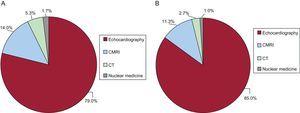
Based on the phrase “imaging technique” specifically appearing in the title (Figure 1A), a search of bibliographic databases and more than 6000 journals (Appendix) shows that 53 864 scientific papers were specifically devoted to imaging techniques as the main topic of the article. Most of these scientific papers were dedicated to echocardiography (79.0%), followed by CMRI (14.0%), CT (5.3%), and nuclear medicine techniques in cardiology (1.7%).

A, distribution of articles on imaging techniques from their first appearance in the literature to the present based on all journals included in Scopus. Total number of articles: 53864. B, distribution of articles on imaging techniques from their first appearance in the literature to the present based on the most important cardiology journals included in Scopus. Total number of articles: 9100. CMRI, cardiac magnetic resonance imaging; CT, computed tomography.
However, a search of the 6 most important scientific journals on cardiology (Circulation, American Journal of Cardiology, Journal of the American College of Cardiology, European Heart Journal, Heart, and American Heart Journal) shows that 167 022 scientific papers were published in the last 60 years of which 9100 (5.44%) were specifically dedicated to cardiac imaging techniques (Figure 1B): echocardiography (85%), CMRI (11.3%), CT (2.7%), and nuclear medicine techniques in cardiology (1%). There are very few differences between the percentage of articles dedicated to each technique in medical journals and those in cardiology journals, although the percentage of articles dedicated to CMRI and multidetector CT is slightly higher in cardiology journals.
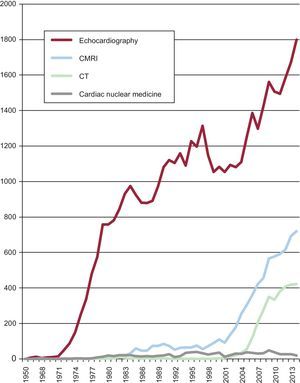
EVOLUTION OF SCIENTIFIC ARTICLES ON CARDIAC IMAGING TECHNIQUESIn addition to determining the number of articles on each of the 4 diagnostic imaging techniques, it is also important to know how these figures have evolved over time. Beginning in the early 1950s, echocardiography was the first cardiac imaging technique to be addressed in the literature. Exponential growth in articles dedicated to this topic began in the 1970s (Figure 2), rising to the current rate of 1800 per year. Articles on CMRI began to increase at the beginning of the new millennium and reached a peak of 700 articles this year (2014) whereas the literature on CT in cardiology began to increase around 2005 and reached > 400 articles this year (2014). However, the number of articles on nuclear medicine has remained steady since the 1980s with little variation.

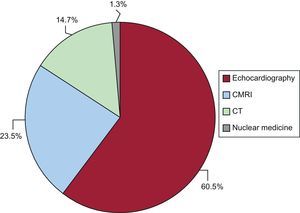
The number of articles on each of the four techniques published in the last 5 years sheds light on the growth in knowledge in these areas. There was a total of 12 958 publications in this period (Figure 3) and although most of the articles were on echocardiography (60.5%) the percentage significantly decreased (Figure 1A), whereas the number of articles on CMRI (23.5%) and CT (14.7%) sharply increased. In contrast, the number of articles on nuclear medicine techniques remained steady (1.3%). Thus, despite the recent decrease, echocardiography remains the most published topic in cardiac imaging techniques.
THE CASE OF ECHOCARDIOGRAPHY
Due to its long development period, echocardiography provides a good example by which to assess the bibliographic evolution of diagnostic procedures and their variants.
Of the articles published in all medical journals, 4.6% (7699 of 167022) were specifically devoted to echocardiography in the leading cardiology journals over their publishing lifetime. This figure shows the importance of the technique within the cardiology literature. It is worth recalling that the technique was the main focus of the studies, rather than simply being a tool referred to in passing, and that the name of the technique formed part of the title.
The percentage of articles published has changed since the technique was introduced in the early 1960s. Thus, an analysis of cardiology journals (Figure 4) shows that after an initial peak in 1983 a maximum plateau was reached between 1991 and 1997, which slowly decreased until the present. Figure 4 shows an example of how scientific knowledge and any new technology in a given area develop over time: a slow beginning followed by rapid development, then a plateau phase of knowledge, and finally a decline in original scientific contributions.

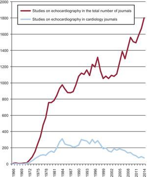
Analysis of echocardiography journals. There has been a steady increase in the number of articles published in the scientific literature worldwide up to the present. In contrast, there has been a steady decrease, particularly from 2000 onward, in the number of articles on imaging techniques published in cardiology journals.
There were 42 571 references to echocardiography in the entire medical literature worldwide. It is noteworthy that there was steady continuous growth in echocardiography articles, which reached a peak of more than 1700 articles in 2013 (Figure 4). The apparent paradox of a steady increase in the number of articles on echocardiography in the medical literature and a decrease in cardiology journals is a clear indication of the diffusion of echocardiography to fields other than cardiology, such as internal medicine, intensive care, and anesthesiology.
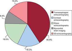
THE PROPORTION OF DIFFERENT ECHOCARDIOGRAPHIC TECHNIQUES IN THE LITERATUREThe number of articles on different echocardiographic techniques has evolved and changed over time (Figure 5). In total, 14 097 articles were devoted to different echocardiographic techniques: transesophageal echocardiography (40.9%), stress echocardiography (18.0%), contrast echocardiography (16.3%), three-dimensional echocardiography (15.4%), and myocardial strain imaging (9.4%). These figures serve as a good indication that in an increase in information is not necessarily equivalent to an increase in new knowledge; thus, although contrast echocardiography and stress echocardiography reach almost the same percentages, the former technique has had little impact on cardiac imaging laboratory practice.

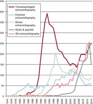
The fastest growth in articles dedicated to these techniques was in transesophageal echocardiography (Figure 6), which reached a peak in 1993 with > 400 articles. This figure reflects the tremendous impact this technique has had on clinical practice since its initial development. There was a gradual decrease from 1993 to 2006 followed by renewed growth that was clearly due to the increase in articles on realtime three-dimensional transesophageal echocardiography; since 2006, this technique has had a great impact on cardiology. The number of articles on myocardial strain imaging reached a peak of 265 articles in 2012 followed by a sharp decrease in the last 2 years. These data not only reflect the level of interest in this technique but also the problems involved in introducing this methodology into clinical practice.

A technological change in diagnostic methods is sometimes easy to introduce and to gain acceptance, whereas at other times even large numbers of scientific publications on the method may not lead to it becoming established in clinical diagnosis.
CARDIAC IMAGING JOURNALSThere are a great many journals dedicated to each of the cardiac imaging techniques. Obviously, the articles focus on each of the techniques. For example, journals specifically dedicated to echocardiography are the Journal of the American Society of Echocardiography and Echocardiography. Although these journals generally offer substantial and valuable information, they are biased toward their topic of interest rather than to more general topics. Three cardiology imaging journals are of special interest: the Journal of the American College of Cardiology Imaging and Circulation Imaging, both first issued in 2008, and the European Heart Journal of Cardiovascular Imaging (formerly the European Journal of Echocardiography), which was issued in 2012. Despite being specialist journals and having a short publishing history, they are representative of a change in the literature indicative of the revolution that has taken place over the last 6 years in the organization of cardiology services: the birth of cardiac imaging laboratories and the close collaboration between different medical specialties such as cardiology and radiology.
These 3 journals have published 4232 scientific articles on cardiac imaging: echocardiography (43%), CMRI (18%), and CT (10%); the remaining articles are dedicated to comparisons of techniques or a variety of other topics.
A BRIEF NOTE ON CARDIAC IMAGING IN REVISTA ESPAÑOLA DE CARDIOLOGÍASince its first issue, Revista Española de Cardiología has published 8151 original articles; 435 articles (5.33%) were specifically devoted to imaging techniques, which is roughly equal to the percentage of articles dedicated to imaging techniques in the literature. Since the Spanish authors publish articles in Revista Española de Cardiología and in other journals, the number of articles on each technique published in this journal was not analyzed as this would have introduced significant bias and the conclusions would not reflect the actual situation.
The “Update” in cardiac imaging section was introduced in 2006,22 which indicates a change in perspective among Spanish cardiologists and reflects a time in which cardiac imaging is beginning to be considered a subspecialty that includes all the non-invasive diagnostic techniques in cardiology.
This brief review of the literature on cardiac imaging techniques provides a glimpse into how much effort is still needed before they become incorporated into clinical practice. It also indicates the different “fashions” which, although sometimes lacking immediate clinical impact, are necessary for the continuing development of imaging techniques such that diagnosis can be improved to the betterment of the cardiac patient.
CONFLICTS OF INTERESTNone declared.
I wish to thank Maria Francisca Abad García from the Department of History of Science and Documentation, Faculty of Medicine, Valencia, Spain for her invaluable help in information search and analysis.
The search for bibliographic information on a specific topic is complex and subject to search strategies that should be well defined to provide a basis for valid scientific conclusions: we used the Scopus database (Elsevier). Scopus includes data on 20 500 journals from 500 international publishers. In total, 33% of these journals are dedicated to biomedicine and health sciences. The database includes 6400 biomedical journals and all the journals included in MEDLINE.23
It is very important to define the search strategy, because the results can change according to the definition. If we wish to analyze the number of publications on a specific technique different methods can be followed:
- 1.
General search based on the name of the technique appearing in the abstract, but not in the title. For example, this type of search locates the studies by Alcíbar et al24 and Ruiz García et al25; the former investigated the usefulness of elective implantation of covered stents for coarctation and recoarctation, and the latter studied percutaneous balloon pericardiectomy. In both cases the echocardiographic technique is systematically used to assess cardiac function or the presence of effusion and is thus mentioned in the abstract; since the technique is secondary to the main topic no new knowledge or information is provided on the imaging technique itself.
- 2.
General search based on the name of the technique appearing the main text, but not in the abstract or title: this type of search locates the study by Morillas et al,26 which investigated inflammation and apoptosis in patients with hypertension. Once again, the echocardiography technique is secondary to the main topic and is referred to in the main text but not in the title, as in the case of Alcibar et al,24 or in the abstract, as in the case of Ruiz García et al.2
- 3.
Search based on the name of the technique appearing in the title. We used this method as it conforms to the aims of our search strategy. For example, this search strategy locates the study by De la Morena et al,27 which specifically investigated exercise Doppler echocardiography in hypertrophic cardiomyopathy patients; obviously, this is the type of search that is of real interest to us because this specific technique was the main topic of the study. In this case, the study was categorized in the echocardiography group and in the subgroup exercise echocardiography. In relation to CMRI, the method located the study by Gran et al,28 which investigated the role of CMRI in the diagnosis of myocarditis in children; the name of the technique appeared in the title, abstract, and main body of the study.
The results of a search may be duplicated if the names of 2 techniques appear in the title, as in the study by Delgado et al,29 which investigated the assessment of myocardial ischemia using CT and CMRI. The duplication of search results could slightly change the percentages described.
In this paper, the search included the period up to June 2014 and an estimate of the second half of the year was made, based on the tendency found in annual bibliographic production.