
ISSN: 1885-5857 Impact factor 2024 4.9
Vol. 78. Num. 2.
Pages 117-126 (February 2025)

Original article
Invasive assessment of coronary microvascular dysfunction and cardiovascular outcomes across the full spectrum of CHD: a meta-analysis

Evaluación invasiva de la disfunción microvascular coronaria y resultados cardiovasculares en todo el espectro de la EC: un metanálisis

Yang XuaXiaochen LiuaYingying GuoaYuyao QiuaYushi ZhangaXiao WangabShaoping Niea
https://doi.org/10.1016/j.rec.2024.05.007

Options

Supplementary data
Imagen extra
Rev Esp Cardiol. 2025;78:117-26
Abstract
Introduction and objectives

Coronary microvascular dysfunction (CMD) is highly prevalent and is recognized as an important clinical entity in patients with coronary heart disease (CHD). Nevertheless, the association of CMD with adverse cardiovascular events in the spectrum of CHD has not been systemically quantified.

Methods

We searched electronic databases for studies on patients with CHD in whom coronary microvascular function was measured invasively, and clinical events were recorded. The primary endpoint was major adverse cardiac events (MACE), and the secondary endpoint was all-cause death. Estimates of effect were calculated using a random-effects model from published risk ratios.

Results

We included 27 studies with 11 404 patients. Patients with CMD assessed by invasive methods had a higher risk of MACE (RR, 2.18; 95%CI, 1.80-2.64; P<.01) and all-cause death (RR, 1.88; 95%CI, 1.55-2.27; P<.01) than those without CMD. There was no significant difference in the impact of CMD on MACE (interaction P value=.95) among different invasive measurement modalities. The magnitude of risk of CMD assessed by invasive measurements for MACE was greater in acute coronary syndrome patients (RR, 2.84, 95%CI, 2.26-3.57; P<.01) than in chronic coronary syndrome patients (RR, 1.77, 95%CI, 1.44-2.18; P<.01) (interaction P value<.01).

Conclusions

CMD based on invasive measurements was associated with a high incidence of MACE and all-cause death in patients with CHD. The magnitude of risk for cardiovascular events in CMD as assessed by invasive measurements was similar among different methods but varied among CHD populations.

Keywords

Coronary heart disease
Coronary microvascular dysfunction
Major adverse cardiovascular events
Death
INTRODUCTION

Over the past few years, a growing body of evidence has underscored the pathophysiological role of coronary microvascular dysfunction (CMD) in a variety of cardiovascular diseases.1 CMD is thought to be a major cause of myocardial ischemia in patients without obstructive coronary heart disease (CHD), as well as several other conditions, such as obstructive CHD and heart failure, especially heart failure with preserved ejection.2 Several noninvasive and invasive techniques can be used to evaluate CMD. CMD assessed by noninvasive imaging is widely used and strongly associated with an increased risk of adverse cardiovascular outcomes in a wide range of pathological processes.3

Invasive functional assessment of CMD requires investigation of vasodilator microvascular response, including coronary flow reserve (CFR), index of microvascular resistance (IMR), and resistive reserve ratio, etc. Recently, coronary angiography-based IMR without the need for a vasodilator has also emerged.4 Assessing coronary microvascular function in catheterization is recommended for patients with CHD,5,6 bit its practical application remains limited. The efficacy of different measurement modalities in predicting adverse cardiovascular outcomes is inconsistent, and there is a lack of comparison among different CHD populations. Thus, a systematic evaluation of the prognostic value of diverse invasive assessments in various populations is warranted.

In this study, we performed a systematic review and meta-analysis of all studies evaluating the association of CMD with cardiovascular events or death in a broad range of CHD.

METHODSData source and search strategies

This meta-analysis was elaborated in accordance with the PRISMA guidelines.7 Two reviewers (Y. Xu and X. Liu) independently performed the online searches. Studies on CHD and cardiovascular events were acquired by searching PubMed, EMBASE, and the Cochrane Library from the inception date to January 2023. The full search strategy is provided in data 1 of the supplementary data. References from reviews, selected articles, and previous meta-analysis were also screened.

Study selection

Two investigators (Y. Xu and X. Liu) reviewed the selected publications to assess their eligibility, with disagreements resolved through discussion with a senior author (X. Wang). We included studies that assessed CMD with invasive modalities and reported a risk ratio (RR) for major adverse cardiac events (MACE) and/or all-cause death. Indices of invasive coronary microvascular function measurement included CFR via Doppler or thermodilution, IMR via thermodilution, hyperemic microvascular resistance via Doppler, resistive reserve ratio, and angiography-based index. The inclusion criteria were as follows: a) evaluation of coronary microvascular function by invasive methods; b) patients with proven or suspected CHD; c) not less than 1 year of follow-up; d) clear definition of the cutoff value of CMD. The exclusion criteria were as follows: a) no follow-up regarding cardiovascular outcomes; b) studies with duplicate publication; c) RR or hazard ratio (HR) were not given in the study.

Data extraction

Two independent reviewers (Y. Xu and X. Liu) extracted the following data from all the eligible studies on a standardized electronic form, including baseline characteristics, risk factors such as hypertension, smoking, diabetes mellitus, dyslipidemia, and the cutoff values of abnormal CMD. We extracted the event rates, RR, and their associated 95% confidence intervals (95%CIs) from the main text and supplementary material. If studies investigated more than 2 measurement methods (eg, CFR and IMR) and provided RR for each, they were considered separate studies for the purpose of the meta-analysis. As far as possible, we chose the adjusted HR as the data source. If there was no HR in the text and supplementary material, the RR value was calculated by the original data.

Risk of bias assessment

Two reviewers (Y. Xu and X. Liu) independently assessed the risk of bias. Based on the categories of selection (4 items: representativeness of the exposed cohort, selection of the nonexposed cohort, ascertainment of exposure, demonstration that outcome was not present at the study start), comparability (2 items: controls for the most important factor and any additional factors), and outcome (3 items: assessment, duration, and adequacy of follow-up), a quality score was calculated for each item (table 1 of the supplementary data).

Outcomes

The primary endpoint was the rate of MACE. The secondary endpoint was the rate of all-cause death. There were several definitions of MACE (table 2 of the supplementary data). We adopted the definition used in each study, including all-cause or cardiac death, myocardial infarction, rehospitalization for heart failure or unstable angina, and revascularization.

Data synthesis and analysis

We performed a random-effects meta-analysis using inverse-variance weighting (expressed as RR with 95%CI), using the natural logarithm of the RR and standard errors. The RR and 95%CI were presented as forest plots. Review Manager (RevMan) 5.4 was used for the statistical analysis. A 2-sided P<.05 was considered statistically significant. Heterogeneity was assessed using the I2 test. An I2 of 0% to 25%, 26% to 50%, and >50% was considered as low, medium, and high statistical heterogeneity, respectively. We performed subgroup analyses by different measurement modalities and CHD populations (eg, chronic coronary syndrome [CCS] or acute coronary syndrome [ACS]). In addition, studies in which most participants presented with stable CHD were also classified as the CCS group. Subgroup analyses according to the study design (retrospective vs prospective), follow-up duration ( ≤ 36 months vs >36 months), and IMR measurement methods (wire-based vs coronary angiography-based) were also conducted. In addition, we conducted meta-regression to explore whether the magnitude of risk would differ across different measurement modalities and CHD populations and the interaction P values were calculated. Sensitivity analyses were conducted to evaluate the effect of each individual study on the pooled estimates. Publication bias was examined using the funnel plot and the Egger's test. If P<.05, the trim-and-fill method was used to evaluate the impact of publication bias on the results of the study. Meta-regression, sensitivity analyses, and publication bias were all performed by Stata SE 17. An interaction P value<.017 after Bonferroni correction was considered statistically significant.

RESULTSStudy selection and characteristics

A total of 27 studies8–34 with 11 404 patients met the inclusion criteria (figure 1 of the supplementary data, figure 1). The characteristics of the included studies are summarized in table 3 of the supplementary data. Coronary microvascular function was assessed by CFR (15 studies), IMR (16 studies), resistive reserve ratio (3 studies), and hyperemic microvascular resistance (1 study). The median length of follow-up ranged from 12 to 135.6 months. The main inclusion criteria for all study populations are summarized in table 4 of the supplementary data. The average age was between 51.4 and 67 years, and there was a preponderance of men. The characteristics of the included patients are summarized in table 5 of the supplementary data.

Figure 1.

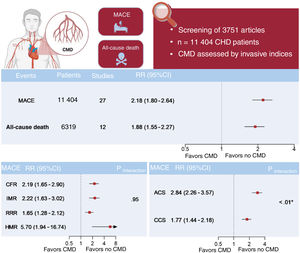
Central illustration. Meta-analysis of invasive assessment of coronary microvascular dysfunction and cardiovascular outcomes. After screening 3751 articles, a meta-analysis of 27 studies with 11 404 CHD patients was performed. CMD was assessed by invasive indices. The primary endpoint was MACE, and the secondary endpoint was all-cause death. CMD was associated with a higher risk of MACE and all-cause death. Subgroup analysis showed that the strength of the impact of CMD on cardiovascular events was similar among various measurement modalities but was greater in ACS patients than in CCS patients. *Significantly different. Created using BioRender.com. 95%CI, 95% confidence interval; ACS, acute coronary syndrome; CCS, chronic coronary syndrome; CFR, coronary flow reserve; CHD, coronary heart disease; CMD, coronary microvascular dysfunction; HMR, hyperamic microvascular resistance; IMR, index of microvascular resistance; IV, inverse-variance; MACE, major adverse cardiac events; RR, risk ratio; RRR, resistive reserve ratio.

(0.32MB).
Impact of CMD on MACE and all-cause death

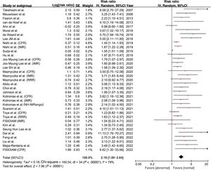
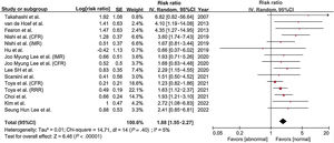
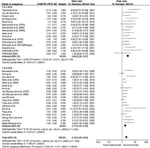
All 27 studies reported the incidence of MACE. Patients with CMD had a higher risk of MACE than those without CMD (RR, 2.18; 95%CI, 1.80-2.64; P<.01) (figure 2). There was wide heterogeneity among the included studies (I2=79%; P<.01). A total of 15 studies reported the incidence of all-cause death. The risk of all-cause death was higher in patients with CMD than in those without CMD (RR, 1.88; 95%CI, 1.55-2.27; P<.01) (figure 3). There was no evidence of heterogeneity among the included studies (I2=5%; P=.40).

Figure 2.

Forest plot for the risk of MACE. Data obtained from 27 studies using a random effects meta-analysis and expressed as risk ratios. 95%CI, 95% confidence intervals; CFR, coronary flow reserve; IMR, index of microvascular resistance; IV, inverse-variance; MACE, major adverse cardiac events; NH-IMRangio, nonhyperemic angiography-derived index of microcirculatory resistance; RRR, resistive reserve ratio; SE, standard error. References: Takahashi et al.20 Meuwissen et al.8 Fearon et al.9 van de Hoef et al.15 Ahn et al.33 Murai et al.23 de Waard et al.32 Lee JM et al.26 Nishi et al.10 Suda et al.21 Hu et al.14 Joo Myung Lee et al.27 Lee SH et al.25 Maznyczka et al.18 Abdu et al.34 Choi et al.17 Johnson et al.30 Kotronias et al.28 Scarsini et al.22 Toya et al.19 FISIOIAM.31 Kim et al.29 Seung Hun Lee et al.24 Dai et al.13 Feng et al.12 Liu et al.11 Mejia-Renteria et al.16

(0.58MB).
Figure 3.

Forest plot of the risk of all-cause death. Data obtained from 12 studies using a random effects meta-analysis and expressed as risk ratio. 95%CI, 95% confidence intervals; CFR, coronary flow reserve; IMR, index of microvascular resistance; IV, inverse-variance; RRR, resistive reserve ratio; SE, standard error. References: Takahashi et al.20 van de Hoef et al.15 Fearon et al.9 Nishi et al.10 Hu et al.14 Joo Myung Lee et al.27 Lee SH et al.25 Scarsini et al.22 Toya et al.19 Choi et al.17 Kim et al.29 Seung Hun Lee et al.24

(0.45MB).
Subgroup analysis by measurement modalities

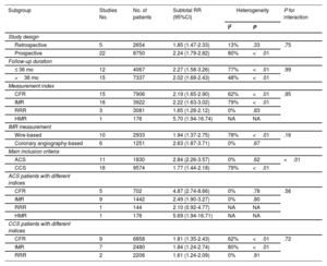
The association of CMD with MACE was consistent among all measurement modalities (figure 4), with significant risk in patients using CFR (RR, 2.19; 95%CI, 1.65-2.90; P<.01), IMR (RR, 2.22; 95%CI, 1.63-3.02; P<.01), resistive reserve ratio (RR, 1.65; 95%CI, 1.28-2.12; P<.01), and hyperemic microvascular resistance (RR, 5.70; 95%CI, 1.94-16.74; P<.01). The heterogeneity among all groups was significant, indicating that the measurement methods were not the reason for the wide heterogeneity of the effect of CMD on MACE. There was no significant difference in the impact of CMD on MACE among different measurement modalities (interaction P value=.95). We observed similar results for the association of CMD with all-cause death. The effect of CMD on all-cause death was consistent among subgroups (figure 2 of the supplementary data). Similarly, there was no significant difference within distinct measurement modalities (interaction P value=.58). Subgroup analysis stratified by the IMR measurement method was conducted (figure 3 of the supplementary data). There was no significant difference in the impact of CMD on MACE among different IMR measurement methods (interaction P value=.16).

Figure 4.

Subgroup analysis of MACE stratified by measurement modalities. 95%CI, 95% confidence intervals; CFR, coronary flow reserve; HMR, hyperemic microvascular resistance; IMR, index of microvascular resistance; IV, inverse-variance; MACE, major adverse cardiac events; NH-IMRangio, nonhyperemic angiography-derived index of microcirculatory resistance; RRR, resistive reserve ratio; SE, standard error. References: Takahashi et al.20 Meuwissen et al.8 Fearon et al.9 van de Hoef et al.15 Ahn et al.33 Murai et al.23 de Waard et al.32 Lee JM et al.26 Nishi et al.10 Suda et al.21 Hu et al.14 Joo Myung Lee et al.27 Lee SH et al.25 Maznyczka et al.18 Abdu et al.34 Choi et al.17 Johnson et al.30 Kotronias et al.28 Scarsini et al.22 Toya et al.19 FISIOIAM.31 Kim et al.29 Seung Hun Lee et al.24 Dai et al.13 Feng et al.12 Liu et al.11 Mejia-Renteria et al.16

(0.95MB).
Subgroup analysis by coronary artery disease populations

In CMD vs no-CMD populations, the RR of MACE was consistent among different CHD populations (figure 5), with greater risk observed in ACS patients (RR, 2.84; 95%CI, 2.26-3.57; P<.01) compared with CCS patients (RR, 1.77; 95%CI, 1.44-2.18; P<.01). Heterogeneity within CCS subgroups was still significant, suggesting that the included population was not the source of the wide heterogeneity of CMD in MACE. The strength of the association of CMD on MACE differed among ACS and CCS subgroups (interaction P value<.01). The RR of all-cause death applied to CMD vs no-CMD was consistent among different CHD populations (figure 4 of the supplementary data), with no significant differences among subgroups (interaction P value=.32).

Figure 5.

Subgroup analysis of MACE stratified by CHD populations. 95%CI, 95% confidence intervals; ACS, acute coronary syndrome; CCS, chronic coronary syndrome; CFR, coronary flow reserve; CHD, coronary heart disease; IMR, index of microvascular resistance; IV, inverse-variance; MACE, major adverse cardiac events; NH-IMRangio, nonhyperemic angiography-derived index of microcirculatory resistance; RRR, resistive reserve ratio; SE, standard error. References: Takahashi et al.20 Meuwissen et al.8 Fearon et al.9 van de Hoef et al.15 Ahn et al.33 Murai et al.23 de Waard et al.32 Lee JM et al.26 Nishi et al.10 Suda et al.21 Hu et al.14 Joo Myung Lee et al.27 Lee SH et al.25 Maznyczka et al.18 Abdu et al.34 Choi et al.17 Johnson et al.30 Kotronias et al.28 Scarsini et al.22 Toya et al.19 FISIOIAM.31 Kim et al.29 Seung Hun Lee et al.24 Dai et al.13 Feng et al.12 Liu et al.11 Mejia-Renteria et al.16

(1.09MB).

We conducted subgroup analysis of different measurement modalities in ACS and CCS populations (figures 5-8 of the supplementary data). There was no significant difference in the impact of CMD on MACE among different measurement modalities in either the ACS (interaction P value=.56) and CCS populations (interaction P value=.72). Similarly, there was no significant difference in the impact of CMD on all-cause death in the ACS (interaction P value=.25) or CCS population (interaction P value=.78).

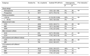
We also performed subgroup analyses with respect to MACE and all-cause death according to study design and length of follow-up (table 1 and table 2; figure 9-12 of the supplementary data). Additionally, we conducted a subgroup analysis based on the diverse definitions of study outcomes. Our findings revealed that the definition of MACE did not account for the heterogeneity observed in the impact of CMD on MACE, with consistent results noted among different subgroups (figure 13 of the supplementary data). Moreover, a subgroup analysis was performed for both the original data source and the HR directly extracted from the original text. Among these, 15 studies calculated RR values based on the original data of the article, while 20 studies directly extracted HR values from the article. Interestingly, we found that the data source did not influence the heterogeneity, with consistent outcomes noted among subgroups (figure 14 of the supplementary data). A summary of the study results is shown in the figure 1.

Table 1.

Subgroup analysis of MACE

Subgroup  Studies No.  No. of patients  Subtotal RR (95%CI)  HeterogeneityP for interaction 
        I2  P   
Study design             
Retrospective  2654  1.85 (1.47-2.33)  13%  .33  .75
Prospective  22  8750  2.24 (1.79-2.82)  80%  <.01 
Follow-up duration             
≤ 36 mo  12  4067  2.27 (1.58-3.26)  77%  <.01  .99
>36 mo  15  7337  2.02 (1.68-2.43)  48%  <.01 
Measurement index             
CFR  15  7906  2.19 (1.65-2.90)  62%  <.01  .95
IMR  16  3922  2.22 (1.63-3.02)  79%  <.01 
RRR  3081  1.65 (1.28-2.12)  0%  .83 
HMR  176  5.70 (1.94-16.74)  NA  NA 
IMR measurement             
Wire-based  10  2933  1.94 (1.37-2.75)  78%  <.01  .16
Coronary angiography-based  1251  2.63 (1.87-3.71)  0%  .67 
Main inclusion criteria             
ACS  11  1830  2.84 (2.26-3.57)  0%  .62  <.01
CCS  16  9574  1.77 (1.44-2.18)  79%  <.01 
ACS patients with different indices             
CFR  702  4.87 (2.74-8.66)  0%  .78  .56
IMR  1442  2.49 (1.90-3.27)  0%  .80 
RRR  144  2.10 (0.92-4.77)  NA  NA 
HMR  176  5.69 (1.94-16.71)  NA  NA 
CCS patients with different indices             
CFR  6858  1.81 (1.35-2.43)  62%  <.01  .72
IMR  2480  1.84 (1.24-2.74)  80%  <.01 
RRR  2206  1.61 (1.24-2.09)  0%  .91 

95%CI, 95% confidence interval; ACS, acute coronary syndrome; CCS, chronic coronary syndrome; CFR, coronary flow reserve; HMR, hyperemic microvascular resistance; IMR, index of microvascular resistance; MACE, major adverse cardiovascular event; mo, months; No., number; RR, risk ratio; RRR, resistive reserve ratio.

The I2 value indicates the percentage of variability across the pooled estimates attributable to heterogeneity beyond chance (0% to 25%, low likelihood; 26% to 50%, moderate likelihood; >50%, high likelihood), and the P value is a test of heterogeneity in each subgroup. After pooling the results from the same study a maximum of 3 times, an interaction P value <.017 after Bonferroni correction was considered statistically significant.

Table 2.

Subgroup analysis of all-cause death

Subgroup  Studies No.  No. of patients  Subtotal RR (95%CI)  HeterogeneityP for interaction 
        I2  P   
Study design             
Retrospective  2001  1.55 (1.22-1.98)  5%  .35  .04
Prospective  10  4317  2.37 (1.79-3.15)  0%  .76 
Follow-up duration             
≤ 36 mo  536  2.13 (0.35-12.84)  53%  .14  .54
>36 mo  10  5782  1.83 (1.52-2.20)  0%  .44 
Measurement index             
CFR  4030  2.35 (1.49-3.71)  45%  .09  .58
IMR  2482  1.87 (1.34-2.60)  0%  .71 
RRR  2937  1.76 (1.27-2.45)  0%  .39 
Main inclusion criteria             
ACS  972  2.27 (1.55-3.32)  0%  .41  .32
CCS  5346  1.76 (1.42-2.18)  4%  .41 
ACS patients with different indices             
CFR  212  4.66 (1.60-13.55)  0%  .68  .25
IMR  760  2.04 (1.36-3.07)  0%  .41 
CCS patients with different indices             
CFR  3818  2.09 (1.29-3.37)  49%  .10  .78
IMR  1722  1.57 (0.90-2.76)  0%  .73 
RRR  2937  1.76 (1.27-2.45)  0%  .39 

95%CI, 95% confidence interval; ACS, acute coronary syndrome; CCS, chronic coronary syndrome; CFR, coronary flow reserve; IMR, index of microvascular resistance; MACE, major adverse cardiovascular event; mo, months; No., number; RR, risk ratio; RRR, resistive reserve ratio.

The I2 value indicates the percentage of variability across the pooled estimates attributable to heterogeneity beyond chance (0% to 25%, low likelihood; 26% to 50%, moderate likelihood; >50%, high likelihood), and the P value is a test of heterogeneity in each subgroup. After pooling results from the same study a maximum of 3 times, an interaction P value <.017 after Bonferroni correction was considered statistically significant.

Publication bias assessment

We observed that the funnel plot for MACE was asymmetrical, suggesting the possible existence of publication bias (figure 15 of the supplementary data). Egger's test proved the possibility of publication bias (P < .01). Considering that publication bias might lead to instability or even reversal of the results, we used the trim-and-fill method to observe the effect of publication bias on this study. Thirteen additional studies were supplemented to control publication bias, the results of which showed that patients with CMD had a higher risk of MACE (RR, 1.69; 95%CI, 1.43-1.99; P<.01). This suggested that publication bias did not have a significant impact on this study. Using the same method, we found that the impact of publication bias of all-cause death was not significant.

Sensitivity analysis

Sensitivity analysis showed that the exclusion of any single study from the analysis for MACE did not alter the overall findings (figure 16 of the supplementary data). The sensitivity analysis of all-cause death showed similar results (figure 17 of the supplementary data).

DISCUSSION

The major findings of the present study are as follows. First, for patients with CHD, invasively-measured CMD was associated with a higher risk of MACE and all-cause death. Second, the predictive value of different measurement modalities, including CFR, IMR, resistive reserve ratio, and hyperemic microvascular resistance, was consistent for MACE and all-cause death. Third, the magnitude of risk for MACE differed among distinct CHD populations, with a higher risk observed in ACS patients than in CCS patients. A summary of the study results is shown in figure 1.

In patients with CHD, myocardial ischemia may be caused by epicardial vascular factors or microvascular factors. Epicardial stenosis contributes to reduced coronary blood flow reserve and progressively impairs myocardial perfusion. Likewise, structural or functional alterations in the microvasculature can lead to CMD, which can also affect myocardial perfusion as a major pathological mechanism of nonobstructive CHD. The development of invasive testing techniques has led to new insights into microvascular function, thus providing a more visual representation of vasodilatory capacity and microvascular resistance in patients with obstructive or nonobstructive CHD. Notably, the RR for mortality were lower than those found in other recent meta-analyses. Current noninvasive functional tests rely on detecting large regional differences in myocardial perfusion or regional wall motion abnormalities in the left ventricle. Most noninvasive functional tests cannot provide anatomical information to exclude obstructive CHD. Furthermore, prior research has focused more on the measurement of CFR, but the evaluation of CFR assessed the entire coronary circulation, only reflecting coronary microvascular function when there was no obvious epicardial vessel obstruction.35 This may have contributed to the discrepancy with our results. Moreover, prior studies noted that physiological assessment based on microvascular resistance was superior to CFR in patients with acute myocardial infarction treated with PCI.18,32,36 A possible explanation for this might be that all of these studies were conducted in the ACS population, in which most patients would develop structural CMD in an acute setting. In contrast, women, young people, and patients with fewer cardiovascular risk factors were more likely to develop functional CMD. Hence, the clinical value of simply comparing the superiority or inferiority of different modalities for the entire CHD population was limited.

Specifically, the present study showed that coronary angiography-based IMR (angio-IMR) had comparable predictive value with guidewire-based IMR. Angio-IMR has been developed recently and has shown good diagnostic accuracy compared with wire-based invasive IMR.37–39 This simple alternative index showed an association with the extent and progression of infarct pathology40,41 and was of prognostic importance.17,28 Our study supports its prognostic implications, but further studies should assess the superiority of this novel technology.

Few studies have evaluated the prognostic value of CMD in different CHD populations. The present meta-analyses shows that the magnitude of risk for MACE was higher in ACS patients (primarily myocardial infarction) than in CCS patients. A structural CMD, namely microvascular obstruction, may occur even after successful revascularization for ST-segment-elevation myocardial infarction.42 This might be attributed to damage to the endothelium by oxygen-free radicals, microvascular spasm, external compression, and accumulation of plaque debris following reperfusion.43 Compared with patients with CCS, ST-segment elevation myocardial infarction patients tended to have a higher degree of CMD, which was manifested as reduced CFR and increased microvascular resistance.44 With reference to cardiac enzymes and cardiac magnetic resonance imaging or positron emission tomography, an elevated IMR predicted a greater degree of myocardial injury in patients with ST-segment elevation myocardial infarction.45 There was less recovery of left ventricular function in patients over time with a high IMR, which in turn led to a worse long-term prognosis.46 In contrast, preserved coronary arteriolar vasodilator capacity and the development of collateral flow might prevent the occurrence of stress-induced myocardial ischemia in CCS.37–48 All of these factors might contribute to a higher risk of MACE in ACS patients with CMD compared with CCS patients. The evaluation of microvascular function after PCI may be more clinically practical in patients with ACS, especially ST-segment elevation myocardial infarction.

Limitations

There was heterogeneity in the assessment of the impact of CMD on MACE. Thus, we performed subgroup analyses based on measurement modalities, study population, study design, length of follow-up, outcome definition, and data sources, etc., and found the results were consistent across different subgroups. Furthermore, publication bias was unavoidable. We used the trim-and-fill method to observe the effect of publication bias and found that the impact of publication bias on this study was not significant. After Bonferroni correction, the risk of invasive CMD assessment in predicting MACE varied among different CHD populations. There is insufficient evidence to demonstrate invasive CMD assessment is associated with all-cause death in the CHD population. The limited number of studies assessing all-cause death compared with MACE, imbalances in the number of patients in the ACS and CCS groups, and the lower overall incidence of all-cause death may contribute to the lack of evidence.

CONCLUSIONS

In this meta-analysis, CMD was associated with an increased risk of MACE and all-cause death in CHD patients. The strength of the impact of CMD on cardiovascular events was similar among various measurement modalities but appeared to different in diverse CHD populations.

WHAT IS KNOWN ABOUT THE TOPIC?

  • CMD assessed by noninvasive imaging is widely used and strongly associated with an increased risk of adverse cardiovascular outcomes in a wide range of pathological processes.

WHAT DOES THIS STUDY ADD?

  • Invasively-measured CMD was associated with a higher risk of MACE and all-cause death in patients with CHD.

  • The predictive value of diverse invasive measurement modalities was consistent for MACE and all-cause death.

  • The magnitude of risk for MACE was greater in ACS patients than in CCS patients.

FUNDING

This study was funded by grants from National Key R&D Program of China (2022YFC2505600), Beijing Outstanding Youth Science Fund Project (JQ24039), Beijing Demonstration Research Ward Construction Project, 2020 Guangdong Provincial Medical Research Fund (A2022364).

ETHICAL CONSIDERATIONS

As this article is a meta-analysis, it did not require approval from an ethics committee or participating institutions. This article does not address the possible variables of sex and gender in accordance with the SAGER guidelines.

STATEMENT ON THE USE OF ARTIFICIAL INTELLIGENCE

No artificial intelligence was used in the preparation of this article.

AUTHORS’ CONTRIBUTIONS

Y. Xu oversaw conceptualization, methodology, software, data curation, and writing—original draft preparation. Y. Guo, Y. Qiu, and Y. Zhang were responsible for visualization and investigation. X. Liu: oversaw software, and validation. X. Wang, and S. Nie were in charge of writing—reviewing and editing.

CONFLICTS OF INTEREST

S. Nie has received research grants from the institution from Boston Scientific, Abbott, Jiangsu Hengrui Pharmaceuticals, China Resources Sanjiu Medical & Pharmaceuticals, and East China Pharmaceuticals. The remaining authors have no relevant relationships to disclose.

REFERENCES
[1]
S. Godo, A. Suda, J. Takahashi, S. Yasuda, H. Shimokawa.
Coronary Microvascular Dysfunction.
Arterioscler Thromb Vasc Biol., (2021), 41 pp. 1625-1637
[2]
M.G. Del Buono, R.A. Montone, M. Camilli, et al.
Coronary Microvascular Dysfunction Across the Spectrum of Cardiovascular Diseases: JACC State-of-the-Art Review.
J Am Coll Cardiol., (2021), 78 pp. 1352-1371
[3]
M.A. Kelshiker, H. Seligman, J.P. Howard, et al.
Coronary flow reserve and cardiovascular outcomes: a systematic review and meta-analysis.
Eur Heart J., (2022), 43 pp. 1582-1593
[4]
H. Ai, Y. Feng, Y. Gong, et al.
Coronary Angiography-Derived Index of Microvascular Resistance.
Front Physiol., (2020), 11 pp. 605356
[5]
J. Knuuti, W. Wijns, A. Saraste, et al.
ESC Scientific Document Group. 2019 ESC Guidelines for the diagnosis and management of chronic coronary syndromes.
Eur Heart J., (2020), 41 pp. 407-477
[6]
M. Gulati, P.D. Levy, D. Mukherjee, et al.
2021 AHA/ACC/ASE/CHEST/SAEM/SCCT/SCMR Guideline for the Evaluation and Diagnosis of Chest Pain: A Report of the American College of Cardiology/American Heart Association Joint Committee on Clinical Practice Guidelines.
Circulation., (2021), 144 pp. e368-e454
[7]
M.J. Page, J.E. McKenzie, P.M. Bossuyt, et al.
The PRISMA 2020 statement: an updated guideline for reporting systematic reviews.
BMJ., (2021), 372 pp. n71
[8]
M. Meuwissen, S.A. Chamuleau, M. Siebes, et al.
The prognostic value of combined intracoronary pressure and blood flow velocity measurements after deferral of percutaneous coronary intervention.
Catheter Cardiovasc Interv., (2008), 71 pp. 291-297
[9]
W.F. Fearon, A.F. Low, A.S. Yong, et al.
Prognostic value of the Index of Microcirculatory Resistance measured after primary percutaneous coronary intervention.
Circulation., (2013), 127 pp. 2436-2441
[10]
T. Nishi, T. Murai, G. Ciccarelli, et al.
Prognostic Value of Coronary Microvascular Function Measured Immediately After Percutaneous Coronary Intervention in Stable Coronary Artery Disease: An International Multicenter Study.
Circ Cardiovasc Interv., (2019), 12 pp. e007889
[11]
L. Liu, N. Dai, G. Yin, et al.
Prognostic value of combined coronary angiography-derived IMR and myocardial perfusion imaging by CZT SPECT in INOCA.
J Nucl Cardiol., (2023), 30 pp. 684-701
[12]
C. Feng, F.A. Abdu, A.Q. Mohammed, et al.
Prognostic impact of coronary microvascular dysfunction assessed by caIMR in overweight with chronic coronary syndrome patients.
Front Endocrinol (Lausanne)., (2022), 13 pp. 922264
[13]
N. Dai, W. Che, L. Liu, et al.
Diagnostic Value of Angiography-Derived IMR for Coronary Microcirculation and Its Prognostic Implication After PCI.
Front Cardiovasc Med., (2021), 8 pp. 735743
[14]
X. Hu, J. Zhang, J.M. Lee, et al.
Prognostic impact of diabetes mellitus and index of microcirculatory resistance in patients undergoing fractional flow reserve-guided revascularization.
Int J Cardiol., (2020), 307 pp. 171-175
[15]
T.P. van de Hoef, M. Bax, M. Meuwissen, et al.
Impact of coronary microvascular function on long-term cardiac mortality in patients with acute ST-segment-elevation myocardial infarction.
Circ Cardiovasc Interv., (2013), 6 pp. 207-215
[16]
H. Mejia-Renteria, D. Faria, J.M. Lee, et al.
Association between patient age, microcirculation, and coronary stenosis assessment with fractional flow reserve and instantaneous wave-free ratio.
Catheter Cardiovasc Interv., (2022), 99 pp. 1104-1114
[17]
K.H. Choi, N. Dai, Y. Li, et al.
Functional Coronary Angiography-Derived Index of Microcirculatory Resistance in Patients With ST-Segment Elevation Myocardial Infarction.
JACC Cardiovasc Interv., (2021), 14 pp. 1670-1684
[18]
A.M. Maznyczka, K.G. Oldroyd, J.P. Greenwood, et al.
Comparative Significance of Invasive Measures of Microvascular Injury in Acute Myocardial Infarction.
Circ Cardiovasc Interv., (2020), 13 pp. e008505
[19]
T. Toya, A. Ahmad, M.T. Corban, et al.
Risk Stratification of Patients With NonObstructive Coronary Artery Disease Using Resistive Reserve Ratio.
J Am Heart Assoc., (2021), 10 pp. e020464
[20]
T. Takahashi, Y. Hiasa, Y. Ohara, et al.
Usefulness of coronary flow reserve immediately after primary coronary angioplasty for acute myocardial infarction in predicting long-term adverse cardiac events.
Am J Cardiol., (2007), 100 pp. 806-811
[21]
A. Suda, J. Takahashi, K. Hao, et al.
Coronary Functional Abnormalities in Patients With Angina and Nonobstructive Coronary Artery Disease.
J Am Coll Cardiol., (2019), 74 pp. 2350-2360
[22]
R. Scarsini, M. Shanmuganathan, G.L. De Maria, et al.
Coronary Microvascular Dysfunction Assessed by Pressure Wire and CMR After STEMI Predicts Long-Term Outcomes.
JACC Cardiovasc Imaging., (2021), 14 pp. 1948-1959
[23]
T. Murai, T. Yonetsu, Y. Kanaji, et al.
Prognostic value of the index of microcirculatory resistance after percutaneous coronary intervention in patients with non-ST-segment elevation acute coronary syndrome.
Catheter Cardiovasc Interv., (2018), 92 pp. 1063-1074
[24]
S.H. Lee, D. Shin, J.M. Lee, et al.
Clinical Relevance of Ischemia with Nonobstructive Coronary Arteries According to Coronary Microvascular Dysfunction.
J Am Heart Assoc., (2022), 11 pp. e025171
[25]
S.H. Lee, J.M. Lee, J. Park, et al.
Prognostic Implications of Resistive Reserve Ratio in Patients With Coronary Artery Disease.
J Am Heart Assoc., (2020), 9 pp. e015846
[26]
J.M. Lee, K.H. Choi, D. Hwang, et al.
Prognostic Implication of Thermodilution Coronary Flow Reserve in Patients Undergoing Fractional Flow Reserve Measurement.
JACC Cardiovasc Interv., (2018), 11 pp. 1423-1433
[27]
J.M. Lee, K.H. Choi, J.H. Doh, et al.
Long-term Patient Prognostication by Coronary Flow Reserve and Index of Microcirculatory Resistance: International Registry of Comprehensive Physiologic Assessment.
Korean Circ J., (2020), 50 pp. 890-903
[28]
R.A. Kotronias, D. Terentes-Printzios, M. Shanmuganathan, et al.
Long-Term Clinical Outcomes in Patients With an Acute ST-Segment-Elevation Myocardial Infarction Stratified by Angiography-Derived Index of Microcirculatory Resistance.
Front Cardiovasc Med., (2021), 8 pp. 717114
[29]
J. Kim, D. Shin, J.M. Lee, et al.
Differential Prognostic Value of Revascularization for Coronary Stenosis With Intermediate FFR by Coronary Flow Reserve.
JACC Cardiovasc Interv., (2022), 15 pp. 1033-1043
[30]
N.P. Johnson, H. Matsuo, M. Nakayama, et al.
Combined Pressure and Flow Measurements to Guide Treatment of Coronary Stenoses.
JACC Cardiovasc Interv., (2021), 14 pp. 1904-1913
[31]
C.H. Flores, F. Diez-Delhoyo, R. Sanz-Ruiz, et al.
Microvascular dysfunction of the non-culprit circulation predicts poor prognosis in patients with ST-segment elevation myocardial infarction.
Int J Cardiol Heart Vasc., (2022), 39 pp. 100997
[32]
G.A. de Waard, G. Fahrni, D. de Wit, et al.
Hyperaemic microvascular resistance predicts clinical outcome and microvascular injury after myocardial infarction.
[33]
J.M. Ahn, F.M. Zimmermann, N.P. Johnson, et al.
Fractional flow reserve and pressure-bounded coronary flow reserve to predict outcomes in coronary artery disease.
Eur Heart J., (2017), 38 pp. 1980-1989
[34]
F.A. Abdu, L. Liu, A.Q. Mohammed, et al.
Prognostic impact of coronary microvascular dysfunction in patients with myocardial infarction with non-obstructive coronary arteries.
Eur J Intern Med., (2021), 92 pp. 79-85
[35]
B. de Bruyne, J. Bartunek, S.U. Sys, N.H. Pijls, G.R. Heyndrickx, W. Wijns.
Simultaneous coronary pressure and flow velocity measurements in humans. Feasibility, reproducibility, and hemodynamic dependence of coronary flow velocity reserve, hyperemic flow versus pressure slope index, and fractional flow reserve.
Circulation., (1996), 94 pp. 1842-1849
[36]
D. Carrick, C. Haig, N. Ahmed, et al.
Comparative Prognostic Utility of Indexes of Microvascular Function Alone or in Combination in Patients With an Acute ST-Segment-Elevation Myocardial Infarction.
Circulation., (2016), 134 pp. 1833-1847
[37]
G.L. De Maria, R. Scarsini, M. Shanmuganathan, et al.
Angiography-derived index of microcirculatory resistance as a novel, pressure-wire-free tool to assess coronary microcirculation in ST elevation myocardial infarction.
Int J Cardiovasc Imaging., (2020), 36 pp. 1395-1406
[38]
D.J. Maron, J.S. Hochman, H.R. Reynolds, et al.
Initial Invasive or Conservative Strategy for Stable Coronary Disease.
N Engl J Med., (2020), 382 pp. 1395-1407
[39]
D. Huang, Y. Gong, Y. Fan, et al.
Coronary Angiography-Derived Index for Assessing Microcirculatory Resistance in Patients with Non-Obstructed Vessels: The FLASH IMR Study.
Am Heart J., (2023), 263 pp. 56-63
[40]
D. Shin, J. Kim, K.H. Choi, et al.
Functional angiography-derived index of microcirculatory resistance validated with microvascular obstruction in cardiac magnetic resonance after STEMI.
Rev Esp Cardiol., (2022), 75 pp. 786-796
[41]
X. Wang, Q. Guo, R. Guo, et al.
Coronary Angiography-Derived Index of Microcirculatory Resistance and Evolution of Infarct Pathology after STEMI.
Eur Heart J Cardiovasc Imaging., (2023), 24 pp. 1640-1652
[42]
G. Niccoli, R.A. Montone, B. Ibanez, et al.
Optimized Treatment of ST-Elevation Myocardial Infarction.
Circ Res., (2019), 125 pp. 245-258
[43]
L.F. Robbers, E.S. Eerenberg, P.F. Teunissen, et al.
Magnetic resonance imaging-defined areas of microvascular obstruction after acute myocardial infarction represent microvascular destruction and haemorrhage.
Eur Heart J., (2013), 34 pp. 2346-2353
[44]
R.P. Betgem, G.A. de Waard, R. Nijveldt, A.M. Beek, J. Escaned, N. van Royen.
Intramyocardial haemorrhage after acute myocardial infarction.
Nat Rev Cardiol., (2015), 12 pp. 156-167
[45]
R. Scarsini, M. Shanmuganathan, R.A. Kotronias, et al.
Angiography-derived index of microcirculatory resistance (IMRangio) as a novel pressure-wire-free tool to assess coronary microvascular dysfunction in acute coronary syndromes and stable coronary artery disease.
Int J Cardiovasc Imaging., (2021), 37 pp. 1801-1813
[46]
R. McGeoch, S. Watkins, C. Berry, et al.
The index of microcirculatory resistance measured acutely predicts the extent and severity of myocardial infarction in patients with ST-segment elevation myocardial infarction.
JACC Cardiovasc Interv., (2010), 3 pp. 715-722
[47]
M. Di Carli, J. Czernin, C.K. Hoh, et al.
Relation among stenosis severity, myocardial blood flow, and flow reserve in patients with coronary artery disease.
Circulation., (1995), 91 pp. 1944-1951
[48]
N.G. Uren, J.A. Melin, B. De Bruyne, W. Wijns, T. Baudhuin, P.G. Camici.
Relation between myocardial blood flow and the severity of coronary-artery stenosis.
N Engl J Med., (1994), 330 pp. 1782-1788
Copyright © 2024. Sociedad Española de Cardiología