ISSN: 1885-5857 Impact factor 2024 4.9
Vol. 78. Num. 4.
Pages 358-367 (April 2025)

Review article
Present and future of aortic risk assessment in patients with heritable thoracic aortic diseases

Presente y futuro de la evaluación del riesgo aórtico en pacientes con enfermedad aórtica hereditaria

Gisela Teixido-TuraabcLydia Dux-SantoybClara BadiaabJavier LimeresabcAndrea GualabcArtur Evangelista MasipdIgnacio Ferreira-GonzálezabefJosé Rodríguez-Palomaresabcf
https://doi.org/10.1016/j.rec.2024.10.009

Options

Imagen extra
Rev Esp Cardiol. 2025;78:358-67
Abstract

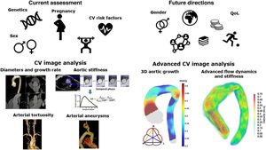
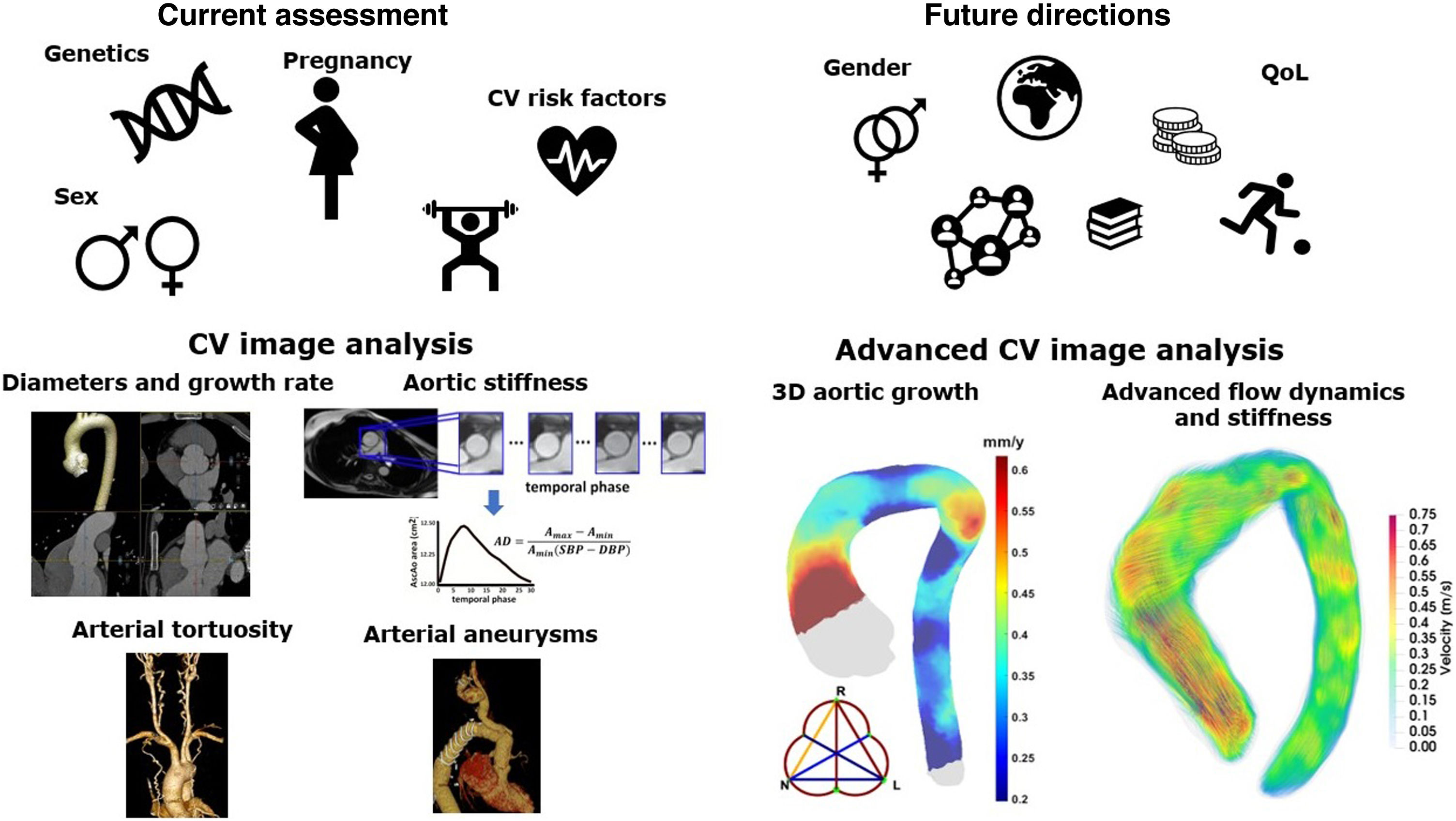
Heritable thoracic aortic diseases (HTAD) are a group of diverse genetic conditions characterized by an increased risk of aortic complications. The standard surveillance of these patients involves monitoring aortic diameters until a defined threshold is reached, at which point preventive aortic surgery is recommended. However, assessing aortic risk in these patients is far more complex and, in many aspects, remains incompletely understood. Several factors contribute to this complexity, including the diversity and low prevalence of the conditions within HTAD and the limited understanding of the factors influencing the progression of aortic dilation and the advent of acute aortic events. This article reviews current knowledge on clinical, genetic, and imaging factors related to aortic risk in HTAD and explores their potential future roles in improving risk assessment. By advancing our understanding of these factors, we aim to enhance the precision of risk stratification and develop more effective, personalized management strategies for HTAD patients, with the final goal of improving clinical outcomes and quality of life in individuals affected by these genetic disorders.

Keywords

Marfan
Aorta
Genetics
INTRODUCTION

Heritable thoracic aortic diseases (HTAD) are a group of genetic conditions with an increased risk of aortic complications. Classically, they have been divided into syndromic and nonsyndromic entities, with Marfan (MFS), Loeys-Dietz (LDS) and vascular Ehlers-Danlos (vEDS) syndromes being the most frequent syndromic forms. The nonsyndromic group is defined as a familial presentation of thoracic aorta disease without extracardiovascular involvement. In this scenario, genetic testing is able to identify a pathogenic genetic variant in around 30% of cases.1

The standard care of HTAD includes periodic aortic imaging, with transthoracic echocardiography being pivotal for this purpose. However, complete thoracoabdominal imaging of the aorta is advised at regular intervals due to the risk of dilation or dissection distal to the ascending aorta, and potential involvement of aortic branch arteries.2,3

Advancements in molecular genetics and familial screening, along with expert monitoring with imaging techniques and aortic surgery, have greatly improved patient prognosis.4,5 In particular, the incidence of type A aortic dissection has decreased among patients receiving an accurate diagnosis and proper follow-up. However, in recent years, type B dissections have been increasingly prevalent, becoming more common than type A in diagnosed patients.6,7

Although aortic root diameter is a reliable risk marker for type A acute aortic syndrome,8,9 resulting in clinical guidelines recommending root replacement based on this parameter,10,11 there is a lack of effective markers for predicting rapid aortic dilation in individual patients. Furthermore, descending aorta diameter is not a good prognostic marker for type B dissection, as it often falls within the range of mild dilation prior to the dissection, limiting its usefulness as a risk discriminator.6,7

The search for optimal aortic diameter thresholds for elective surgery and other risk markers for aortic dissection beyond diameter remains ongoing. This review will examine the limitations of using aortic diameters as risk markers for complications in HTAD and expand the perspective of risk assessment to include additional biomarkers that may refine the aortic prognosis in these patients (figure 1).

Figure 1.

Central illustration. Present and future of aortic risk assessment in patients with heritable thoracic aortic diseases. Current risk stratification relies on genetic analysis and conventional risk factors, such as hypertension, sex, and pregnancy-related risks. Although the aorta is a dynamic 3D structure, imaging assessment is typically limited to a few anatomical levels for determining diameters and stiffness. Future risk stratification could be enhanced by incorporating factors such as gender, socioeconomic status, QoL, and advanced imaging analysis. Three-dimensional geometrical mapping, 4D flow-derived descriptors, and extraaortic vascular involvement can provide valuable insights. A, area; AD, aortic distensibility; CV, cardiovascular; DBP, diastolic blood pressure; QoL, quality of life; SBP, systolic blood pressure.

(0.38MB).
AORTIC DIAMETERS

Aortic root diameters have been shown to be reliable risk biomarkers for type A dissection in MFS.8,9 Milleron et al.9 investigated the occurrence of aortic events in 954 patients with MFS who were followed up at specialized centers and had no prior aortic surgery or dissection. They found an annual risk of type A dissection below 0.05% for individuals with aortic root diameters <50mm. Less information is available for LDS patients. In a multicenter study12 that included patients with TGFBR1 and TGFBR2 variants, aortic diameter was below 45mm in 7 out of 31 patients (23%) 1 year before or at the time of type A dissection. Interestingly, all these patients were female, and 6 out of the 7 had TGFBR2 variants. In addition, in SMAD3-related LDS, dissections have been reported at diameters <50mm.13 For nonsyndromic HTAD, in which ACTA2 variants are the most frequently identified, preventive root replacement is indicated at 45mm diameter,11,14 since dissections have also been reported at diameters below 50mm.15

Therefore, HTAD patients are monitored with aortic imaging, and aortic root replacement is recommended once a specific diameter threshold is reached.10,11 Despite this, predicting the onset and rate of aortic dilation remains challenging, making it difficult to anticipate when aortic root replacement will be necessary. Furthermore, caution is required in the evaluation of aortic diameters, and specific guidelines should be followed to prevent measurement error or misinterpretation of rapid growth.16 Specifically, for transthoracic echocardiography, ascending aortic diameters should be measured in the parasternal long axis, using the leading-edge to leading-edge method in telediastole.16 For cardiac computed tomography (CCT) or magnetic resonance (CMR) angiography, electrocardiographic gating is recommended, especially for measuring the aortic root. Additionally, double-oblique planning is advised to obtain images perpendicular to the aortic centerline, and the inner-to-inner edge approach should be used for better correlation with echocardiography.17 Particular care is required when measuring aortic root diameters due to their nontubular shape, and it is recommended to measure the 3 cusp-to-cusp diameters.16 General caution is advisable when comparing diameters from different evaluations (ie, different time points or imaging techniques), since rapid aortic growth (> 3mm/y) is a risk marker that might prompt aortic root replacement.10,11

The close surveillance and indication for elective root/ascending aortic replacement have resulted in a scenario where type B dissection is becoming more common than type A.7 This is also influenced by the fact that aortic diameter is a poor predictor of type B dissection, rendering surgical prevention strategies unfeasible. Several studies reported that aortic diameters preceding type B dissection are in the range of mild dilation.6,7 Therefore, our understanding of predicting type B dissection in HTAD patients remains limited, leaving ample room for further investigation.

RISK FACTORS AND CLINICAL CONDITIONSSex and gender

Few studies have explored the influence of sex and gender on cardiovascular events among MFS patients. Initial reports of peripartum ascending aorta dissection or rupture suggested that women with MFS might face a higher risk of aortic dilation and events. However, subsequent publications have consistently shown that men exhibit more aggressive aortic manifestations, with a higher occurrence of aortic events (including type A and B dissection and aortic surgery), typically at a younger age than women.18–20 Further studies on genotype/phenotype correlations suggest that the observed sex differences are unrelated to specific genotype.21,22

The pathophysiological mechanisms underlying these differences remain incompletely understood, making it currently impossible to disentangle the effects of sex from gender. However, a few experimental studies using FBN1 mutant mouse models have suggested a protective role for elevated estrogen levels,23 which have been inversely related to aortic diameters, growth, and fragmentation of aortic wall elastic lamellae. This effect may be mediated by the inhibition of proteoglycan deposition and reduction in collagen and matrix metalloproteinase expression.

Less information is available about the impact of sex and gender on cardiovascular involvement in other syndromic and nonsyndromic HTAD. In LDS associated with TGFBR1 variants, male sex has been reported to be a risk factor for adverse aortic events. Conversely, female LDS patients with TGFBR2 variants experience a higher occurrence of type A dissections at diameters <45mm compared with men.12 Despite these findings, the underlying cause of this sex disparity remains elusive. Additionally, while male LDS patients with TGFB2 variants seem to have a higher aortic risk, there is no evidence of sex or gender differences in LDS patients carrying SMAD3 variants.24 In vEDS, male sex has been related to higher mortality due to vascular complications compared with women, especially among the youngest patients.25

In nonsyndromic HTAD patients carrying an ACTA2 variant, adverse aortic events were more prevalent in men than in women; however, the median age at the first event did not differ between sexes, and the type of aortic dissection (A vs B) was unaffected by sex.24 Sex or gender differences have not been reported in nonsyndromic HTAD attributed to SMAD3, MYLK, MYH11 and LOX, although studies on these genes are limited.24

Pregnancy in women with HTAD increases the risk of aortic complications compared with the general population and to the nonpregnant period.26–28 Type A dissections during pregnancy mostly occur in patients unaware of their MFS diagnosis,26,29 highlighting the critical importance of diagnosis and prepregnancy counseling. Type B dissections can occur in diagnosed patients, and their onset remains unpredictable.26,28,29 Furthermore, MFS women with prepartum aortic diameters between 40 and 45mm demonstrated stable aortic dimensions throughout pregnancy.26 Importantly, outside the peripartum period, MFS women who have ever been pregnant do not exhibit an increased aortic risk compared with those who have never been pregnant.26,30 Limited information exists on pregnancy-related risks in LDS,31,32ACTA233 and vEDS.34 The risk is likely higher in these syndromes than in MFS, especially in vEDS, which includes not only aortic but also nonaortic vascular complications and uterine rupture. Specialized prepregnancy counseling and shared decision-making are advised in women with HTAD who are planning pregnancy.

Quality of life and socioeconomic status

A diagnosis of HTAD can affect patients’ functional, mental, and social well-being.35,36 Although there is a growing body of literature examining quality of life (QoL) in MFS patients,37,38 studies on LDS and vEDS remain scarce.35,39,40 Most studies, typically based on patient-reported outcome measures, indicate that QoL is generally impaired in these patients,35,37–39 especially in women.36 Several socioeconomic factors, such as lower income and educational level,41,42 unemployment,36,41,42 and limited social support,42 have been associated with decreased QoL in MFS. This highlights the importance of assessing QoL alongside socioeconomic status to gain insights into the overall well-being of HTAD patients. Assessments should encompass symptom burden, functional status, psychological well-being, social and occupational functioning, treatment adverse effects, health care utilization, and patient satisfaction to enhance the understanding and management of HTAD.10

Physical activity

A sedentary lifestyle is a known cardiovascular risk factor in the general population, but evidence of its effects on HTAD is scarce. Similarly, the impact of physical exercise on the progression of aortic diameters and the risk of acute aortic syndrome in HTAD patients is unclear, preventing health care professionals from recommending exercise to these patients. However, 2 studies in murine models have suggested a potential benefit of low- to moderate-intensity aerobic exercise, which may reduce aortic elastin fiber fragmentation and growth.43,44

Few studies have focused on evaluating the impact of physical exercise on aortic structure and function in HTAD patients. Recent data on children and young adults with MFS suggest that daily physical activity, such as walking 10 000 steps a day, limits aortic root growth,45 and 2 studies have evaluated exercise rehabilitation programs in MFS.46,47 However, larger cohort studies are needed to validate these findings, and to ultimately guide more precise recommendations.

European clinical practice guidelines currently recommend that patients with HTAD avoid high- or very high-intensity exercise, as well as contact and power sports, even in the absence of aortic dilation.48 In contrast, skill sports, mixed sports, and low-intensity endurance sports are not considered risky, even in patients with moderate aortic dilation (40-45mm) or previous proximal aorta replacement.

Cardiovascular risk factors

Several cardiovascular risk factors, such as ageing, obesity,49 hypertension, diabetes, and dyslipidaemia; 11,50,51 as well as lifestyle characteristics, such as smoking,11 have been related to the presence and progression of ascending aortic aneurysms. As a result, current clinical guidelines10,11 recommend controlling modifiable risk factors. Specifically, the most recent guidelines10 advise aiming for a systolic blood pressure of ≤ 129mmHg (≤ 120mmHg if tolerated), a diastolic blood pressure of ≤ 79mmHg, and low-density lipoprotein cholesterol levels of ≤ 55mg/dL. They also emphasize the importance of optimizing diabetes treatment and smoking cessation in patients with aortic aneurysms, but specific recommendations for HTAD patients are lacking.

Blood biomarkers

Despite promising studies on the role of certain serum biomarkers52,53 in assessing aortic risk in MFS patients, these biomarkers have yet to be integrated into clinical practice due to the need for larger, more comprehensive studies to support their use. Nevertheless, this area of research holds significant promise and potential clinical utility.54

GENETIC PROGNOSTIC FACTORS

Significant progress has been made in understanding the genetic determinants of cardiovascular disease. This advancement has led to the integration of genetics into risk stratification for HTAD patients, influencing therapeutic decision-making and promoting the concept of precision medicine.

MFS is caused by pathogenic variants in the FBN1 gene, with over 3000 mutations identified to date. A clinically relevant aspect of FBN1 variants is their extremely high penetrance. However, significant phenotypic variability exists, even among family members sharing the same genetic variant. This variability is likely influenced by concurrent environmental or genetic modifying factors.

Advances in sequencing technology have enabled the genotyping of an increasing number of MFS patients, facilitating the establishment of genotype-phenotype correlations in FBN1 variants. Among the most recognized associations are: a) loss of cysteine, which is associated with a higher propensity for developing ectopia lentis55; b) variants generating premature stop codons, which are associated with lower rates of ectopia lentis, but a higher incidence of hyperlaxity, skin striae, and aortic events21; c) variants located within the first 15 exons, which show a higher incidence of isolated ectopia lentis compared with those located in other regions of the gene; and d) variants located in the last exons (59-65) that tend to be associated with milder forms of MFS, while those in the central region (exons 24-32) have been associated with severe forms.56

Not all FBN1 variants that cause MFS affect the fibrillin-1 protein in the same way. Missense variants can create or disrupt nonfunctioning domains, resulting in an abnormal protein, with these variants behaving as dominant negatives. Indel variants can lead to the expression of a shorter protein, also acting as a dominant negative, or may cause haploinsufficiency, where all encoded fibrillins are normal, but the available gene dosage is insufficient for normal connective tissue function. It is highly likely that different alterations occur depending on the type of variant, its location, and the disruption of regulatory mechanisms.

The classification of FBN1 variants based on their biological effects holds prognostic value: haploinsufficiency has been correlated with larger aortic diameters and growth, as well as an elevated risk of dissection and death.22,57 Haploinsufficient variants and missense variants that lose a cysteine residue have been associated with higher aortic risk compared with missense variants that gain a cysteine (low risk) or those that do not alter a cysteine residue (intermediate risk).21

Genotype-phenotype correlations may also influence medical management in LDS. The causal variants of LDS are primarily missense mutations, located mainly in the intracellular region of the TGFBR1 and TGFBR2 genes (specifically within the serine-threonine kinase domain). Few mutations have been described in the extracellular domains or in other genes affecting the TGF-β pathway, such as SMAD3, or the receptor ligands TGFB2 and TGFB3.58

An international multicenter registry that included 441 patients with TGFBR1 and TGFBR2 variants found that survival and aortic risk were similar for both variants.12 However, males with TGFBR1 had a higher risk than females, while these differences did not appear in TGFBR2 variants. However, women with TGFBR2 variants, marked systemic features, and low body surface area experienced aortic complications at lower aortic diameters (< 45mm), warranting earlier recommendation of aortic root replacement in this scenario.11 In vEDS, haploinsufficient (null) variants of COL3A1 have been associated with a better prognosis.25

Furthermore, a recent study24 found no significant differences in the cumulative risk of aortic events between patients with smooth muscle cell (SMC) variants and those with TGFβ-related variants. However, among patients with SMC variants, the highest risk was observed in PRKG1, followed by ACTA2 and MYLK variants. Similarly, in the TGFβ-related group, the cumulative risk was highest for TGFBR2 and lowest for SMAD3 variants. Interestingly, the pattern of the first aortic event also varied among HTAD genes: type A dissection was more frequent than elective surgery in patients with SMC genes (ACTA2, MYLK, and PRKG1) and SMAD3 variants, while elective surgery was more common in patients with TGFBR2 and TGFB2 variants. The frequency of both events was similar for TGFBR1 variants. Despite the limited number of patients with each variant studied, specific variants such as Arg179 in ACTA2 and Arg528 in TGFBR2 may be associated with a higher aortic risk.24

These genotype-phenotype correlations highlight the influence of genetic factors on the clinical and therapeutic management of patients with HTAD, enabling personalized aortic risk assessment and surveillance.

ADVANCED IMAGING FOR ANATOMIC, BIOMECHANIC AND HEMODYNAMIC AORTIC RISK ASSESSMENTAortic geometry beyond diameters

The shape of the diseased aorta is more intricate than previously thought, exhibiting not only a transverse growth but also elongation and more complex geometric alterations. Ascending aortic length has been identified as a risk factor for dissection in patients with ascending aortic aneurysms of all etiologies,59 with lengths greater than 11cm predicting adverse aortic events beyond diameter. In addition, aortic tortuosity has been linked to an increased risk of aortic events, including type B dissection, in MFS patients.6,60

Of note, vascular involvement in these patients extends beyond the aorta. In patients with MFS, LDS, or vEDS, increased vertebral artery tortuosity has been associated with a poorer aortic prognosis.61 Moreover, while uncommon in MFS, aortic branch aneurysms have been associated with a higher risk of aortic growth, even after adjusting for aortic diameter.2

Understanding and monitoring changes in aortic geometry and extraaortic vascular features could provide valuable insights into overall cardiovascular involvement in patients with HTAD and may aid in the stratification of aortic risk.

Advanced aortic growth quantification

Precision in measuring aortic diameter growth rates is crucial due to its pivotal role in patient management. Nonetheless, current assessments of aortic dimensions are subject to various sources of variability, affecting their reproducibility.62 Although reproducibility for aortic diameter measurements has been reported as excellent for both CCT and CMR,17,63,64 systematic studies on growth rate reproducibility are lacking.65 Only 2 studies have reported limited reproducibility of manual growth rate measurements on CCT and CMR angiography, especially in the aortic root.66,67 Such variability is expected due to potential inaccuracies in both baseline and follow-up diameter measurements, as well as comparisons of nonequivalent segments. Moreover, manual assessment restricts the number of levels at which aortic diameters are assessed, typically relying on predefined anatomical landmarks for reference. This limitation, combined with the failure of manual assessment to accurately capture aortic geometry in 3 dimensions, could lead to risk misestimation.

To address these limitations, various strategies have been proposed. Methods based on aortic segmentation have demonstrated excellent accuracy and reproducibility for measuring both tubular aorta68–71 and aortic root72 diameters. Aortic diameter growth can be further evaluated by applying deformable image registration to serial CCT67,73 or CMR66 angiography, enabling the calculation of 3D aortic diameter and growth maps (figure 2) with limited observer dependence.66,67

Figure 2.

Aortic geometry mapping in a patient with Marfan syndrome. A: baseline aortic mesh and aortic root plane with detected commissures in green. B: diameter maps and aortic root diameters at baseline and follow-up. C: growth rate map and root growth from automatically computed diameters.

(0.36MB).

In patients with aortic disease, including MFS, image registration-based measurements of aortic growth rates have proven to be substantially more reproducible than the current standard, both in the aortic root and in the tubular aorta.66,67 Notably, when applied to CCT angiography, the reproducibility at 6 months of follow-up was comparable to that achieved by manual assessment at 3 years of follow-up, opening the possibility of a shorter follow-up period for the assessment of growth.67 Moreover, the maximum rate of aortic growth is rarely located at the level of the pulmonary artery bifurcation, a typical anatomical landmark,67 and does not always occur at the site of maximum diameter.73,74 This highlights the usefulness of mapping 3D growth to estimate risk. Therefore, accurate local assessment of aortic growth can improve clinical management and enhance the understanding of aneurysm physiopathology.74 However, the precise value of this method in assessing the risk of aortic events in HTAD patients has yet to be determined.

Aortic biomechanics

The aorta serves not only as a conduit for blood flow but also as a buffer to reduce the pulsatile flow and pressure generated by left ventricular contraction, ensuring a steadier flow rate to peripheral circulation. Aortic elasticity is crucial for cardiovascular function, but it diminishes with age, which in turn affects cardiovascular health.50,75 Basic research demonstrating the underlying structural abnormalities of the aorta in patients with MFS76,77 prompted imaging studies aiming to identify whether these abnormalities were reflected in measurable elastic properties.78,79 Positive findings stimulated further research, particularly with the advent of CMR,80–85 which provides vast possibilities for assessing aortic biomechanics.86 While much attention has focused on MFS, efforts are not limited to this condition.82,87,88

Various stiffness parameters can be assessed using transthoracic echocardiography or CMR. Circumferential strain measures changes in aortic diameter or area throughout the cardiac cycle at a specific aortic level. When normalized by local pulse pressure, it yields aortic distensibility. This measurement is normally obtained via CMR but can also be measured by echocardiography, provided that there is no aortic motion through the imaging plane, and assuming that the aorta is circular. Of note, aortic distensibility measurement should use local pulse pressure, which is rarely available.89 Ascending aorta longitudinal strain evaluates the longitudinal stretch of the proximal aorta during the cardiac cycle due to left ventricular contraction.90,91 Pulse wave velocity, which refers to the velocity of propagation of pressure or flow waves, is typically measured with 2D phase-contrast CMR or tonometry, and can be regionally evaluated with 4D flow CMR. CMR-derived biomechanical descriptors have been validated against ex vivo mechanical tissue properties.92 Additionally, recent studies have linked distensibility with specific genetic loci, supporting their role in predicting cardiovascular outcomes.93,94

Research has consistently demonstrated that patients with MFS have increased aortic stiffness compared with normal controls at all aortic levels,80,81,83,88,95,96 at an early age82,88 and even in the absence of aortic dilation.81,95 This increased stiffness has also been reported in patients with various connective tissue disorders,82 and in patients with LDS87 or Turner syndrome.88 Longitudinal studies in MFS97 and other connective tissue disorders98 indicate a progressive increase in aortic stiffness with age, with rates of change similar to those observed in healthy individuals.98

Remarkably, increased aortic stiffness predicts progressive ascending91,99 and descending aorta dilation,85 need for preventive surgery,82 and adverse aortic events91 in these patients. In MFS, elevated baseline aortic root stiffness, assessed by echocardiography, has been associated with faster aortic root growth, and adverse aortic events at young ages.99 Local descending aorta distensibility by CMR has also been shown to predict colocalized progressive dilation.85 Additionally, CMR-derived proximal aorta longitudinal strain independently predicts aortic root growth rate and adverse aortic events in MFS patients,91 underscoring the importance of measuring the often-neglected longitudinal deformation. In children and young patients with connective tissue disorders, retrospective analyses of CMR revealed lower aortic root strain as an independent predictor of aortic root replacement when excluding root diameter (the primary surgical indicator) from the model.82

Assessing aortic biomechanics provides valuable insights for risk prediction in HTAD patients. Using 4D flow CMR allows for a comprehensive characterization of aortic biomechanics and fluid dynamics, as described in the following section.

Aortic flow patterns and aortic risk

Patients with HTAD have distinct hemodynamic profiles throughout the thoracic aorta, which may be related to progressive dilation and dissection.100 Moreover, flow patterns before and after aortic surgery can be used to assess the efficacy of the intervention (figure 3), although no study has yet tested the long-term implications of such evaluations.

Figure 3.

Peak systolic velocity streamlines in a patient with Marfan syndrome before (A) and after (B) prophylactic root replacement. After surgery, higher velocity is observed in the proximal ascending aorta, as well as increased rotational flow at the distal ascending aorta.

(0.44MB).

Hemodynamic profiles are characterized by various fluid descriptors. Among the most studied is rotational flow, which can be defined as the coherent rotation of blood flow: this rotation can occur around an axis parallel to the aortic centerline, a physiological feature of blood flow in the thoracic aorta often known as “helical flow,” or perpendicular to it, which is often referred to as “vortex flow.” Vortex flow results in backward flow during systole and has not been reported in healthy individuals. In addition, flow can be characterized by its interaction with the aortic wall. Specifically, wall shear stress (WSS) is the tangential force per unit of wall area exerted by the moving blood on the aortic wall. WSS is a vector, normally reported with its magnitude, which can be decomposed into circumferential (along the aortic circumference) and axial (along the longitudinal direction) components.

In the proximal ascending aorta, patients with MFS have lower axial and magnitude WSS, as well as increased vortexes compared with healthy individuals.101,102 These alterations have been related to local dilation,103 and normalized after surgical exclusion of the dilation.102 Several studies have identified abnormal flow patterns in the proximal descending aorta of MFS patients, consistently showing abnormally low helical flow103 and circumferential WSS,104 especially in the inner part of the proximal descending aorta.101,105 These abnormalities were related to the severity of local dilation,101,105 but were present even in patients without dilation,103 and in young patients,105 suggesting a potential causal relationship with local dilation. Furthermore, these alterations worsened during follow-up105 and were not restored by proximal aorta surgery.100,106 Abnormally intense vortical flow has also been reported in the proximal descending aorta,107 correlating with local diameter.103 However, this flow abnormality was not present in absence of local dilation,103 and was not restored by proximal aortic surgery,102 indicating that it may result from dilation. Longitudinal studies are required to determine whether these abnormalities are associated with future type B aortic dissections.

Notably, only a limited number of studies have measured abnormal flow patterns in other HTAD entities. To our knowledge, only one study has reported flow patterns in LDS, showing similar aortic flow abnormalities as those found in MFS patients.87

CONCLUSIONS AND FUTURE DIRECTIONS

A comprehensive review of clinical, genetic, and imaging factors related to aortic risk in patients with HTAD is summarized in table 1. This thorough assessment may facilitate more accurate risk stratification and personalized management, ultimately improving clinical outcomes. Advancements in genetic research and imaging techniques will play a crucial role in refining these strategies.

Table 1.

Types of heritable thoracic aortic diseases, clinical manifestations, and known risk factors related to poorer aortic outcome

Gene  Manifestations  Factors related to poorer aortic outcome  Predicts aortic dilation  Predicts aortic events
        Elective aortic surgery  Acute events 
Marfan syndrome
FBN1CardiovascularAortic disease, MVP, LV dysfunction, aortic branch aneurysmsAdditionalLens luxation, skeletal features*, striae, pneumothorax, dural ectasiaAging8 
Male sex18–20 
Aortic diameter8,9 
Gene variants: 21,22,57Haploinsufficient/premature termination codon variants.In-frame with cysteine loss variants worse than other in-frame.Variants in the central region (exons 24-32). 
Aortic branch aneurysms2     
Aortic tortuosity/vertebral artery tortuosity6,60,61,82     
Increased aortic stiffness82,91,99   
Loeys-Dietz syndrome
TGFBR1 TGFBR2 SMAD3 TGFB2 TGFB3 SMAD2CardiovascularAortic disease, MVP, aortic branch aneurysms and tortuosity, PDAAdditionalBifid uvula, cleft palate hypertelorism skeletal features*, pneumothorax, osteoarthritis (SMAD3), blue scleraeAging12,24 
Male worse in TGFBR1, SMAD312,24,108 
Aortic diameter12 
Gene/variants:12,24TGFBR2 in women.Arg528 in TGFBR2. SMAD3 better prognosis. 
Aortic/SAT tortuosity61,82     
Increased aortic stiffness82   
Ehlers-Danlos syndrome
COL3A1CardiovascularAortic/arterial dissectionAdditionalThin and translucent skin, facial characteristics, club feet, uterine and gastrointestinal ruptureAging 25,109 
Male sex25 
Non-null variants25 
Increased aortic stiffness82   
ACTA2-realated non-syndromic HTAD
ACTA2CardiovascularThoracic aneurysms/dissections, PDA, CAD, cerebrovascular diseaseAdditionalThin and translucent skin, facial characteristics, club feet, uterine and gastrointestinal ruptureAging15,24 
Male sex24 
Aortic diameter15 
Gene variants:24Variants in Arg179 

CAD, coronary artery disease; MVP, mitral valve prolapse; LV, left ventricle; PDA, patent ductus arteriosus; SAT, supra-aortic trunks.

*

Skeletal features includes arachnodactylia, pectus deformities, scoliosis, feet deformities, increased armspan, dolichocephalia.

However, further progress is needed to deepen our understanding of the impact of the various factors influencing aortic risk and disease progression. Enhanced knowledge may enable clinicians to tailor interventions more effectively, including earlier implementation of medical therapies, timely surgical interventions, and more frequent monitoring to detect potential complications before they become critical. This approach aims to improve the QoL and long-term health outcomes in individuals with these genetic conditions.

To better understand aortic risk in HTAD, multicenter research through registries and clinical trials is essential. Initiatives such as the Red Española de Patología Aórtica Genética (REPAG) and the European Reference Network on Rare Multisystemic Vascular Diseases (VASCERN) provide valuable settings for advancing knowledge. Prioritizing patient-centered care and embracing scientific advancements will significantly improve outcomes for those affected by these complex genetic disorders.

FUNDING

The authors have not received any specific funding for this work.

STATEMENT ON THE USE OF ARTIFICIAL INTELLIGENCE

No artificial intelligence was used in the preparation of this article.

AUTHORS’ CONTRIBUTIONS

All authors have contributed to drafting and revising the content of the work and have approved the final version of the manuscript.

References
[1]
D.M. Milewicz, A.C. Cecchi.
Heritable Thoracic Aortic Disease Overview.
GeneReviews [Internet], University of Washington, Seattle,
[2]
A. Lopez-Sainz, L. Mila, J. Rodriguez-Palomares, et al.
Aortic Branch Aneurysms and Vascular Risk in Patients With Marfan Syndrome.
J Am Coll Cardiol., (2021), 77 pp. 3005-3012
[3]
Q. Pellenc, A. Boitet, A. Roussel, et al.
Front Cardiovasc Med., (2022), 9 pp. 827357
[4]
R.E. Pyeritz.
Marfan syndrome: 30 years of research equals 30 years of additional life expectancy.
Heart., (2009), 95 pp. 173-175
[5]
D.I. Silverman, K.J. Burton, J. Gray, et al.
Life expectancy in the Marfan syndrome.
Am J Cardiol., (1995), 75 pp. 157-160
[6]
A.W. den Hartog, R. Franken, A.H. Zwinderman, et al.
The risk for type B aortic dissection in Marfan syndrome.
J Am Coll Cardiol., (2015), 65 pp. 246-254
[7]
N. Narula, R.B. Devereux, E. Arbustini, et al.
Risk of Type B Dissection in Marfan Syndrome: The Cornell Aortic Aneurysm Registry.
J Am Coll Cardiol., (2023), 82 pp. 2009-2017
[8]
G. Jondeau, D. Detaint, F. Tubach, et al.
Aortic event rate in the Marfan population: a cohort study.
Circulation., (2012), 125 pp. 226-232
[9]
O. Milleron, F. Arnoult, G. Delorme, et al.
Pathogenic FBN1 Genetic Variation and Aortic Dissection in Patients With Marfan Syndrome.
J Am Coll Cardiol., (2020), 75 pp. 843-853
[10]
L. Mazzolai, G. Teixido-Tura, S. Lanzi, et al.
2024 ESC Guidelines for the management of peripheral arterial and aortic diseases.
Eur Heart J., (2024), 45 pp. 3538-3700
[11]
E.M. Isselbacher, O. Preventza, J. Hamilton Black, 3rd, et al.
2022 ACC/AHA Guideline for the Diagnosis and Management of Aortic Disease: A Report of the American Heart Association/American College of Cardiology Joint Committee on Clinical Practice Guidelines.
Circulation., (2022), 146 pp. e334-e482
[12]
G. Jondeau, J. Ropers, E. Regalado, et al.
International Registry of Patients Carrying TGFBR1 or TGFBR2 Mutations: Results of the MAC (Montalcino Aortic Consortium).
Circ Cardiovasc Genet., (2016), 9 pp. 548-558
[13]
E.M. Hostetler, E.S. Regalado, D.C. Guo, et al.
SMAD3 pathogenic variants: risk for thoracic aortic disease and associated complications from the Montalcino Aortic Consortium.
J Med Genet., (2019), 56 pp. 252-260
[14]
I. van de Laar, E. Arbustini, B. Loeys, et al.
European reference network for rare vascular diseases (VASCERN) consensus statement for the screening and management of patients with pathogenic ACTA2 variants.
Orphanet J Rare Dis., (2019), 14 pp. 264
[15]
E.S. Regalado, D.C. Guo, S. Prakash, et al.
Aortic Disease Presentation and Outcome Associated With ACTA2 Mutations.
Circ Cardiovasc Genet., (2015), 8 pp. 457-464
[16]
A. Evangelista, M. Sitges, G. Jondeau, et al.
Multimodality imaging in thoracic aortic diseases: a clinical consensus statement from the European Association of Cardiovascular Imaging and the European Society of Cardiology working group on aorta and peripheral vascular diseases.
Eur Heart J Cardiovasc Imaging., (2023), 24 pp. e65-e85
[17]
J.F. Rodriguez-Palomares, G. Teixido-Tura, V. Galuppo, et al.
Multimodality Assessment of Ascending Aortic Diameters: Comparison of Different Measurement Methods.
J Am Soc Echocardiogr., (2016), 29 pp. 819-826
[18]
D. Detaint, L. Faivre, G. Collod-Beroud, et al.
Cardiovascular manifestations in men and women carrying a FBN1 mutation.
Eur Heart J., (2010), 31 pp. 2223-2229
[19]
K.A. Groth, K. Stochholm, H. Hove, et al.
Aortic events in a nationwide Marfan syndrome cohort.
Clin Res Cardiol., (2017), 106 pp. 105-112
[20]
M.J. Roman, R.B. Devereux, L.R. Preiss, et al.
Associations of Age and Sex With Marfan Phenotype: The National Heart.
Lung, and Blood Institute GenTAC (Genetically Triggered Thoracic Aortic Aneurysms and Cardiovascular Conditions) Registry. Circ Cardiovasc Genet., (2017), 10
[21]
P. Arnaud, O. Milleron, N. Hanna, et al.
Clinical relevance of genotype-phenotype correlations beyond vascular events in a cohort study of 1500 Marfan syndrome patients with FBN1 pathogenic variants.
Genet Med., (2021), 23 pp. 1296-1304
[22]
R. Franken, M. Groenink, V. de Waard, et al.
Genotype impacts survival in Marfan syndrome.
Eur Heart J., (2016), 37 pp. 3285-3290
[23]
L.Y. Sakai, J. De Backer.
An Appealing Story of Sex Differences in Marfan Syndrome.
Can J Cardiol., (2023), 39 pp. 1568-1570
[24]
E.S. Regalado, S.A. Morris, A.C. Braverman, et al.
Comparative Risks of Initial Aortic Events Associated With Genetic Thoracic Aortic Disease.
J Am Coll Cardiol., (2022), 80 pp. 857-869
[25]
M.G. Pepin, U. Schwarze, K.M. Rice, M. Liu, D. Leistritz, P.H. Byers.
Survival is affected by mutation type and molecular mechanism in vascular Ehlers-Danlos syndrome (EDS type IV).
Genet Med., (2014), 16 pp. 881-888
[26]
N. Narula, R.B. Devereux, G.P. Malonga, I. Hriljac, M.J. Roman.
Pregnancy-Related Aortic Complications in Women With Marfan Syndrome.
J Am Coll Cardiol., (2021), 78 pp. 870-879
[27]
L. Campens, L. Baris, N.S. Scott, et al.
Pregnancy outcome in thoracic aortic disease data from the Registry Of Pregnancy And Cardiac disease.
Heart., (2021), 107 pp. 1704-1709
[28]
A.C. Braverman, E. Mittauer, K.M. Harris, et al.
Clinical Features and Outcomes of Pregnancy-Related Acute Aortic Dissection.
JAMA Cardiol., (2021), 6 pp. 58-66
[29]
M.J. Roman, N.L. Pugh, T.P. Hendershot, et al.
Aortic Complications Associated With Pregnancy in Marfan Syndrome: The NHLBI National Registry of Genetically Triggered Thoracic Aortic Aneurysms and Cardiovascular Conditions (GenTAC).
J Am Heart Assoc., (2016), 5
[30]
L.J. Meijboom, F.E. Vos, J. Timmermans, G.H. Boers, A.H. Zwinderman, B.J. Mulder.
Pregnancy and aortic root growth in the Marfan syndrome: a prospective study.
Eur Heart J., (2005), 26 pp. 914-920
[31]
M. Cauldwell, P.J. Steer, S. Curtis, et al.
Maternal and fetal outcomes in pregnancies complicated by the inherited aortopathy Loeys-Dietz syndrome.
BJOG., (2019), 126 pp. 1025-1031
[32]
C.J. Frise, A. Pitcher, L. Mackillop.
Loeys-Dietz syndrome and pregnancy: The first ten years.
Int J Cardiol., (2017), 226 pp. 21-25
[33]
E.S. Regalado, D.C. Guo, A.L. Estrera, L.M. Buja, D.M. Milewicz.
Acute aortic dissections with pregnancy in women with ACTA2 mutations.
Am J Med Genet A., (2014), 164A pp. 106-112
[34]
M.L. Murray, M. Pepin, S. Peterson, P.H. Byers.
Pregnancy-related deaths and complications in women with vascular Ehlers-Danlos syndrome.
Genet Med., (2014), 16 pp. 874-880
[35]
G. Velvin, J.E. Wilhelmsen, H. Johansen, T. Bathen, A.O. Geirdal.
Systematic review of quality of life in persons with hereditary thoracic aortic aneurysm and dissection diagnoses.
Clin Genet., (2019), 95 pp. 661-676
[36]
C.G.E. Thijssen, D.E. Doze, A.L. Gokalp, et al.
Male-female differences in quality of life and coping style in patients with Marfan syndrome and hereditary thoracic aortic diseases.
J Genet Couns., (2020), 29 pp. 1259-1269
[37]
C. Andonian, S. Freilinger, S. Achenbach, et al.
Quality of life in patients with Marfan syndrome: a cross-sectional study of 102 adult patients.
Cardiovasc Diagn Ther., (2021), 11 pp. 602-610
[38]
T. Edouard, M.C. Picot, F. Bajanca, et al.
Health-related quality of life in children and adolescents with Marfan syndrome or related disorders: a controlled cross-sectional study.
Orphanet J Rare Dis., (2024), 19 pp. 180
[39]
C. Baeza-Velasco, N. Rodriguez, L. Parra, T. Gutierrez-Rosado.
Adjustment to disease and quality of life in people with vascular Ehlers-Danlos and Loeys-Dietz syndromes: A mixed-method study.
Front Psychol., (2023), 14
[40]
G. Velvin, H. Johansen, A. Ostertun-Geirdal, T. Bathen.
Fatigue in patients with syndromic heritable thoracic aortic disease: a systematic review of the literature and a qualitative study of patients’ experiences and perceptions.
Orphanet J Rare Dis., (2023), 18 pp. 119
[41]
J.Z. Goldfinger, L.R. Preiss, R.B. Devereux, et al.
Marfan Syndrome and Quality of Life in the GenTAC Registry.
J Am Coll Cardiol., (2017), 69 pp. 2821-2830
[42]
J.R. Moon, Y.A. Cho, J. Huh, I.S. Kang, D.K. Kim.
Structural equation modeling of the quality of life for patients with marfan syndrome.
Health Qual Life Outcomes., (2016), 14 pp. 83
[43]
A. Mas-Stachurska, A.M. Siegert, M. Batlle, et al.
Cardiovascular Benefits of Moderate Exercise Training in Marfan Syndrome: Insights From an Animal Model.
J Am Heart Assoc., (2017), 6
[44]
C. Gibson, C. Nielsen, R. Alex, et al.
Mild aerobic exercise blocks elastin fiber fragmentation and aortic dilatation in a mouse model of Marfan syndrome associated aortic aneurysm.
J Appl Physiol., (2017), 123 pp. 147-160
[45]
E.S. Selamet Tierney, S. Chung, K.J. Stauffer, et al.
Can 10 000 Healthy Steps a Day Slow Aortic Root Dilation in Pediatric Patients With Marfan Syndrome?.
J Am Heart Assoc., (2022), 11
[46]
S. Jouini, O. Milleron, L. Eliahou, G. Jondeau, D. Vitiello.
Effects of a personalized home-based training program among patients suffering from Marfan syndrome: a pilot randomized and controlled study.
Intractable Rare Dis Res., (2021), 10 pp. 263-268
[47]
D. Benninghoven, D. Hamann, Y. von Kodolitsch, et al.
Inpatient rehabilitation for adult patients with Marfan syndrome: an observational pilot study.
Orphanet J Rare Dis., (2017), 12 pp. 127
[48]
A. Pelliccia, S. Sharma, S. Gati, et al.
2020 ESC Guidelines on sports cardiology and exercise in patients with cardiovascular disease.
Eur Heart J., (2021), 42 pp. 17-96
[49]
A.T. Yetman, B.W. McCrindle.
The prevalence and clinical impact of obesity in adults with Marfan syndrome.
Can J Cardiol., (2010), 26 pp. 137-139
[50]
K.E. Boczar, M. Boodhwani, L. Beauchesne, et al.
Aortic Stiffness, Central Blood Pressure, and Pulsatile Arterial Load Predict Future Thoracic Aortic Aneurysm Expansion.
Hypertension., (2021), 77 pp. 126-134
[51]
J. DePaolo, M.G. Levin, C. Tcheandjieu, et al.
Relationship Between Ascending Thoracic Aortic Diameter and Blood Pressure: A Mendelian Randomization Study.
Arterioscler Thromb Vasc Biol., (2023), 43 pp. 359-366
[52]
Z. Iskandar, M. Dodd, J. Huang, et al.
Exaggerated elastin turnover in young individuals with Marfan syndrome: new insights from the AIMS trial.
Eur Heart J Open., (2023), 3
[53]
E. Garcia-Izquierdo, S. Mingo-Santos, C. Olivo-Rodriguez, et al.
Exploring the potential relationship between collagen cross-linking and impaired myocardial relaxation in Marfan syndrome: An observational study using serum biomarkers.
Int J Cardiol., (2022), 352 pp. 125-130
[54]
N.S. Udugampolage, S. Frolova, J. Taurino, A. Pini, F. Martelli, C. Voellenkle.
Coding and Non-Coding Transcriptomic Landscape of Aortic Complications in Marfan Syndrome.
Int J Mol Sci., (2024), 25 pp. 7367
[55]
L. Faivre, G. Collod-Beroud, B.L. Loeys, et al.
Effect of mutation type and location on clinical outcome in 1,013 probands with Marfan syndrome or related phenotypes and FBN1 mutations: an international study.
Am J Hum Genet., (2007), 81 pp. 454-466
[56]
L. Faivre, A. Masurel-Paulet, G. Collod-Beroud, et al.
Clinical and molecular study of 320 children with Marfan syndrome and related type I fibrillinopathies in a series of 1009 probands with pathogenic FBN1 mutations.
Pediatrics., (2009), 123 pp. 391-398
[57]
R. Franken, G. Teixido-Tura, M. Brion, et al.
Relationship between fibrillin-1 genotype and severity of cardiovascular involvement in Marfan syndrome.
Heart., (2017), 103 pp. 1795-1799
[58]
D. Schepers, G. Tortora, H. Morisaki, et al.
A mutation update on the LDS-associated genes TGFB2/3 and SMAD2/3.
Hum Mutat., (2018), 39 pp. 621-634
[59]
J. Wu, M.A. Zafar, Y. Li, et al.
Ascending Aortic Length and Risk of Aortic Adverse Events: The Neglected Dimension.
J Am Coll Cardiol., (2019), 74 pp. 1883-1894
[60]
R. Franken, A. El Morabit, V. de Waard, et al.
Increased aortic tortuosity indicates a more severe aortic phenotype in adults with Marfan syndrome.
Int J Cardiol., (2015), 194 pp. 7-12
[61]
S.A. Morris, D.B. Orbach, T. Geva, M.N. Singh, K. Gauvreau, R.V. Lacro.
Increased vertebral artery tortuosity index is associated with adverse outcomes in children and young adults with connective tissue disorders.
Circulation., (2011), 26 pp. 388-396
[62]
J.A. Elefteriades, S.K. Mukherjee, H. Mojibian.
Discrepancies in Measurement of the Thoracic Aorta: JACC Review Topic of the Week.
J Am Coll Cardiol., (2020), 76 pp. 201-217
[63]
C. Nussbaumer, J. Bouchardy, C. Blanche, et al.
2D cine vs. 3D self-navigated free-breathing high-resolution whole heart cardiovascular magnetic resonance for aortic root measurements in congenital heart disease.
J Cardiovasc Magn Reson., (2021), 23 pp. 65
[64]
S. Potthast, L. Mitsumori, L.A. Stanescu, et al.
Measuring aortic diameter with different MR techniques: comparison of three-dimensional (3D) navigated steady-state free-precession (SSFP), 3D contrast-enhanced magnetic resonance angiography (CE-MRA), 2D T2 black blood, and 2D cine SSFP.
J Magn Reson Imaging., (2010), 31 pp. 177-184
[65]
D. Oladokun, B.O. Patterson, J. Sobocinski, et al.
Systematic Review of the Growth Rates and Influencing Factors in Thoracic Aortic Aneurysms.
Eur J Vasc Endovasc Surg., (2016), 51 pp. 674-681
[66]
L. Dux-Santoy, J.F. Rodriguez-Palomares, G. Teixido-Tura, et al.
J Cardiovasc Magn Reson., (2024), 26 pp. 100992
[67]
L. Dux-Santoy, J.F. Rodriguez-Palomares, G. Teixido-Tura, et al.
Registration-based semi-automatic assessment of aortic diameter growth rate from contrast-enhanced computed tomography outperforms manual quantification.
Eur Radiol., (2022), 32 pp. 1997-2009
[68]
M. Muller-Eschner, T. Muller, A. Biesdorf, et al.
3D morphometry using automated aortic segmentation in native MR angiography: an alternative to contrast enhanced MRA?.
Cardiovasc Diagn Ther., (2014), 4 pp. 80-87
[69]
Z. Sedghi Gamechi, L.R. Bons, M. Giordano, et al.
Automated 3D segmentation and diameter measurement of the thoracic aorta on non-contrast enhanced CT.
Eur Radiol., (2019), 29 pp. 4613-4623
[70]
P. Entezari, A. Kino, A.R. Honarmand, et al.
AnalysisSost processing tool.
Eur J Radiol., (2013), 82 pp. 1558-1564
[71]
T. Hepp, M. Fischer, M.T. Winkelmann, et al.
Fully Automated Segmentation and Shape Analysis of the Thoracic Aorta in Non-contrast-enhanced Magnetic Resonance Images of the German National Cohort Study.
J Thorac Imaging., (2020), 35 pp. 389-398
[72]
X. Gao, S. Boccalini, P.H. Kitslaar, et al.
Quantification of aortic annulus in computed tomography angiography: Validation of a fully automatic methodology.
Eur J Radiol., (2017), 93 pp. 1-8
[73]
N.S. Burris, Z. Bian, J. Dominic, et al.
Vascular Deformation Mapping for CT Surveillance of Thoracic Aortic Aneurysm Growth.
Radiology., (2022), 302 pp. 218-225
[74]
A. Guala, L. Dux-Santoy, G. Teixido-Tura, et al.
Wall Shear Stress Predicts Aortic Dilation in Patients With Bicuspid Aortic Valve.
JACC Cardiovasc Imaging., (2022), 15 pp. 46-56
[75]
R.E. Climie, J. Alastruey, C.C. Mayer, et al.
Vascular ageing: moving from bench towards bedside.
Eur J Prev Cardiol., (2023), 30 pp. 1101-1117
[76]
T. Halme, T. Savunen, H. Aho, T. Vihersaari, R. Penttinen.
Elastin and collagen in the aortic wall: changes in the Marfan syndrome and annuloaortic ectasia.
Exp Mol Pathol., (1985), 43 pp. 1-12
[77]
A.J. Perejda, P.A. Abraham, W.H. Carnes, W.F. Coulson, J. Uitto.
Marfan's syndrome: structural, biochemical, and mechanical studies of the aortic media.
J Lab Clin Med., (1985), 106 pp. 376-383
[78]
K. Hirata, F. Triposkiadis, E. Sparks, J. Bowen, C.F. Wooley, H. Boudoulas.
The Marfan syndrome: abnormal aortic elastic properties.
J Am Coll Cardiol., (1991), 18 pp. 57-63
[79]
A. Savolainen, P. Keto, P. Hekali, et al.
Aortic distensibility in children with the Marfan syndrome.
Am J Cardiol. Sep, (1992), 1 pp. 691-693
[80]
M. Groenink, A. de Roos, B.J. Mulder, et al.
Biophysical properties of the normal-sized aorta in patients with Marfan syndrome: evaluation with MR flow mapping.
[81]
G. Teixido-Tura, A. Redheuil, J. Rodriguez-Palomares, et al.
Aortic biomechanics by magnetic resonance: early markers of aortic disease in Marfan syndrome regardless of aortic dilatation?.
Int J Cardiol., (2014), 171 pp. 56-61
[82]
A. Prakash, H. Adlakha, N. Rabideau, et al.
Segmental Aortic Stiffness in Children and Young Adults With Connective Tissue Disorders: Relationships With Age, Aortic Size, Rate of Dilation, and Surgical Root Replacement.
Circulation., (2015), 132 pp. 595-602
[83]
J.J. Westenberg, A.J. Scholte, Z. Vaskova, et al.
Age-related and regional changes of aortic stiffness in the Marfan syndrome: assessment with velocity-encoded MRI.
J Magn Reson Imaging., (2011), 34 pp. 526-531
[84]
R. Fattori, L. Bacchi Reggiani, G. Pepe, et al.
Magnetic resonance imaging evaluation of aortic elastic properties as early expression of Marfan syndrome.
J Cardiovasc Magn Reson., (2000), 2 pp. 251-256
[85]
G.J. Nollen, M. Groenink, J.G. Tijssen, E.E. Van Der Wall, B.J. Mulder.
Aortic stiffness and diameter predict progressive aortic dilatation in patients with Marfan syndrome.
Eur Heart J., (2004), 25 pp. 1146-1152
[86]
E. Bianchini, M.T. Lonnebakken, P. Wohlfahrt, et al.
Magnetic Resonance Imaging and Computed Tomography for the Noninvasive Assessment of Arterial Aging: A Review by the VascAgeNet COST Action.
J Am Heart Assoc., (2023), 12
[87]
A. Ruiz-Munoz, A. Guala, J. Rodriguez-Palomares, et al.
Aortic flow dynamics and stiffness in Loeys-Dietz syndrome patients: a comparison with healthy volunteers and Marfan syndrome patients.
Eur Heart J Cardiovasc Imaging., (2022), 23 pp. 641-649
[88]
M. Schafer, L.P. Browne, U. Truong, et al.
Aortic stiffness in adolescent Turner and Marfan syndrome patients.
Eur J Cardiothorac Surg., (2018), 54 pp. 926-932
[89]
A. Guala, F. Tosello, D. Leone, et al.
Multiscale mathematical modeling vs. the generalized transfer function approach for aortic pressure estimation: a comparison with invasive data.
Hypertens Res., (2019), 42 pp. 690-698
[90]
V. Bell, W.A. Mitchell, S. Sigurethsson, et al.
Longitudinal and circumferential strain of the proximal aorta.
J Am Heart Assoc., (2014), 3
[91]
A. Guala, G. Teixido-Tura, J. Rodriguez-Palomares, et al.
Proximal aorta longitudinal strain predicts aortic root dilation rate and aortic events in Marfan syndrome.
Eur Heart J., (2019), 40 pp. 2047-2055
[92]
A. Guala, M. Cilla, G. Teixido-Tura, et al.
Validating aortic stiffness parameters by cardiac magnetic resonance against mechanical testing of ex-vivo aortic aneurysms specimens.
Eur Heart J Cardiovasc Imaging., (2023), 24
[93]
C.M. Francis, M.E. Futschik, J. Huang, et al.
Genome-wide associations of aortic distensibility suggest causality for aortic aneurysms and brain white matter hyperintensities.
Nat Commun., (2022), 13 pp. 4505
[94]
J.P. Pirruccello, J.T. Ramo, S.H. Choi, et al.
The Genetic Determinants of Aortic Distention.
J Am Coll Cardiol., (2023), 81 pp. 1320-1335
[95]
A. Guala, J. Rodriguez-Palomares, L. Dux-Santoy, et al.
Influence of Aortic Dilation on the Regional Aortic Stiffness of Bicuspid Aortic Valve Assessed by 4-Dimensional Flow Cardiac Magnetic Resonance: Comparison With Marfan Syndrome and Degenerative Aortic Aneurysm.
JACC Cardiovasc Imaging., (2019), 12 pp. 1020-1029
[96]
P. Salvi, A. Grillo, S. Marelli, et al.
Aortic dilatation in Marfan syndrome: role of arterial stiffness and fibrillin-1 variants.
J Hypertens., (2018), 36 pp. 77-84
[97]
M.M. van Andel, V. de Waard, J. Timmermans, et al.
Aortic distensibility in Marfan syndrome: a potential predictor of aortic events?.
Open Heart., (2021), 8
[98]
A. Merlocco, R.V. Lacro, K. Gauvreau, N. Rabideau, M.N. Singh, A. Prakash.
Longitudinal Changes in Segmental Aortic Stiffness Determined by Cardiac Magnetic Resonance in Children and Young Adults With Connective Tissue Disorders (the Marfan, Loeys-Dietz, and Ehlers-Danlos Syndromes, and Nonspecific Connective Tissue Disorders).
Am J Cardiol., (2017), 120 pp. 1214-1219
[99]
E.S. Selamet Tierney, J.C. Levine, L.A. Sleeper, et al.
Influence of Aortic Stiffness on Aortic-Root Growth Rate and Outcome in Patients With the Marfan Syndrome.
Am J Cardiol., (2018), 121 pp. 1094-1101
[100]
T.A. Hope, J.P. Kvitting, M.D. Hope, D.C. Miller, M. Markl, R.J. Herfkens.
Evaluation of Marfan patients status post valve-sparing aortic root replacement with 4D flow.
Magn Reson Imaging., (2013), 31 pp. 1479-1484
[101]
R.L. van der Palen, A.J. Barker, E. Bollache, et al.
Altered aortic 3D hemodynamics and geometry in pediatric Marfan syndrome patients.
J Cardiovasc Magn Reson., (2017), 19 pp. 30
[102]
L. Dux-Santoy, A. Ruiz-Munoz, A. Guala, et al.
Impact of valve-sparing aortic root replacement on aortic fluid dynamics and biomechanics in patients with syndromic heritable thoracic aortic disease.
J Cardiovasc Magn Reson., (2024),
[103]
A. Guala, G. Teixido-Tura, L. Dux-Santoy, et al.
Decreased rotational flow and circumferential wall shear stress as early markers of descending aorta dilation in Marfan syndrome: a 4D flow CMR study.
J Cardiovasc Magn Reson., (2019), 21 pp. 63
[104]
H.H. Wang, H.H. Chiu, W.Y. Tseng, H.H. Peng.
Does altered aortic flow in marfan syndrome relate to aortic root dilatation?.
J Magn Reson Imaging., (2016), 44 pp. 500-508
[105]
J. Geiger, D. Hirtler, K. Gottfried, et al.
Longitudinal Evaluation of Aortic Hemodynamics in Marfan Syndrome: New Insights from a 4D Flow Cardiovascular Magnetic Resonance Multi-Year Follow-Up Study.
J Cardiovasc Magn Reson., (2017), 19 pp. 33
[106]
M.M. van Andel, P. van Ooij, V. de Waard, et al.
Abnormal aortic hemodynamics are associated with risk factors for aortic complications in patients with marfan syndrome.
Int J Cardiol Heart Vasc., (2022), 43 pp. 101128
[107]
J. Geiger, M. Markl, L. Herzer, et al.
Aortic flow patterns in patients with Marfan syndrome assessed by flow-sensitive four-dimensional MRI.
J Magn Reson Imaging., (2012), 35 pp. 594-600
[108]
A.T. van den Hoven, L.R. Bons, S.J. Baart, et al.
Aortic Dimensions and Clinical Outcome in Patients With SMAD3 Mutations.
Circ Genom Precis Med., (2018), 11
[109]
M. Frank, S. Adham, S. Seigle, et al.
Vascular Ehlers-Danlos Syndrome: Long-Term Observational Study.
J Am Coll Cardiol., (2019), 73 pp. 1948-1957
Copyright © 2024. Sociedad Española de Cardiología