
The CHA2DS2-VASc score, used to assess the risk of left atrial appendage thrombus (LAAT) formation in patients with atrial fibrillation (AF), has limited predictive value. Moreover, transesophageal echocardiography imaging, the gold standard diagnostic method to identify thrombi, is semi-invasive. Consequently, there is a need for alternative and noninvasive diagnostic methods for LAAT risk assessment.
MethodsDeep proteomic analysis was conducted in plasma samples from 8 patients with nonvalvular AF, divided into thrombus and control groups (4 patients in each group) based on the presence or absence of LAAT. Biomarkers associated with LAAT were validated using an enzyme-linked immunosorbent assay in a cohort of 179 patients with available clinical, transthoracic, and transesophageal echocardiography data. Predictive models were developed to assess the improvement in LAAT identification.
ResultsThe LAAT group had higher CHA2DS2-VASc scores, larger LA diameter, and lower LAA flow velocities. Deep proteomic analysis identified 30 differentially expressed proteins, including myosin light chain 4, prenylcysteine oxidase 1 (PCYOX1), and decorin as potential diagnostic biomarkers of LAAT. The model showed that PCYOX1 and decorin provided an area under the curve (AUC) of 0.970 for LAAT prediction compared with 0.672 in a model including the CHA2DS2-VASc score and LAA cauliflower morphology. The incremental value of proteomic biomarkers for LAAT in patients with nonvalvular AF was further confirmed with the net reclassification improvement and integrated discrimination improvement indices.
ConclusionsProtein levels of PCYOX1 and decorin improve the predictive performance for LAAT in patients with nonvalvular AF.
Keywords
Atrial fibrillation (AF), a common heart disorder that causes irregular rhythms, has shown a 4-fold increase in prevalence over the past 5 decades.1 A Danish study found that AF can reduce lifespan by up to 10 years, primarily owing to stroke within the 2 decades following the initial AF diagnosis.2 Disability-adjusted life years attributed to AF have shown a notable escalation, soaring from 3.8 million in 1990 to 8.4 million in 2019.3 Stroke is one of the most serious complications of AF, accounting for a minimum of 25% of ischemic strokes.4 Evidence-based research has indicated that left atrial appendage thrombus (LAAT) is the predominant source of stroke etiology among nonvalvular AF patients.5
While the CHA2DS2-VASc score serves as a convenient and widely used tool for LAAT prediction, its predictive value is somewhat limited, with a modest c-statistic of just 0.606.6 Transesophageal echocardiography (TEE) remains the gold standard for LAAT diagnosis; however, because it is semi-invasive and requires specialized expertise, it is not widely used. Prior research has shed light on the promising role of biomarkers and imaging parameters such as plasma homocysteine levels in accurately identifying LAAT in nonvalvular AF populations.7 Furthermore, imaging markers identified through TEE, such as smoking, sludge, and reduced left atrial appendage flow velocity, have been correlated with left atrial appendage thrombosis.8 Some studies have considered spontaneous echo contrast (SEC) as a surrogate marker for assessing stroke risk.9
However, the usefulness of traditional single-biomarker screening approaches is restricted because of their limited sensitivity and specificity, making them less suitable for large-scale population-based screening. Omics technologies, characterized by their high-throughput and automated capabilities, provide an avenue for concurrently assessing a wide range of high-sensitivity and high-specificity biomarkers.10–12 This study aimed to harness deep proteomics techniques to systematically identify biomarkers correlated with LAAT formation and high thrombotic risk states, with the ultimate objective of formulating a robust and universally applicable LAAT prediction model.
METHODSStudy cohortNonvalvular AF patients were prospectively and consecutively enrolled at the Chinese People's Liberation Army General Hospital between January 2021 and December 2022. Enrollment eligibility was determined based on the following criteria: patients had to be aged at least 18 years and had to have been diagnosed with AF. Individuals were excluded if they exhibited any of the following: mitral stenosis, severe mitral or tricuspid regurgitation, aortic stenosis or regurgitation, the presence of an artificial valve, a history of LAAT resection, congenital heart disease (eg, atrial septal defect, ventricular septal defect, tetralogy of Fallot), a history of catheter ablation, contraindications to TEE such as esophageal stenosis or varices, refusal to undergo TEE, or poor TEE image quality. A fasting period of at least 10hours was mandated before the TEE examination. Ethical approval for this study protocol was granted by the Ethics Committee of the Chinese People's Liberation Army General Hospital, and informed consent was obtained from all participants.
Blood sample collectionBlood samples were collected early in the morning using ethylene diamine tetra-acetic acid (EDTA) anticoagulant tubes. After centrifugation at 1500g for 15minutes, the supernatant was stored at -80°C for subsequent proteomic detection and enzyme-linked immunosorbent assay (ELISA).
ProteomicsSample processingPlasma samples were processed to remove highly abundant proteins using the Agilent Human 14/Mouse 3 Multiple Affinity Removal System (Agilent, United States). Proteins were desalted using Sartorius ultrafiltration tubes and quantified using the BCA assay (Bio-Rad, United States). For denaturation and digestion, we treated the samples with dithiothreitol at a final concentration of 10mM, agitated at 600rpm for 1.5hours at 37°C for reduction, followed by alkylation with 20mM iodoacetamide (IAA) in the dark for 30minutes. The digestion was then performed using trypsin at an enzyme-to-protein ratio of 1:50 for 15 to 18hours at 37°C. The resulting peptides were fractionated into 10 fractions using a high-pH reversed-phase fractionation kit (Thermo Fisher Scientific, United States). Each fraction was desalted on C18 Cartridges (Empore SPE Cartridges C18 standard density, bed I.D. 7mm, 3mL volume, Sigma-Aldrich, United States) and reconstituted in 40μL of 0.1% v/v formic acid.
LC-MS/MS analysisThe digested samples were analyzed using a TIMSTOF mass spectrometer connected to an Evosep One system for liquid chromatography (Bruker Corporation, Denmark). LC-MS/MS was run in a data-dependent mode, with ionization in positive electrospray mode and an m/z range of 100-1700, supplemented by internal retention time calibration peptides for data-independent acquisition. The dynamic exclusion was set to 24.0seconds. Ion source voltage was set at 1500V; temperature, 180° C; and dry gas flow, 3 L/min. Ion mobility separation, with a range of 0.75-1.35 Vs/cm2, was employed, followed by 8 cycles of Parallel Accumulation-Serial Fragmentation MS/MS. This technique enhances the throughput and sensitivity of tandem mass spectrometry by accumulating multiple precursor ions simultaneously before fragmenting them in a serial manner. It allows for more efficient and faster analysis by enabling the simultaneous analysis of multiple ions, thereby significantly increasing data acquisition speed and sensitivity.
Mass spectrometry data analysisThe Spectronaut 14.4.200727.47784 software (Biognosiys, Switzerland) was used to analyze the DIA data. This process involved a FASTA sequence downloaded from UniProt (UniProt Consortium, Switzerland), to which iRT peptide sequences from the Biognosys iRT kit were added. The search parameters were meticulously set with trypsin as the enzyme, allowing a maximum of 1 missed cleavage, fixed modification of carbamidomethyl, and dynamic modifications including oxidation and acetylation at the protein N-terminus. To ensure high accuracy in protein identification, all data were based on a 99% confidence level, maintaining a false discovery rate (FDR) of ≤ 1%. In the software analysis; dynamic iRT was used for retention time prediction with interference correction enabled at the MS2 level and cross-run normalization. To guarantee the quality of the results, a stringent filtering criterion was applied using a Q-value cutoff of 0.01, which is equivalent to an FDR of<1%.
Raw data repository detailsIn compliance with open data standards and to facilitate the reproducibility and further analysis of our research, mass spectrometry proteomics data were deposited in the ProteomeXchange Consortium13 via the iProX partner repository14,15 with the dataset identifier PXD047778. This repository provides detailed information on the experimental setup, data acquisition parameters, and raw data files for all protein identification and quantification experiments conducted in this study.
ELISA kitsPlasma samples were centrifuged at 12 000g, 4°C for 10minutes to remove cellular debris. Quantitative Sandwich ELISA kits were used to measure the levels of myosin light chain 4 (MYL4), prenylcysteine oxidase 1 (PCYOX1), and decorin in patients’ plasma using MYL4, PCYOX1, and decorin ELISA kits (LunChangShuo Biotechnology Co, Ltd, China, ED-200171, ED-200172, and ED-10690, respectively). The coating antibodies for the 3 proteins were standardized at a concentration of 2μg/mL, with 100μL applied per well, and the labeled antibodies were initially at a concentration of 1mg/mL and were then diluted 1000-fold before use. Measurements were performed using an ELISA reader (Rayto, RT-6100, Shenzhen Rayto Life Science Co, Ltd, China) at a wavelength of 450nm within 15minutes, and the mean value of duplicate wells was selected as the optical density value for the assay.
Transthoracic and transesophageal echocardiographyCardiac assessments were performed using an EPIC 7C ultrasound system (Philips, The Netherlands) for transthoracic echocardiography and TEE. Transthoracic echocardiography measurements, such as left atrial diameter, interventricular septum thickness, left ventricular end-diastolic diameter, and left ventricular ejection fraction, were recorded. TEE, which was conducted after ensuring that there were no contraindications, focused on scanning the LAA for thrombi or SEC. An X7-2t adult 3D probe (Philips, Netherlands) at 2-5MHz frequency was used with synchronous electrocardiograph waveforms displayed. Ahead of the 8-hour fasted examination, probe adjustments were made to enable comprehensive 0 to 180 degrees scanning of the LAA to detect thrombi or SEC. Thrombus diagnosis was confirmed upon detection of an echo-dense mass in the left atrium or appendage, which was distinguished from the pectinate muscles through multiplane scanning (especially at 110-135 degrees). SEC, defined as characteristic swirling echoes indicative of impending thrombus formation and stroke, was differentiated from left atrial white noise artifacts. Patients with SEC were categorized as high risk for LAAT and grouped based on their imaging characteristics. Imaging was performed by experienced cardiologist sonographers, and discrepancies were resolved by a third expert.
Statistical analysisContinuous variables are presented as mean ± standard deviation (x̅ ± s) or median (Q3-Q1), depending on their distribution. Categorical variables are expressed as frequencies (percentages). We used the independent sample t-test or Mann-Whitney U test for continuous variables and the chi-square test for categorical variables. Univariate logistic regression was used to assess risk factors for LAAT. Lasso regression refined variable selection, and model performance was evaluated via concordance c-statistics, receiver operating characteristic curves, and area under the curve (AUC). Net reclassification improvement (NRI) and integrated discrimination improvement (IDI) indices were used to determine the predictive capacity of the model. Calibration and clinical utility were examined using bootstrap-based calibration plots and nomograms, respectively. Analyses were performed in R version 4.3.0 (R Foundation, Austria), employing packages including “stats,” “MannWhitneyU,” “MASS,” “car,” “glmnet,” “pROC,” “rms,” “clusterProfiler,” and “org.Hs.eg.db.”
RESULTSProteomic resultsPlasma samples from 8 patients were analyzed and grouped based on the presence or absence of LAAT/SEC into the thrombosis and control groups, with 4 patients each. Higher levels of NT-proBNP were observed in the thrombosis group than those in the control group (764.3±489.2 pg/mL vs 63.5±27.9 pg/mL; P=.029). Significantly larger left ventricular end-diastolic diameter (47.3±8.5mm vs 33.8±3.2mm; P=.024) and notably lower LAA flow velocities (0.273±0.030 m/s vs 0.643±0.051 m/s; P<.001) were also observed in the thrombosis group. No significant differences were found between the thrombosis and control groups in terms of age, sex, CHA2DS2-VASc score, history of stroke, and anticoagulant treatment (table 1 of the supplementary data).
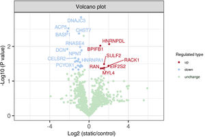
In our study, 8087 peptides were detected in all 8 samples. However, the number of proteins identified in each sample varied slightly, ranging from 1349 to 1353, with a total of 1351 proteins overlapping between the 2 groups. Using an absolute value of fold change greater than 1.5 and a P value less than .05 as criteria for significant difference, 7 upregulated and 23 downregulated proteins were identified between the 2 groups, totaling 30 (figure 1; table 2 of the supplementary data). The screening process, guided by proteomic results, human gene databases, and relevant literature, for these 30 differentially expressed proteins specifically targeted AF and thrombosis. Orr et al.16 discovered that the E11K-MYL4 mutation in humans and zebrafish can destabilize F-actin-Z-disk complexes, potentially impairing calcium signaling and causing atrial myopathy leading to arrhythmias. Gudbjartsson et al.17 reported that frameshift deletions in the MYL4 gene (c.234delC) can cause early-onset AF. A deficiency in PCYOX1 can lead to diminished platelet reactivity and impaired arterial thrombus formation.18 Structural features of decorin can facilitate its interaction with platelet-reactive proteins. MYL4, PCYOX1, and decorin were ultimately selected as potential biomarkers for predicting LAAT.
Volcano plot comparing significant protein differences between the thrombus group and control group, based on fold change and statistical P value. Proteins with significant downregulation (fold change less than -1.5) are marked in blue, significantly upregulated proteins (fold change greater than 1.5) in red, and proteins without significant difference in green.
The enrichment analysis revealed that the identified pathways and GO functions, as presented in figures 1 and 2 of the supplementary data, might not be directly relevant to the disease under study, or, in cases such as those with KEGG analysis, did not yield substantial enrichment. This observation could be attributed to the nature of plasma as a fluid medium, which is not cellular or tissue-specific. Plasma primarily contains a mixture of proteins secreted by various tissues and cells; thus, the pathways and functions identified in plasma samples may not accurately reflect intracellular or tissue-specific processes.
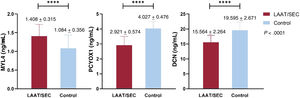
ELISA validationThe CHA2DS2-VASc score was higher in the thrombosis group than in the control group (3.2±1.2 vs 2.7±1.6; P=.019). The left atrial anteroposterior diameter in the thrombosis group was 47.0±9.5mm, which was significantly larger than that in the control group, 42.3±7.6mm (P=.001). The LAA flow velocities in the thrombosis group (0.315±0.07 m/s) were smaller than in the control group (0.510±0.228 m/s). The morphology of the LAA differed significantly between the 2 groups (P=.005). The cauliflower-shaped lesion was predominant in the thrombosis group (53.4%). In contrast, the cactus shape was the most common in the control group (42.1%). However, the type of LAA opening did not significantly differ between the groups (P=.291). No significant differences in age, sex, or opening type were observed between thrombosis and control groups. A comparison of other baseline data is shown in table 3 of the supplementary data. Three biomarkers (MYL4, PCYOX1, decorin), validated using ELISA, showed significant differences between the thrombosis and control groups (1.408±0.315 ng/mL vs 1.084±0.356 ng/mL; 2.921±0.574 ng/mL vs 4.027±0.476 ng/mL; 15.564±2.264 ng/mL vs 19.595±2.671 ng/mL, respectively; P<.0001), which is in line with proteomics testing results (figure 2).
Levels of MYL4, PCYOX1, and decorin between groups. The x-axis categorizes the samples into control (blue bars) and LAAT/SEC (red bars) groups. Asterisks indicate a highly significant difference with a P value <.0001. DCN, decorin; LAAT, left atrial appendage thrombus; MYL4, myosin light chain 4; PCYOX1, prenylcysteine oxidase 1; SEC, spontaneous echo contrast.
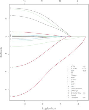
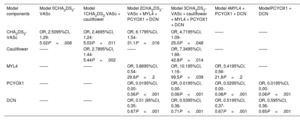
Univariate logistic regression was performed in the collected variables to analyze the risk factors for LAAT and high-risk status. CHA2DS2-VASc, left atrial anteroposterior diameter, chicken wing, cauliflower morphology of the LAA, LAA orifice blood flow velocity, fibrinogen, prothrombin time, MYL4, PCYOX1, and decorin were associated with LAAT (figure 3 of the supplementary data). Lasso regression was performed for variable selection, yielding 5 variables (λ=0.03587), as shown in figure 3. Multivariate logistic regression analysis was performed to assess the predictive factors for LAAT. The models demonstrated that while the CHA2DS2-VASc score and cauliflower morphology of the LAA were consistently identified as independent predictors in multiple models, the inclusion of biomarkers MYL4, PCYOX1, and decorin varied, with PCYOX1 and decorin emerging as significant independent risk factors in the final models (table 1). No significant multicollinearity was found among the 5 variables used to construct the prediction model, as indicated by a variance inflation factor of less than 5.
Results of Lasso regression. APTT, activated partial thromboplastin time; CRP, C-reactive protein; DCN, decorin; LAA, left atrial appendage; LAD, left atrial diameter; LVEF, left ventricular ejection fraction; MYL4, myosin light chain 4; PCYOX1, prenylcysteine oxidase 1; PT, prothrombin time.
Integrated model for left atrial appendage thrombus prediction
| Model components | Model 0CHA2DS2-VASc | Model 1CHA2DS2-VASc + cauliflower | Model 2CHA2DS2-VASc + MYL4 + PCYOX1 + DCN | Model 3CHA2DS2-VASc + cauliflower + MYL4 + PCYOX1 + DCN | Model 4MYL4 + PCYOX1 + DCN | ModelPCYOX1 + DCN |
|---|---|---|---|---|---|---|
| CHA2DS2-VASc | OR, 2.5095%CI, 1.29-5.02P=.008 | OR, 2.4695%CI, 1.24-5.03P=.011 | OR, 6.1795%CI, 1.54-31.1P=.016 | OR, 4.7195%CI, 1.09-25.0P=.048 | —— | —— |
| Cauliflower | —— | OR, 2.7895%CI, 1.44-5.44P=.002 | —— | OR, 7.3495%CI, 1.66-42.8P=.014 | —— | —— |
| MYL4 | —— | —— | OR, 3.8695%CI, 0.54-29.8P=.2 | OR, 10.195%CI, 1.16-99.5P=.039 | OR, 3.4195%CI, 0.56-21.8P=.2 | —— |
| PCYOX1 | —— | —— | OR, 0.0195%CI, 0.00-0.06P<.001 | OR, 0.0195%CI, 0.00-0.06P<.001 | OR, 0.0295%CI, 0.00-0.08P<.001 | OR, 0.0195%CI, 0.00-0.06P<.001 |
| DCN | —— | —— | OR, 0.51 |95%CI, 0.35-0.67P<.001 | OR, 0.5395%CI, 0.36-0.71P<.001 | OR, 0.5195%CI, 0.37-0.67P<.001 | OR, 0.595%CI, 0.36-0.65P<.001 |
95%CI, 95% confidence interval; CHA2DS2-VASc, clinical prediction tool for estimating the risk of stroke in patients with non-rheumatic atrial fibrillation; DCN, decorin; MYL4, myosin light chain 4; OR, odds ratio; PCYOX1, prenylcysteine oxidase 1.
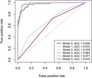
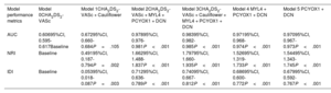
To further appraise the model's performance, a comparison was made with the CHA2DS2-VASc score. The baseline AUC for the CHA2DS2-VASc score was 0.606. The AUC increased modestly to 0.672 with the inclusion of the LAA cauliflower morphology. Substantial enhancement was observed when biomarkers were added, with the AUC reaching 0.983 in model 3, which included LAA cauliflower morphology, MYL4, PCYOX1, and decorin. Models 4 and 5, which used different combinations of these biomarkers (MYL4+PCYOX1+decorin for model 4 and PCYOX1+ decorin for model 5), yielded AUCs of 0.971 and 0.970, respectively, indicating that while the inclusion of MYL4, PCYOX1, and decorin provided the highest AUC, the differences between models 2, 3, 4, and 5 were not statistically significant (figure 4). Model 5, which did not include MYL4, was deemed the most parsimonious and was proposed as the final model. Compared with the baseline CHA2DS2-VASc score, this final model (model 5) demonstrated an NRI of 1.544 (95%CI, 1.343-1.745; P<.001) and an IDI of 0.679 (95% confidence interval [95%CI], 0.592-0.767; P<.001), as illustrated in table 2.
Comparison of Receiver Operating Characteristic (ROC) curves for the 3 models. The figure compares the ROC curves for 3 predictive models, illustrating their respective sensitivities and specificities. Model 0: CHA2DS2-VASc; Model 1: CHA2DS2-VASc + cauliflower; Model 2: CHA2DS2-VASc + MYL4 + PCYOX1 + decorin; Model 3: CHA2DS2-VASc + cauliflower + MYL4 + PCYOX1 + decorin; Model 4: MYL4 + PCYOX1 + decorin; Model 5: PCYOX1 + decorin; AUC, area under curve; MYL4, myosin light chain 4; PCYOX1, prenylcysteine oxidase 1.
Comparison of the performance of left atrial appendage thrombus prediction models
| Model performance metrics | Model 0CHA2DS2-VASc | Model 1CHA2DS2-VASc + Cauliflower | Model 2CHA2DS2-VASc + MYL4 + PCYOX1 + DCN | Model 3CHA2DS2-VASc + Cauliflower + MYL4 + PCYOX1 + DCN | Model 4 MYL4 + PCYOX1 + DCN | Model 5 PCYOX1 + DCN |
|---|---|---|---|---|---|---|
| AUC | 0.60695%CI, 0.595-0.617Baseline | 0.67295%CI, 0.660-0.684P=.105 | 0.97895%CI, 0.976-0.981P<.001 | 0.98395%CI, 0.982-0.985P<.001 | 0.97195%CI, 0.968-0.974P<.001 | 0.97095%CI, 0.967-0.973P<.001 |
| NRI | Baseline | 0.49195%CI, 0.187-0.794P=.002 | 1.66295%CI, 1.488-1.837P<.001 | 1.79795%CI, 1.660-1.935P<.001 | 1.52695%CI, 1.319-1.733P<.001 | 1.54495%CI, 1.343-1.745P<.001 |
| IDI | Baseline | 0.05395%CI, 0.018-0.087P=.003 | 0.71295%CI, 0.636-0.789P<.001 | 0.74095%CI, 0.667-0.812P<.001 | 0.68695%CI, 0.600-0.772P<.001 | 0.67995%CI, 0.592-0.767P<.001 |
95%CI, 95% confidence interval; AUC, area under the curve; CHA2DS2-VASc, clinical prediction tool for estimating the risk of stroke in patients with non-rheumatic atrial fibrillation; DCN, decorin; IDI, integrated discrimination improvement; MYL4, myosin light chain 4; NRI, net reclassification improvement; OR, odds ratio; PCYOX1, prenylcysteine oxidase 1.
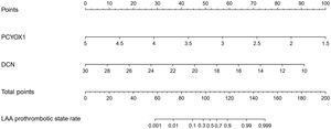
A nomogram was developed to integrate and quantify independent predictors of LAAT/SEC (figure 5). The nomogram exhibited excellent discrimination with a c-statistic of 0.970. We performed 1000 bootstrap repetitions for internal validation of the performance of the nomogram. The calibration plot showed that the standard curve of the LAAT/SEC probability model was closely aligned with the 45° diagonal line, indicating good calibration of the new model (figure 4 of the supplementary data). The bootstrap c-statistic was 0.9418, with a bias of −0.0043.
Nomogram for LAAT/SEC. The ‘Points’ axis at the top assigns numerical values for each variable. The ‘Total Points’ axis sums these individual points, which is then mapped to the ‘Probability’ axis to estimate the likelihood of LAAT/SEC. DCN, decorin; LAA, left atrial appendage; LAAT, left atrial appendage thrombus; PCYOX1, prenylcysteine oxidase 1; SEC, spontaneous echo contrast.
LAAT is a common complication in AF patients, with an incidence of up to 27%.19 It is a primary cause of ischemic stroke and significantly increases mortality and disability rates among AF patients. Therefore, early diagnosis of LAAT is of vital clinical significance for patients and can substantially alleviate the public health burden. In our study, we confirm that not only the CHA2DS2-VASc score and cauliflower LAA morphology but also MYL4, PCYOX1, and decorin—identified for the first time through deep proteomic techniques and ELISA—are independent risk factors for LAAT/high-risk states. Subsequent analysis demonstrated that the predictive accuracy remained high when only PCYOX1 and decorin were used, providing a more accurate and efficient diagnosis of LAAT.
PCYOX1 is a flavin adenine dinucleotide-dependent enzyme that generates free cysteine, allicin aldehyde, and hydrogen peroxide.20 It is a multifunctional protein that affects systems ranging from peptidase activity modulation to platelet degranulation and stress response21 PCYOX1 can influence platelet activity by generating H2O2,22 increasing thrombus susceptibility,23 or by oxidizing the lipoprotein apoB100.24 Jung et al.25 reported reduced plasminogen activator inhibitor-1 (PAI-1) activity in Pcyox1−/−/Apoe−/− mice, suggesting enhanced fibrinolysis. Recently, a contrasting study by Banfi et al.18 using Pcyox1−/− mice revealed downregulation of platelet activation and lower platelet/leukocyte aggregation. Platelet/leukocyte aggregates are involved in both thrombus formation and inflammation, implicating PCYOX1 as a mediator of inflammation and thrombogenesis.26 Recent research has emphasized that PCYOX1 may play a role in inflammation and coagulation by being transported in the human plasma through lipoproteins.27 Our study found significantly lower plasma levels of PCYOX1 in AF thrombosis patients than in controls. Notably, we focused on human participants with nonvalvular AF receiving anticoagulant therapy, unlike the study by Banfi et al.18 in knockout mice. The observed discrepancies may stem from these differences, including the variation in the location and type of thrombus under examination—ours addressing LAAT, while the study by Banfi et al.18 focused on carotid artery thrombus.
Decorin is a major matrix proteoglycans found in bones. In persistent AF patients, N-terminal cleavage of decorin promotes atrial hypertrophy through antimyostatin activity, whereas in patients in sinus rhythm, a more prevalent C-terminal truncation in the atria releases a CTGF-binding domain that inhibits atrial fibrosis.28 Decorin immunostaining is associated with certain microvessels in thrombi and late-stage atherosclerotic plaques.29 Moreover, reduced decorin levels in placental tissue increase thrombin generation.30 Interestingly, extracellular matrix-associated decorin encourages the formation of thin, curved fibrin fibers. This accelerates tissue-type plasminogen activator-dependent fibrinolysis, reducing the likelihood of thrombosis.31 Hyaluosome-loaded decorin alleviates inflammation in the body by reducing levels of tumor necrosis factor-α and interleukin (IL)-1b.32 Consistent with our findings, we found a significant decrease in plasma decorin levels within the AF thrombosis group compared with the AF control group, which may be associated with an increase in inflammation, thereby contributing to thrombus formation. However, the mechanism and significance of this observation warrant further exploration.
MYL4 is crucial for maintaining the structural integrity of myosin and is predominantly expressed in the atria after birth.33 Mutations in the MYL4 gene disrupt sarcomeric structure, leading to atrial enlargement and electrical abnormalities, thus contributing to AF.34 This indicates that the dysregulation of MYL4, whether at the genetic or protein level, may influence AF pathophysiology. By analyzing AF patients and controls, Liu et al.35 found that MYL4 levels were inversely correlated with AF severity and the CHA2DS2 score. The presence of MYL4 in cardiac tissue may correct the dysregulation of autophagic flux, thereby attenuating atrial fibrosis.36 Additionally, a deficiency in MYL4 has been linked to increased CX43 phosphorylation, leading to conduction abnormalities.37 However, we observed higher plasma MYL4 levels in AF patients with LAAT than in those without LAAT. These diverse findings may arise from our focus on thrombotic vs nonthrombotic patients within the AF population, and elevated serum MYL4 levels may reflect myocardial membrane damage, leading to localized reduction and subsequent exacerbation of atrial myopathy.
Previous studies have classified LAA morphologies into 4 types with varying stroke risks: cactus, chicken wing, windsock, and cauliflower. Di Biase et al.11 found that chicken wing morphologies were less embolic, whereas other types posed higher risks. Meta-analyses have reinforced the protective nature of chicken wing morphology against embolic events, irrespective of CHA2DS2-VASc scores.38,39 In our study, we confirmed these classifications, identifying cauliflower as a risk factor and chicken wing as a protective factor against LAAT/high-risk thrombotic states. In their study, Lee et al.40 linked larger LAA diameters to cardioembolic stroke and transient ischemic attacks, whereas Huang et al.41 and Khurram et al.42 highlighted other factors, such as smaller diameter and extensive trabeculations. In our study, we did not observe any significant differences in the depths or opening diameters of the LAA. While Huang et al. focused on patients with a low CHA2DS2-VASc score and the study by Khurram et al. was US-centric, our study involved a Chinese population. In summary, the inconsistencies among studies may stem from varied patient demographics but do not negate the importance of LAA morphology in stroke risk assessment.
The CHA2DS2-VASc score is currently recommended by the European Society of Cardiology and American Heart Association for assessing stroke risk. While it is straightforward and offers some predictive capabilities for endpoints, including LAAT and SEC,43 its limitations are significant. Specifically, it overlooks crucial factors, such as LAA morphology9 and biomarkers.44,45 Therefore, the stroke discrimination ability of the CHA2DS2-VASc score is limited.6 Moreover, the lack of follow-up data for 31% of the patients and the short 1-year follow-up period further undermine its reliability.46 In our study, we found its c-statistic to be similarly low at 0.606 but still recognized it as an independent risk factor for LAAT or high-risk thrombotic states. In summary, the CHA2DS2-VASc score is a starting point but is not sufficient for comprehensive risk assessment. Our findings suggest that integrating imaging indicators, such as cauliflower LAA morphology, with biomarkers can refine the model's predictive accuracy for LAAT/high-risk states and that using only 2 proteins, PCYOX1 and decorin, can yield a substantially effective prediction.
LimitationsThis study had several limitations. First, it was based on preliminary findings from a single-center dataset with a modest sample size. Second, although existing models consider various factors, such as clinical history, LAA morphology, and blood biomarkers, they still fall short in terms of detailed LAA morphological evaluation and the incorporation of hemodynamic parameters. Third, our study did not include further mechanistic research on the identified biomarkers. In future studies, we plan to increase the sample size and collect a more comprehensive dataset. We aim to holistically assess the risk factors of LAAT formation, thereby refining and developing new diagnostic and predictive models. Subsequent validation of these models for clinical relevance and accuracy will be performed through large-sample cohort studies and randomized controlled trials, thereby fostering the translation of our scientific findings into clinical practice.
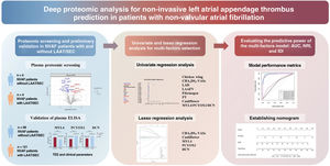
CONCLUSIONSThis study shows that, alongside cauliflower LAA morphology, the proteins MYL4, PCYOX1, and decorin could act as independent risk factors for LAAT and high-risk thrombotic states in nonvalvular AF patients without prior catheter ablation. While our research supports the enhancement of the CHA2DS2-VASc scoring system by including these biomarkers and morphological indicators, we also found that using PCYOX1 and decorin alone considerably improved predictive accuracy (figure 6). Further research is warranted to elucidate the roles of these biomarkers in the development of LAAT.
- -
Research on left atrial appendage thrombus (LAAT) aims to identify reliable biomarkers for early detection and stroke prevention.
- -
Traditional models, such as CHA2DS2-VASc, provide risk assessments but may miss nuanced indicators essential for more accurate identification of at-risk individuals.
- -
Proteomic screening has emerged as a pivotal tool for identifying such biomarkers, offering insights into the molecular underpinnings of LAAT.
- -
Through deep proteomic analysis, this study revealed novel potential biomarkers (MYL4, PCYOX1, decorin) for LAAT, expanding the range of detectable physiological indicators.
- -
We introduce a novel predictive model based on Lasso regression, integrating PCYOX1 and decorin, which demonstrates exceptional predictive accuracy (AUC of 0.970).
- -
This model markedly enhances risk prediction and classification beyond the capabilities of the conventional CHA2DS2-VASc model, offering a more accurate, noninvasive method for preventing LAAT and subsequent stroke events.
Central illustration. Comprehensive overview of the key findings of the study. Novel biomarkers, PCYOX1 and decorin, were identified through deep proteomic analysis of 8 initial samples and later validated in a larger cohort of 179 patients. This led to the creation of a predictive model with an AUC of 0.970, markedly improving upon the conventional CHA2DS2-VASc model. AUC, area under curve; DCN, decorin; IDI, integrated discrimination improvement; LAA, left atrial appendage; LAAT, left atrial appendage thrombus; MYL4, myosin light chain 4; NRI, NRI, net reclassification improvement; NVAF, nonvalvular atrial fibrillation; PCYOX1, prenylcysteine oxidase 1; PT, prothrombin time. SEC, spontaneous echo contrast; TEE, transesophageal echocardiography.
This research was funded by the National Natural Science Foundation of China, grant number 82070328, to Yang Li. Yang Li serves as the financial beneficiary for this research, overseeing project supervision, conducting reviews, and spearheading funding acquisition.
ETHICAL CONSIDERATIONSEthical approval for this study protocol was granted by the Ethics Committee of the Chinese People's Liberation Army General Hospital, and informed consent was obtained from all participants. In this study, we ensured a balanced representation of male and female participants. Given the equal distribution of genders and the homogeneous nature of the sample with respect to the outcomes measured, we determined that a sex-disaggregated analysis was not necessary. We believe this approach maintains the integrity of the study while focusing on the generalizability of the results between sexes without the need for separate analyses.
STATEMENT ON THE USE OF ARTIFICIAL INTELLIGENCENo artificial intelligence tools or services were used during the preparation of this study. The authors take full responsibility for the contents of this manuscript.
AUTHORS’ CONTRIBUTIONSZ. Xie, T. Chen, and X. Lu: conceptualization, methodology, software usage, writing, and visualization; M. Zhao: resources, validation, and investigation; Y. Chen, X. Wang, H. Zhou, and J. Shen: resources, data curation; J. Guo and Y. Li: supervision, review, and funding acquisition. All authors have read and approved the final version of the manuscript.
CONFLICTS OF INTERESTThe authors declare no conflict of interest.