To the Editor,
The Ross procedure has emerged as a good alternative for aortic valve disease, particularly in children and young people in whom repair is not possible.1 Although the procedure is safe, complications related to the pulmonary autograft have been described.2 One of these complications has been reported on only two occasions,3,4 but was potentially severe: pseudoaneurysm of the autograft.
A total of 200 patients have undergone the Ross procedure at our hospital; we present two who were asymptomatic and diagnosed with a false aneurysm of the pulmonary autograft at 2 years of follow-up, with a different cause in each case.
The first was a 27-year-old man who underwent the Ross procedure for severe postendocarditis aortic incompetence. He was admitted one month later for endocarditis on the autograft suture. The patient's progress was favorable following antibiotic therapy, with the pre-discharge echocardiogram showing that the endocardial vegetation had disappeared and both graft valves were functioning normally. The patient was followed as an outpatient, with 6-month echocardiographic controls. At 24 months postoperatively, the patient was asymptomatic; however, follow-up transthoracic echocardiography revealed severe aortic regurgitation due to dilation of the pulmonary autograft root. In addition, a pulsatile retroaortic outpouching with interior flow was seen at the autograft suture, consistent with a pseudoaneurysm (Figure 1). Ultimately, the pulmonary autograft was replaced with an aortic valve (Bono-Bentall) with good results.

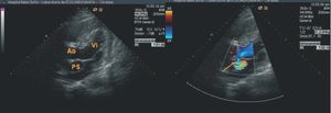
Figure 1. Two transthoracic echocardiography images in a longitudinal parasternal view. The first shows a pulsatile, retroaortic outpouching consistent with pseudoaneurysm; in the second, color Doppler ultrasound reveals flow through the point of entry (arrow). Ao indicates aorta; PS, pseudoaneurysm; LV, left ventricle.
The second case was a 37-year-old man who successfully underwent the Ross procedure for severe rheumatic aortic regurgitation. He received regular follow-up, with the two-year transthoracic echocardiogram showing a pulsatile retroaortic outpouching with interior flow, that was not present in the echocardiogram the previous year and was consistent with a pseudoaneurysm of the autograft. In addition, he had mild dilation of the aortic ring that caused mild aortic regurgitation. Transesophageal echocardiography disclosed the point of entry in the inferior suture of the autograft in the noncoronary sinus, with extension to the left coronary sinus, compromising the origin of the left coronary artery (Figure 2). Ultimately, the patient underwent surgery with aortic valve placement (Bono-Bentall).

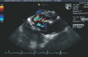
Figure 2. Transesophageal echocardiographic image at 90o, in which color Doppler reveals the shunt of the pseudoaneurysm with the left ventricular outflow tract through two points of entry.
The cause of pseudoaneurysm of the pulmonary autograft has been discussed in recent years. It has been attributed to structural weakness of the pulmonary autograft which, when exposed to systemic pressures, experiences progressive dilation and localized rupture, thereby originating the point of entry of the pseudoaneurysm.4 In our first case, the pseudoaneurysm could be explained by the presence of postinfection friable tissue, whereas the other could be due to weakness of the pulmonary annulus, despite reinforcement via annuloplasty.
Since both pseudoaneurysms were retroaortic, a relatively inaccessible area for surgical control, we suggest monitoring by intraoperative transesophageal echocardiography. A review of our series showed that these 2 patients had no intraoperative study, and therefore this complication might have been avoided.
In conclusion, the 2 patients were diagnosed 2 years after the surgery, despite being asymptomatic, confirming that close clinical and echocardiographic follow-up is important. We believe that transthoracic echocardiography, because it is a simple, noninvasive technique readily available at most hospitals, would be sufficient for follow-up, whereas other techniques, such as transesophageal echocardiography, would be used to complete the diagnosis.