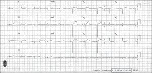
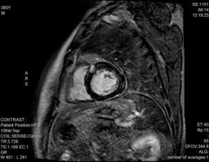
Initially, the condition was considered an ST-elevation acute coronary syndrome and the Infarction Code was implemented. Coronary angiography, no lesions; troponin T by ultrasensitive assay, 252-185 ng/L; creatine kinase, 196-287 U/L; echocardiography, left ventricular ejection fraction 55%, with no regional contractility defects. Electrocardiography was performed at 48 h following hospital admittance (Figure 1). Enhanced cardiac magnetic resonance imaging demonstrated heterogeneous gadolinium uptake, with several foci at the midline of the mediobasal portion of the septum and the basal anteroseptal segment of the epicardium (Figure 2), consistent with acute myocarditis.


Copyright ©
2014. Sociedad Española de Cardiología