ISSN: 1885-5857 Impact factor 2024 4.9
Vol. 77. Num. 9.
Pages 723-733 (September 2024)

Original article
Subjective identification and ablation of drivers improves rhythm control in patients with persistent atrial fibrillation. The CHAOS-AF study

La identificación subjetiva y ablación de impulsores auriculares mejora el control del ritmo en la fibrilación auricular persistente. Estudio CHAOS-AF

Eduardo FrancoabCristina Lozano-GraneroabRoberto Matía FrancésaAntonio Hernández-MadridaInmaculada SánchezcJosé Luis ZamoranoabJavier Morenoab
https://doi.org/10.1016/j.rec.2024.01.005

Options

Supplementary data
Imagen extra
Rev Esp Cardiol. 2024;77:723-33
Abstract
Introduction and objectives

The optimal approach for persistent atrial fibrillation (AF) ablation remains unknown. In patients with persistent AF, we compared an ablation strategy based on pulmonary vein isolation (PVI) plus ablation of drivers (PVI+D), with a conventional PVI-only approach performed in a 1:1 propensity score-matched cohort.
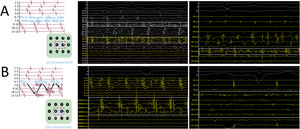
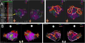
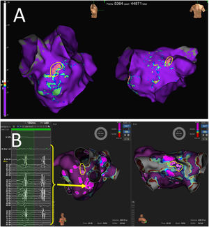
Methods

Drivers were subjectively identified using conventional high-density mapping catheters (IntellaMap ORION, PentaRay NAV or Advisor HD Grid), without dedicated software, as fractionated continuous or quasicontinuous electrograms on 1 to 2 adjacent bipoles, which were ablated first; and as sites with spatiotemporal dispersion (the entire cycle length comprised within the mapping catheter) plus noncontinuous fractionation, which were only targeted in patients without fractionated continuous electrograms, or without AF conversion after ablation of fractionated continuous electrograms. Ablation included PVI plus focal or linear ablation targeting drivers.
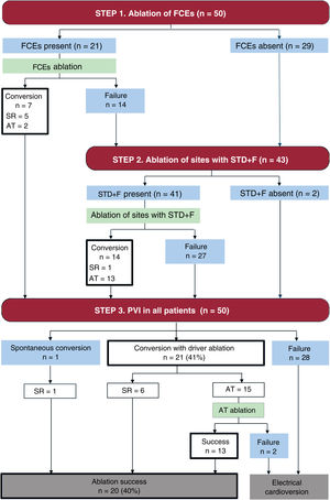
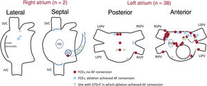
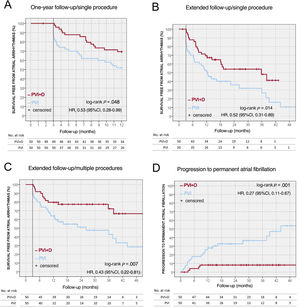
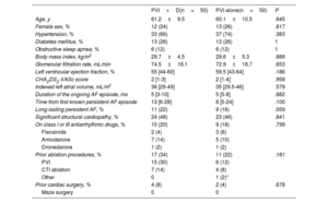
Results

A total of 50 patients were included in each group (61±10 years, 25% women). Fractionated continuous electrograms were found and ablated in 21 patients from the PVI+D group (42%), leading to AF conversion in 7 patients. In the remaining 43 patients, 143 sites with spatiotemporal dispersion plus noncontinuous fractionation were targeted. Globally, AF conversion was achieved in 21 patients (42%). The PVI+D group showed lower atrial arrhythmia recurrences at 1 year of follow-up (30.6% vs 48%; P=.048) and at the last follow-up (46% vs 72%; P=.013), and less progression to permanent AF (10% vs 40%; P=.001).

Conclusions

Subjective identification and ablation of drivers, added to PVI, increased 1-year freedom from atrial arrhythmia and decreased long-term recurrences and progression to permanent AF.

Keywords

Atrial fibrillation
Catheter ablation
High-density mapping
Cardiac electrophysiologic technics
INTRODUCTION

The optimal approach to persistent atrial fibrillation (AF) ablation remains unknown. Pulmonary vein isolation (PVI) alone has shown limited efficacy,1,2 and consequently other strategies have been developed. These strategies can be summarized in 3 categories: category I: ablation of the atrial fibrotic substrate, identified by voltage maps or cardiac magnetic resonance3–5; category II: ablation of the atrial electrical substrate, targeting complex fragmented atrial electrograms (CFAE) and/or performing empirical sets of lines6–10; and category III: ablation of drivers (sites responsible for AF perpetuation), which are generally identified using dedicated software, and whose ablation aims to terminate or control AF recurrences.11–15 The most promising strategies may be the latest, as the best rates of survival free from atrial arrhythmias (up to 80%) have been reported in case series of driver ablation.13 However, controlled studies using such strategies compared with PVI have shown controversial results.11,12

The conversion of hardly mappable atrial tachycardias via rotor ablation into sinus rhythm-atrial fibrillation (CHAOS-AF) study was a nonrandomized, prospective single-center study, designed to test an ablation strategy based on PVI plus ablation of drivers (PVI+D) and compare it with a PVI-only approach in patients with persistent AF and ongoing AF. Drivers were subjectively identified with conventional high-density mapping catheters.

METHODS

The single-center CHAOS-AF study prospectively included 50 consecutive patients with persistent AF scheduled for ablation with PVI+D and compared them with a 1:1 propensity score-matched cohort of patients with persistent AF treated with PVI alone during the same period. A secondary comparative analysis was also performed between patients undergoing PVI+D ablation and all patients undergoing PVI-only regardless of the propensity matching score. Recruitment lasted from May 2017 to September 2021.

In both the PVI+D and PVI alone groups, all patients were in AF at the recruitment visit and continued with the same ongoing AF episode at the beginning of the ablation procedure, unless spontaneous cardioversion and new onset of another AF episode appeared without being noticed at intermediate visits. The duration of the ongoing AF episode was estimated from the first ECG recording in AF prior to the ablation procedure. Long-lasting persistent AF was defined if the ongoing episode lasted>12 months.

Persistent AF patients considered for ablation were submitted in an unselected administrative process irrespective of medical reports to 4 different outpatient clinics. To reduce the risk of selection bias, given the nonrandomized nature of this study, all consecutive patients referred to one of these clinics were offered PVI+D and included if accepted to undergo that procedure. Patients from the other 3 outpatient clinics were offered a PVI-only approach.

The primary endpoint was 1-year survival free from atrial arrhythmias lasting >30seconds after the index procedure. Secondary endpoints were survival free from atrial arrhythmias during extended follow-up, survival free from atrial arrhythmias after multiple ablation procedures, progression to permanent AF, and need of chronic antiarrhythmic medication for patients without progression to permanent AF.

All patients gave written informed consent for their participation in the study and patient data were collected in a dedicated database. The study complied with the Declaration of Helsinki and received prior approval by the ethics committee of our institution.

Additional information on procedural details is available in the supplementary data.

Procedures. Pulmonary vein isolation plus ablation of drivers groupMapping protocol and driver identification

Mapping was performed using conventional high-density electroanatomical mapping catheters (IntellaMap ORION, Boston Scientific; PentaRay NAV, Biosense Webster; or Advisor HD Grid, Abbott Medical, all from the United States) and their respective mapping system (Rhythmia, Carto3, EnSite Precision or EnSite X).

Drivers were subjectively identified, without the use of dedicated software, using bipolar signals from the mapping catheters on a digital recorder (LabSystem Pro, Boston Scientific) at 200mm/s speed. To identify drivers, operators visually addressed the pattern of all electrograms simultaneously registered by the mapping catheter. Two different patterns, which have been previously hypothesized as surrogates or rotational activity,12,16,17 were identified as drivers (figure 1).

Figure 1.

Schematic rationale of driver identification using a hypothetical 12-pole mapping catheter and representative examples of drivers. A: a stable rotor would theoretically show fractionated continuous or quasicontinuous electrograms on the bipoles placed on the rotor core (6-7), as those bipoles would detect electrical activity during almost the entire the cycle length. Blue arrow represents rotor spiral wavefront. Examples using HD Grid catheter (middle panel; driver detected in A2-A3 and A3-A4, also visible in A3-B3) and PentaRay catheter (right panel; driver detected in a single bipole) are shown. B: spatiotemporal dispersion plus noncontinuous fractionation. If the rotor core meanders around the neighboring tissue, the fractionation will move between different bipoles through time, resulting in noncontinuous fractionation. Blue arrow represents meandering of the rotor core. Examples using ORION catheter (middle panel) and PentaRay catheter (right panel) are shown. Paper speed: 200mm/s. PR, PentaRay catheter.

(0.9MB).

a) Fractionated continuous (or quasicontinuous) electrograms (FCE) on single bipoles of the mapping catheter. Two adjacent bipoles were considered to fulfill continuous fractionation criteria if not completely met using 1 bipole. Drivers with this pattern were the first target of ablation.

b) Sites with spatiotemporal dispersion (STD) (ie, all the cycle length comprised within the mapping catheter) plus noncontinuous fractionation (STD+F) on single bipoles, arbitrarily defined as continuous bipolar EGM with >4 deflections and total duration >70ms. Drivers with this pattern were only targeted with ablation if no conversion to sinus rhythm or atrial tachycardia (AT) occurred during ablation of FCE, or in patients without FCE.

At each location, mapping catheters were maintained stable for 2 to 3seconds. When one of these patterns were suspected, the mapping catheter was kept still for 10seconds to confirm temporal stability of the driver, which was then annotated with a manual marker on bipolar voltage maps. In sites with STD+F, only bipoles with fractionation were tagged and targeted for ablation. The mapping catheter was repositioned in sites with potential drivers to confirm temporal stability of EGM pattern before ablation. Temporally unstable drivers were not ablated.

Ablation protocol

Radiofrequency ablation was performed using open-tip irrigated catheters (IntellaNav MIFI, and IntellaNav Stablepoint, Boston Scientific; Thermocool SmartTouch, Biosense Webster; or TactiCath, Abbott Medical, all from the United States). Power settings were decided at the discretion of operators.

First, the left atrium (LA) was mapped and ablated. If no conversion to sinus rhythm or atrial tachycardia (AT) was documented during ablation and AF cycle length was faster in the right atrium (RA) than in the LA (based on the cycle lengths at the left and right atrial appendages), the RA and coronary sinus were subsequently mapped, and ablated if appropriate. If the AF cycle length remained faster in the LA, electrical cardioversion was performed, and the procedure was finished without RA mapping or ablation.

Drivers were targeted by focal ablation with 1 to 4 adjacent lesions, delivered at the previously annotated markers, or by ablation lines between unexcitable structures passing through the driver location. First, ablation of FCE was attempted; if no conversion to sinus rhythm or AT occurred, sites with STD+F were targeted. For drivers at the LA, ablation lines were delivered including the location of the drivers through the line in some circumstances (see supplementary data). Drivers at any other location were ablated focally, but if lesions were deployed <1cm from a scar or another radiofrequency application, an ablation line between them was performed. Circumferential PVI was performed in all patients.

Ablation success was defined as conversion to sinus rhythm or an AT during driver ablation, and additional successful ablation of any AT, if present, to finish the procedure in sinus rhythm.

Procedures. Pulmonary vein isolation-only group

Ablation consisted in PVI using cryoballoon ablation (Arctic Front Cardiac Cryoablation System, Medtronic Medical, United States, or POLARx Cryoablation System, Boston Scientific, United States) or circumferential radiofrequency ablation. Cryoablation cases with failure to achieve bidirectional block at any PV received touch-up radiofrequency applications.

Follow-up

Patients were followed up with clinical visits with 12-lead ECG and 24-hour Holter monitoring at 3, 6, and 12 months. Subsequently, follow-up visits were scheduled according to physicians’ preference. Any arrhythmia lasting >30seconds, according to patients’ symptoms or documented on Holter monitoring, was considered a recurrence.

Antiarrhythmic drugs were maintained during the initial 3-month blanking period after the procedure. Subsequently, antiarrhythmic drugs could also be maintained up to completing 6 months at the criterion of the attending physician.

Statistical analysis

Categorical variables are described as number (percentage) and were compared with the chi-square or Fisher exact tests, as appropriate. Continuous variables are described as mean±standard deviation for variables with normal distributions or as median [interquartile range] for variables not normally distributed. The Student t test and the Mann-Whitney U test were used as appropriate. Bilateral P values <.05 were considered statistically significant. For survival analysis (recurrence of atrial arrhythmias), the Kaplan-Meier method was used. Multiple logistic regression was used to identify predictors of ablation success, and the Cox proportional-hazards model was used to identify find predictors of atrial arrhythmia recurrence.

Propensity scores were calculated using multinomial logistic regression according to the following baseline characteristics: age, sex, body mass index, indexed LA volume, duration of the ongoing AF episode, glomerular filtration rate, hypertension, diabetes mellitus, obstructive sleep apnea, structural cardiopathy, prior ablation procedures and prior cardiac surgeries. Patients from the PVI+D group were matched 1:1 with patients from the PVI-only group according to their propensity scores.

The statistical analysis was performed using SPSS Statistics Base version 22.0 package (IBM Inc., United States).

RESULTSStudy population

A total of 239 patients were included: 50 patients received ablation with the PVI+D approach, and 189 patients with PVI alone. After 1:1 propensity-score matching, the 50 patients from the PVI+D group were compared with 50 patients with a PVI-only approach. Baseline characteristics were comparable between groups (table 1).

Table 1.

Baseline characteristics in the 1:1 propensity score-matched cohort

  PVI+D(n=50)  PVI alone(n=50)  P 
Age, y  61.2±9.5  60.1±10.5  .845 
Female sex, %  12 (24)  13 (26)  .817 
Hypertension, %  33 (66)  37 (74)  .383 
Diabetes mellitus, %  13 (26)  13 (26) 
Obstructive sleep apnea, %  6 (12)  6 (12) 
Body mass index, kg/m2  29.7±4.5  29.6±5.3  .888 
Glomerular filtration rate, mL/min  74.5±18.1  72.9±18.7  .653 
Left ventricular ejection fraction, %  55 [44-60]  59.5 [43-64]  .186 
CHA2DS2-VASc score  2 [1-3]  2 [1-4]  .958 
Indexed left atrial volume, mL/m2  36 [29-49]  35 [29.5-46]  .579 
Duration of the ongoing AF episode, mo  5 [3-10]  5 [3-8]  .882 
Time from first known persistent AF episode  13 [6-28]  8 [5-24]  .100 
Long-lasting persistent AF, %  11 (22)  9 (18)  .059 
Significant structural cardiopathy, %  24 (48)  23 (46)  .841 
On class I or III antiarrhythmic drugs, %  10 (20)  9 (18)  .799 
Flecainide  2 (4)  3 (6)   
Amiodarone  7 (14)  5 (10)   
Dronedarone  1 (2)  1 (2)   
Prior ablation procedures, %  17 (34)  11 (22)  .181 
PVI  15 (30)  6 (12)   
CTI ablation  7 (14)  4 (8)   
Other  1 (2)*   
Prior cardiac surgery, %  4 (8)  2 (4)  .678 
Maze surgery   

AT, atrial tachycardia; CTI, cavotricuspid isthmus; PVI, pulmonary vein isolation; PVI+D, pulmonary vein isolation plus driver ablation.

Data are shown as mean±standard deviation, median [interquartile range] or No. (%).

*

Focal AT ablation (n=1).

In the analysis including all 189 patients with PVI alone, the baseline characteristics were also similar, although the percentage of patients with prior atrial arrhythmia ablations was higher in the PVI+D group, mostly driven by a higher percentage of patients with prior PVI procedures (30% vs 11%; see table 1 of the supplementary data).

Procedural results

Procedural time was higher in the PVI+D group than in the PVI-only group median -244 [187-275] vs 116 [82-149] minutes; P<.001). Fluoroscopy time was also higher (41 [28-65] vs 29 [20-41] minutes; P<.001), but radiation dose, expressed as dose-product area, was lower (18 515 [10 625-32 930] vs 42 446 [21 595-79 952] mGy cm2; P<.001), due to the different acquisition protocol used in cases with or without electroanatomic navigation systems. The complication rate was similar between groups (6% vs 2%; P=.617). However, in the PVI+D group, 1 patient had an atrioesophageal fistula (related to posterior right PVI, without posterior wall lesions related to driver ablation). Complications are shown in table 2 of the supplementary data: 1 patient had transient neuropathic pain located in the right thigh, and 1 patient had a low-risk acute pulmonary embolism during in-hospital stay. In the PVI-only group, a phrenic nerve palsy related to cryoablation was noted.

Driver identification and ablation success in the PVI+D group

Mapping was performed with PentaRay NAV (Carto3) in 35 patients (70%), with IntellaMap ORION (Rhythmia) in 9 (18%), and with Advisor HD Grid (EnSite Precision or EnSite X) in 6 (12%). Figure 1 shows representative examples of drivers. Results comparing performance of mapping catheters and navigation systems for driver identification are shown in the supplementary data.

Procedures were restricted to the LA in 27 patients (54%). In the remaining 23 patients (46%), the RA and the coronary sinus were subsequently mapped; of these patients, 18/23 (78%) showed drivers in the RA. Mean LA ablated surface was 14.9±9.9%. Considering only patients with RA mapping, the mean RA ablated surface was 3.4±2.3% and biatrial ablated surface was 9.0±4.7%. Figure 1 of the supplementary data shows examples of ablated surface measurements and explains the calculation method.

Figure 2 shows the driver ablation approach in the PVI+D group. First, sites with FCE, present in 21 patients (42%), were targeted; their location is shown in figure 3. Forty sites were identified, with a median of 2 [1-2] sites per patient, and 18 (45% of them) were related to the pulmonary vein antra (most frequently to the anterior antrum of the right superior pulmonary vein, n=9). In all cases, sites with FCE also exhibited local or adjacent STD.

Figure 2.

Detailed stepwise ablation approach in the PVI+D group. AT, atrial tachycardia; FCE, fractionated continuous or quasi-continuous electrograms; PVI, pulmonary vein isolation; SR, sinus rhythm; STD+F, spatiotemporal dispersion plus noncontinuous fractionation.

(0.52MB).
Figure 3.

Location of sites with FCE, highlighting sites in which ablation achieved atrial fibrillation (AF) conversion. Sites with STD+F in which ablation achieved AF conversion are also shown. In 1 patient, (asterisk) AF conversion was achieved in the left atrium, but AF reappeared; AF conversion was again achieved in the right atrium. AF, atrial fibrillation; CS, coronary sinus; FCE, fractionated continuous or quasi-continuous EGMs FO, foramen ovale; IVC, inferior vena cava; LIPV, left inferior pulmonary vein; LSPV, left superior pulmonary vein; RIPV, right inferior pulmonary vein; RSPV, right superior pulmonary vein; STD+F, spatiotemporal dispersion+noncontinuous fractionation; SVC, superior vena cava.

(0.19MB).

Sites with STD+F were ablated in the 43 patients without FCE or with failed FCE ablation; 41/43 patients (95%) showed 143 sites, with a median of 4 [3–4] per patient. No drivers were found in 2 patients, who received only PVI.

Globally, driver ablation achieved AF conversion in 21 patients (42%). In 5 patients (24%), driver ablation leading to AF conversion was related to the pulmonary vein antra; in 3 of them, the PVI circumference was significantly modified to include the drivers. Ablation success was achieved in 20 patients (40%).

The presence of FCE, the main target of ablation, was not associated with a higher ablation success rate (P=.154) or conversion rate during driver ablation (P=.099). No baseline characteristics, including indexed LA volume (P=.439) or the duration of the index AF episode (P=.164), predicted AF conversion. In the 15 patients with prior PVI, 12 had at least 1 pulmonary vein reconnected; the AF conversion rate (P=.805) was similar to that in the remaining patients. In addition, total radiofrequency times (P=.540) and the extent of atrial ablated tissue (P=.426) did not predict AF conversion.

Table 3 of the supplementary data shows the number of drivers and the ablation set performed in each patient. Most patients (n=44, 88%) received ablation lines in the LA besides PVI (median 2 [1–3] lines per patient; figure 4): anterior mitral lines were performed in 34 patients (68%), 14 of whom received 2 lines; 28 patients (56%) received a roof line, in 15 cases as a part of a posterior box. Coronary sinus defragmentation was performed in 15 patients (30%). Cavotricuspid isthmus ablation, due to AF conversion to common atrial flutter (n=12) or prior history of common atrial flutter (n=3), was performed in 15 patients (30%).

Figure 4.

Examples of driver identification and ablation approach. A: 1 site with FCE (green dots; dotted yellow line encloses electrograms at that site) and 4 sites with STD+F (pink dots) were marked. Ablation consisted in PVI, a roof line, an anteroseptal mitral line and focal ablation at the inferoanterior septum. B: no sites with FCE and 3 sites with STD+F at the LA anterior wall, in a patient with prior PVI with gaps. Ablation included ablation of gaps and a Y-shaped anterior mitral line.

(0.63MB).

Finally, data on the AF cycle length recorded at sites with drivers and its relationship with the cycle length recorded in neighboring regions without drivers are presented in the supplementary data. Briefly, a trend toward a cycle length gradient (faster at sites with drivers compared with neighboring regions) was noted.

Comparison of automatic fractionation maps and subjectively identified drivers in the PVI+D group

We performed a comparison between subjectively identified drivers and areas of fractionation automatically detected with the EnSite and Rhythmia navigation systems (the Carto3 CFAE module was not available in our institution). As our driver definition implied the presence of fragmented EGMs plus additional criteria (STD or continuous fractionation), we hypothesized that automatic fractionation detection algorithms should identify most drivers (ie, high sensitivity for detecting drivers), but would also detect areas with fractionation that did not fulfil driver criteria (lower positive predictive value). The methodology used for automatic fractionation maps is described in the supplementary data.

For cases performed with EnSite, the automatic fractionation module was used. In these 6 cases, 15 sites with drivers were subjectively identified, whereas automatic fractionation maps showed 33 areas with fractionated EGM. The automatic fractionation module achieved 87% sensitivity (13/15 drivers detected) and 39% positive predictive value (13/33 areas with automatically detected fractionation contained drivers; figure 5A).

Figure 5.

Representative examples of the performance of automatic fractionation maps for detecting drivers. Manual markers indicate subjectively identified drivers (enclosed by yellow lines). With EnSite (A), most drivers were included in areas with automatically detected fractionation, shown with colors other than purple; in the example, 2/2 visualized drivers are detected. With Rhythmia, only 33% of the drivers were included in areas with fractionation, which are highlighted using the LUMIPOINT module; in the example, only 1/2 visualized drivers is detected; the electrograms present at the probe position, located into a highlighted area, show some fractionation but without spatiotemporal dispersion.

(0.32MB).

For cases performed with Rhythmia, the LUMIPOINT module for detecting fractionated EGM was used. In these 9 cases, 36 drivers were identified. LUMIPOINT showed 46 areas with fragmented EGM. The LUMIPOINT module showed only 33% sensitivity (12/36 drivers detected), and 26% positive predictive value (only 12/46 areas with automatically detected fractionated EGM included drivers; figure 5B).

Procedural results in the pulmonary vein isolation-only group

PVI was performed with cryoballoon ablation (47 patients, 94%) or circumferential radiofrequency ablation (3 patients, 6%). Results according to the different ablation energy (cryoablation or radiofrequency) are shown in the supplementary data. Deep sedation was used in 42 patients (84%) and general anesthesia in 8 patients (16%). All pulmonary veins were effectively isolated; in the cryoballoon ablation group, 3 patients needed bailout radiofrequency applications to achieve PVI. AF conversion to sinus rhythm with ablation was observed in 1 patient (2%); conversion occurred during the ablation of the right superior pulmonary vein.

Clinical resultsPrimary endpoint

A total of 99 patients completed the 1-year follow-up. Excluding the 3-month blanking period, patients in the PVI+D group showed a lower rate of atrial arrhythmia recurrences than the PVI-only group (30.6% vs 48%; log-rank P=.048; figure 6). Recurrences in the PVI-only group consisted mainly of AF episodes (92% of all recurrences), whereas the PVI+D group had a higher percentage of re-entrant AT (47% of all recurrences; P=.015).

Figure 6.

Central illustration. Kaplan-Meier plots of primary and secondary endpoints. A: survival free from atrial arrhythmias at 1 year of follow-up. B: survival free from atrial arrhythmia recurrences at extended follow-up. C: survival free from atrial arrhythmia recurrences at extended follow-up after multiple ablation procedures. D: progression to permanent AF. 95%CI, 95% confidence interval; HR, hazard ratio; PVI: pulmonary vein isolation-only group; PVI+D, pulmonary vein isolation plus driver ablation group.

(0.71MB).

In the PVI-D group, no baseline characteristics, including indexed LA volume (P=.179) or the presence of long-standing persistent AF (P=.225), predicted arrhythmia recurrences. Patients with ablation success at the index procedure showed a nonsignificant tendency toward fewer recurrences (15.8% vs 40%; log-rank P=.078; figure 2 of the supplementary data). Total radiofrequency times (P=.112) and the extent of LA and biatrial ablated surface (P=.753 and P=.937, respectively) did not predict arrhythmia recurrences.

The results of the secondary analysis that included all 189 patients treated with a PVI-only approach were similar and are shown in the supplementary data. In addition, results according to the operator responsible for each procedure are included.

Secondary endpoints

Mean follow-up was 33.4±14.5 months. Atrial arrhythmia recurrences during extended follow-up were lower in the PVI+D group than in the PVI-only group (46% vs 72%; log-rank P=.013; figure 6). The percentage of patients who received redo procedures was similar between the 2 groups (PVI+D: 47% of the patients with recurrences; PVI-only: 33%; P=.266); arrhythmia recurrences after multiple ablation procedures were also lower in the PVI+D group (28% vs 56%; log-rank P=.007). Finally, progression to permanent AF was lower in the PVI+D group (10% vs 40%; log-rank P=.001).

Similar class I or III antiarrhythmic drug use was noted between groups prior to the index procedure (20% vs 18%, P=.799), and also at the last follow-up (11% vs 27%, P=.081).

DISCUSSION

We tested an ablation strategy based on the subjective identification and ablation of sites with suspected rotational activity (ie, rotors) in patients with persistent AF. This strategy has also proven its usefulness in patients with unstable re-entrant ATs (ie, those with frequent circuit modification or conversion to AF), to convert them into sinus rhythm or a stable AT that could be subsequently ablated.18

Rotor identification is generally based on the use of dedicated catheters and/or software.11–15,19 These systems offer automatic identification of drivers, which would probably be more reproducible than subjective identification subject to operator bias, but usually imply a financial investment that limits their accessibility. In contrast, STD detection as a surrogate of rotational activity can be performed with conventional high-density mapping catheters and can be subjectively performed by operators according to visually detectable electrical patterns which show very high (94.3%) interobserver concordance.12

Our driver identification protocol focused on STD, similarly to that described by Seitz et al.,12 in which regions with STD were subjectively identified and received cluster ablation, resulting in a mean 17% of LA surface ablation. Our protocol tried to be more selective, considering the 2 following criteria:

  • a)

    Only regions displaying FCE were first targeted, in order to avoid extensive atrial ablation if AF conversion was achieved. We hypothesize that FCE should be visualized within stable rotor cores on bipolar signals, as true functional microre-entries. FCE were found inside regions with STD in >90% of cases in the work by Seitz et al.,12 and in all cases in our work. Moreover, AF conversion occurred at a site with FCE in ≈ 90% of cases in the work by Seitz et al.12 Therefore, we believe this pattern might be considered a subset of STD that more specifically represents the position of a meaningful driver.

  • b)

    In patients without AF conversion by FCE ablation, only regions with STD that also exhibited fractionation were targeted. If a rotor core is not stable but slightly meanders locally, we hypothesize that fractionated but not continuous activity could be visualized within the travelling trajectory of the rotor core, which would result in the STD+F pattern. Regions with STD but without fractionation were not ablated in this study.

The AF conversion rate in our study (42%) was similar to published rates in driver ablation studies, which range between one third11 and two thirds of cases in most reports,14 and highlights the fact that the true nature of AF perpetuation is not fully understood. However, our AF conversion rate was considerably lower than that reported by Seitz et al.12 (which ranged between 66%-95%) despite using a similar rationale and ablation protocol; our lower conversion rate might be due to a stricter driver definition which did not consider STD regions without fractionation, and a different ablation protocol which preferred lines rather than cluster ablation. Total radiofrequency times and the percentage of ablated atrial surface were comparable, although Seitz et al. did not systematically perform PVI; hence, as an important fraction of radiofrequency time (table 2 of the supplementary data) and percentage of LA surface ablated in our group was related to PVI, a lower proportion of total ablation time might have been dedicated to driver ablation instead of PVI in our group.

A high proportion of drivers were found within the antra of the pulmonary veins, most frequently at the anterior antrum of the right superior pulmonary vein, which is the location of the right anterior ganglionated plexus. The role of the parasympathetic nervous system is well established in AF pathogenesis and cardioneuroablation has been proposed as a potential therapeutic tool.20 Interestingly, in the 4 patients in the PVI-only group in whom AF conversion occurred, it happened during right superior pulmonary vein ablation. Hence, some degree of cardioneuroablation might be partially responsible for the clinical results in our study, but this was not prospectively addressed and is only hypothetical.

A differential aspect of our driver ablation protocol is the creation of lines between unexcitable tissue transecting sites with drivers, instead of performing wide focal ablation covering the full area of drivers, which is the most usual approach.11–15 We hypothesized that lines might prevent re-entrant ATs using corridors between ablation sites, as long as durable bidirectional block is obtained. Conversely, gaps at those lines might result in the opposite endpoint. The individual role of driver ablation vs creation of lines on clinical results in our cohort cannot be elucidated. In the work by Seitz et al.,12 who performed cluster ablation without lines, 34% of patients experienced AT recurrences at 18 months of follow-up, and AT comprised 75% of arrhythmia (AF or AT) recurrences. In our series, 9 patients (18%) in the PVI+D group experienced AT recurrences at 18 months, and AT comprised 53% of total recurrences; 6 of these 9 patients with AT recurrence underwent a redo procedure, and a gap-related re-entrant AT was found in 2 patients. The best driver ablation approach (lines or cluster ablation) remains unknown.

Finally, 2 serious complications appeared in the PVI+D group: 1 patient had an atrioesophageal fistula; although no posterior-wall lesions related to driver ablation were performed in this patient, radiofrequency energy was most probably responsible for this complication. Another patient had a low-risk acute pulmonary embolism during in-hospital stay, which might be related to longer procedural times associated with the PVI+D protocol (181minutes in this particular case). Larger series are, however, needed to correctly address complication rates and compare them with a PVI-only approach.

Study limitations

The main limitation of this study is its nonrandomized nature; to overcome this issue, a propensity score-matched analysis was designed, but randomized trials are encouraged to validate our findings. This was a single-center study, and the sample size was limited. Multicenter studies would be valuable to test the reproducibility of the ablation technique, which is operator-dependent as identification of drivers was subjective and based on visual analysis. In addition, the ablation protocol included lines in most patients, and therefore the results reflect the effect of an ablation strategy (drivers+lines) in which the individual effect of driver ablation cannot be elucidated.

Drivers were identified and annotated by only 1 operator. No intra- or interobserver agreement analysis addressing driver identification was performed, although an interobserver concordance of 94% has been previously described.12

In addition, our mapping protocol can theoretically detect only static drivers, or those with a slightly meandering core (within the mapping area of the mapping catheter). Meandering or drifting drivers, which have also been described in persistent AF, could not be identified. Moreover, to avoid excessively long procedural times, the RA was not systematically mapped in all patients, and mapping catheters were initially kept still only for 2 to 3seconds at each point for screening of drivers. Therefore, many RA drivers and temporally but not spatially stable (drifting) drivers displaying only intermittent and short duration fractionation at specific electrode locations may have been completely missed. All of the above limits the sensitivity of the presented mapping strategy for driver detection.

Finally, the more frequent use of general anesthesia in the PVI+D group (100%) than in the PVI-only group (16%) might have influenced the higher AF conversion rate found.

CONCLUSIONS

Subjective identification and ablation of drivers, added to PVI, increased 1-year freedom from atrial arrhythmias and decreased long-term recurrences and progression to permanent AF.

WHAT IS KNOWN ABOUT THE TOPIC?

  • -

    The optimal approach to persistent AF ablation remains unknown.

  • -

    PVI has shown limited efficacy. Among other strategies, ablation of drivers (sites responsible for AF perpetuation) has shown better results.

  • -

    The definition of drivers is not universal, and their detection usually needs dedicated catheters or software, which limits driver ablation in clinical practice.

WHAT DOES THIS STUDY ADD?

  • -

    We used conventional high-density mapping catheters, without dedicated software, to subjectively detect drivers in patients with persistent AF, as 1 of these 2 patterns: a) sites with continuous (or quasi-continuous) fragmented electrograms; and b) spatiotemporal dispersion plus noncontinuous fractionation.

  • -

    Ablation of these drivers, added to PVI, achieved AF conversion to atrial flutter or sinus rhythm in 42% of patients.

  • -

    Compared with a PVI alone strategy, PVI plus ablation of drivers increased 1-year freedom from atrial arrhythmias and decreased long-term recurrences and progression to permanent AF.

FUNDING

None.

ETHICAL CONSIDERATIONS

All patients gave written informed consent for their participation in the study, which complied with the Declaration of Helsinki and received prior approval by the ethics committee of Hospital Universitario Ramón y Cajal. SAGER guidelines were taken into account and no relevant sex-related differences in the results were noted.

STATEMENT ON THE USE OF ARTIFICIAL INTELLIGENCE

No artificial intelligence was used in the preparation of this article.

AUTHORS’ CONTRIBUTIONS

E. Franco has designed the work, acquired and analyzed the data, drafted the work, gave final approval of the version to be published, and has agreed to be accountable for all aspects of the work. C. Lozano-Granero acquired and analyzed data, revised the work critically, gave final approval of the version to be published, and has agreed to be accountable for all aspects of the work. R. Matía Francés revised the work critically, gave final approval of the version to be published, and has agreed to be accountable for all aspects of the work. A. Hernández-Madrid revised the work critically, gave final approval of the version to be published, and has agreed to be accountable for all aspects of the work. I. Sánchez revised the work critically, gave final approval of the version to be published, and has agreed to be accountable for all aspects of the work. J.L. Zamorano revised the work critically, gave final approval of the version to be published, and has agreed to be accountable for all aspects of the work. J. Moreno contributed to the design of the work, revised the work critically, gave final approval of the version to be published, and has agreed to be accountable for all aspects of the work.

CONFLICTS OF INTEREST

E. Franco has received consulting fees from Biosense Webster, Abbott Medical, and Boston Scientific. J. Moreno has received consulting fees from Biosense Webster, Abbott Medical, and Boston Scientific. The rest of the authors declare no conflicts of interest.

REFERENCES
[1]
J.A. Clarnette, A.G. Brooks, R. Mahajan, et al.
Outcomes of persistent and long-standing persistent atrial fibrillation ablation: a systematic review and meta-analysis.
Europace., (2018), 20 pp. f366-f376
[2]
W.W. Su, V.Y. Reddy, K. Bhasin, et al.
Cryoballoon ablation of pulmonary veins for persistent atrial fibrillation: Results from the multicenter STOP Persistent AF trial.
Heart Rhythm., (2020), 17 pp. 1841-1847
[3]
D. Schreiber, A. Rieger, F. Moser, H. Kottkamp.
Catheter ablation of atrial fibrillation with box isolation of fibrotic areas: Lessons on fibrosis distribution and extent, clinical characteristics, and their impact on long-term outcome.
J Cardiovasc Electrophysiol., (2017), 28 pp. 971-983
[4]
G. Yang, L. Zheng, C. Jiang, et al.
Circumferential Pulmonary Vein Isolation Plus Low-Voltage Area Modification in Persistent Atrial Fibrillation: The STABLE-SR-II Trial.
JACC Clin Electrophysiol., (2022), 8 pp. 882-891
[5]
N.F. Marrouche, O. Wazni, C. McGann, et al.
Effect of MRI-Guided Fibrosis Ablation vs Conventional Catheter Ablation on Atrial Arrhythmia Recurrence in Patients With Persistent Atrial Fibrillation: The DECAAF II Randomized Clinical Trial.
JAMA., (2022), 327 pp. 2296-2305
[6]
J. Vogler, S. Willems, A. Sultan, et al.
Pulmonary Vein Isolation Versus Defragmentation: The CHASE-AF Clinical Trial.
J Am Coll Cardiol., (2015), 66 pp. 2743-2752
[7]
A. Verma, C.Y. Jiang, T.R. Betts, et al.
Approaches to catheter ablation for persistent atrial fibrillation.
N Engl J Med., (2015), 372 pp. 1812-1822
[8]
K.C. Wong, J.R. Paisey, M. Sopher, et al.
No Benefit of Complex Fractionated Atrial Electrogram Ablation in Addition to Circumferential Pulmonary Vein Ablation and Linear Ablation: Benefit of Complex Ablation Study.
Circ Arrhythm Electrophysiol., (2015), 8 pp. 1316-1324
[9]
K. Inoue, S. Hikoso, M. Masuda, et al.
Pulmonary vein isolation alone vs. more extensive ablation with defragmentation and linear ablation of persistent atrial fibrillation: the EARNEST-PVI trial.
Europace., (2021), 23 pp. 565-574
[10]
N. Derval, J. Duchateau, A. Denis, et al.
Marshall bundle elimination, Pulmonary vein isolation, and Line completion for ANatomical ablation of persistent atrial fibrillation (Marshall-PLAN): Prospective, single-center study.
Heart Rhythm., (2021), 18 pp. 529-537
[11]
F. Atienza, J. Almendral, J.M. Ormaetxe, et al.
Comparison of radiofrequency catheter ablation of drivers and circumferential pulmonary vein isolation in atrial fibrillation: a noninferiority randomized multicenter RADAR-AF trial.
J Am Coll Cardiol., (2014), 64 pp. 2455-2467
[12]
J. Seitz, C. Bars, G. Théodore, et al.
AF ablation guided by spatiotemporal electrogram dispersion without pulmonary vein isolation: a wholly patient-tailored approach.
J Am Coll Cardiol., (2017), 69 pp. 303-321
[13]
T. Baykaner, A.J. Rogers, G.L. Meckler, et al.
Clinical implications of ablation of drivers for atrial fibrillation: a systematic review and meta-analysis.
Circ Arrhythm Electrophysiol., (2018), 11 pp. e006119
[14]
J.G. Quintanilla, J.M. Alfonso-Almazán, N. Pérez-Castellano, et al.
Instantaneous amplitude and frequency modulations detect the footprint of rotational activity and reveal stable driver regions as targets for persistent atrial fibrillation ablation.
Circ Res., (2019), 125 pp. 609-627
[15]
S. Honarbakhsh, R.J. Schilling, M. Finlay, E. Keating, R.J. Hunter.
Prospective STAR-guided ablation in persistent atrial fibrillation using sequential mapping with multipolar catheters.
Circ Arrhythm Electrophysiol., (2020), 13 pp. e008824
[16]
M. Haissaguerre, M. Hocini, A. Denis, et al.
Driver domains in persistent atrial fibrillation.
Circulation., (2014), 130 pp. 530-538
[17]
S. Zlochiver, M. Yamazaki, J. Kalifa, O. Berenfeld.
Rotor meandering contributes to irregularity in electrograms during atrial fibrillation.
Heart Rhythm., (2008), 5 pp. 846-854
[18]
E. Franco, C. Lozano Granero, R. Matía, et al.
Stabilization of unstable reentrant atrial tachycardias via fractionated continuous electrical activity ablation (CHAOS study).
Cardiol J., (2023), 30 pp. 799-809
[19]
D. Calvo, J. Rubín, D. Pérez, et al.
Ablation of rotor domains effectively modulates dynamics of human long-standing persistent atrial fibrillation.
Circ Arrhythm Electrophysiol., (2017), 10 pp. e005740
[20]
P.S. Chen, L.S. Chen, M.C. Fishbein, et al.
Role of the autonomic nervous system in atrial fibrillation: pathophysiology and therapy.
Circ Res., (2014), 114 pp. 1500-1515
Copyright © 2024. Sociedad Española de Cardiología