Publish in this journal
Journal Information
Vol. 73. Issue 9.
Pages 758-762 (September 2020)
Visits
Not available
Vol. 73. Issue 9.
Pages 758-762 (September 2020)
Review article
Full text access
What every clinician should know about Bayés syndrome
Síndrome de Bayés. Lo que todo clínico debe conocer
Visits
4785
Antoni Bayés de Lunaa, Manuel Martínez-Sellésb,
Corresponding author
mmselles@secardiologia.es

Corresponding author: Servicio de Cardiología, Hospital Universitario Gregorio Marañón, Dr. Esquerdo 46, 28007 Madrid, Spain.
, Antoni Bayés-Genísc, Roberto Elosuad, Adrián Baranchuke
a Fundación de Investigación Cardiovascular ICCC, Instituto de Investigación Hospital de la Santa Creu i Sant Pau, IIB-Sant Pau, Barcelona, Spain
b Servicio de Cardiología, Hospital General Universitario Gregorio Marañón, CIBERCV, Universidad Europea, Universidad Complutense, Madrid, Spain
c Instituto del Corazón, Hospital Universitario Germans Trias i Pujol, Universidad Autónoma de Barcelona, Badalona, Barcelona, Spain
d Instituto Hospital del Mar de Investigaciones Médicas (IMIM), CIBERCV, Facultad de Medicina, Universidad de Vic-Universidad Central de Cataluña, Vic, Barcelona, Spain
e Department of Medicine, Queens University, Kingston, Ontario, Canada
This item has received
Article information
Abstract
Full Text
Bibliography
Download PDF
Statistics
Figures (5)
Show moreShow less
Tables (1)
Table 1. Studies reporting an association between advanced interatrial block and prognosis (atrial fibrillation, stroke, and mortality)
Abstract

Bayés syndrome is a new clinical entity, characterized by the association of advanced interatrial block (IAB) on surface electrocardiogram with atrial fibrillation (AF) and other atrial arrhythmias. This syndrome is associated with an increased risk of stroke, dementia, and mortality. Advanced IAB is diagnosed by the presence of a P-wave ≥ 120ms with biphasic morphology (±) in inferior leads. The cause of IAB is complete Bachmann bundle blockade, leading to retrograde depolarization of the left atrium from areas near the atrioventricular junction. The anatomic substrate of advanced IAB is fibrotic atrial cardiomyopathy. Dyssynchrony induced by advanced IAB is a trigger and maintenance mechanism of AF. This alteration of the atrial architecture produces atrial remodeling, blood stasis and hypercoagulability, triggering the thrombogenic cascade. The presence of advanced IAB, even in patients without documented atrial arrhythmias, has also been associated with AF, stroke, dementia, and mortality. However, in these patients, there is no evidence to support the use of anticoagulation. Therefore, in patients with advanced IAB, a proactive search for AF is recommended.

Keywords:
Interatrial block
Bayés syndrome
Atrial fibrillation
Stroke
Abbreviations:
AF
IAB
Resumen

El síndrome de Bayés es una nueva entidad clínica, caracterizada por la combinación de bloqueo interauricular (BIA) avanzado en el electrocardiograma de superficie con fibrilación auricular (FA) y otras arritmias auriculares. Este síndrome se asocia con un riesgo incrementado de ictus, demencia y mortalidad. El BIA avanzado se diagnostica con la presencia de una onda P ≥ 120ms de morfología bifásica (±) en derivaciones de cara inferior. Se produce por un bloqueo completo del haz de Bachmann que causa una despolarización retrógrada de la aurícula izquierda desde zonas cercanas a la unión auriculoventricular. La miocardiopatía auricular fibrótica es el sustrato anatómico del BIA avanzado. La disincronía inducida por el BIA avanzado funciona como desencadenante y mecanismo de mantenimiento de la FA. Esta alteración de la arquitectura auricular produce remodelado auricular, estasis sanguínea e hipercoagulabilidad, lo cual desencadena la cascada trombogénica. El BIA avanzado, incluso sin arritmias auriculares documentadas, también se ha relacionado con FA, ictus, demencia y mortalidad. Sin embargo, todavía no se ha demostrado el beneficio de la anticoagulación para los pacientes sin FA documentada. Por lo tanto, es recomendable una búsqueda proactiva de FA en los pacientes con BIA avanzado.

Palabras clave:
Bloqueo interauricular
Síndrome de Bayés
Fibrilación auricular
Ictus
Full Text
BAYÉS SYNDROME

In 1988, Bayés de Luna et al.1 reported that patients with advanced interatrial block (IAB) presented with supraventricular arrhythmia more often than patients with partial IAB. However, until a consensus article was published on IAB in 2012,2 only a few authors had shown interest in the subject, mainly the groups led by Spodick,3 García-Cosío,4 and Platonov,5 as well as our own group.6,7 Since this consensus article was published, there has been growing interest, and Conde and Baranchuk8 named this combination “Bayés syndrome,” a term quickly accepted by the scientific community.9–11

ATRIAL ACTIVATION

The P wave originates in the sinus node, producing an atrial activation sequence that is directed downward and leftward in the frontal plane, with counterclockwise rotation, producing a P wave that is positive in leads II and aVF, variable in III and aVL, and negative in aVR. Impulses are conducted from the sinus node to the atrioventricular node without genuine bundles, whereas impulses are conducted from the right to the left atrium mainly through the upper portion of the atrium through the Bachmann bundle or region, the main conduction route between the 2 atria. The final portion of this bundle bifurcates, then encircling the neck of the atrial appendage.12,13 In the posterior and inferior aspects of the septum, almost 30% of patients also show fibers that can conduct the impulse from the right to the left atrium.5 However, the rest of the septum consists of connective tissue and does not allow impulse transmission. Consequently, if there is complete AV block of the Bachmann region, left atrial activation is retrograde from the area near the atrioventricular junction (coronary sinus and fossa ovalis), with the resulting abnormality seen on electrocardiography (P±in lower leads). As in other types of cardiac blocks, IAB may be transient, may be induced experimentally,14,15 and may be present in the absence of structural heart disease, in which case it is seen as left atrial enlargement.

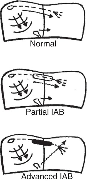
TYPES OF INTERATRIAL BLOCK AND ELECTROCARDIOGRAPHY CRITERIA

The type of atrial pacing determines the type of IAB (figure 1). Therefore, we have:

  • Partial IAB. Impulses are conducted from the right atrium to the left atrium by the Bachmann bundle, but with a delay.

  • Advanced IAB. Impulses are not conducted by the Bachmann bundle. Left atrial activation is retrograde through the coronary sinus musculature and the fossa ovalis.

Figure 1.

Atrial activation in healthy (normal) patients and in patients with partial and advanced interatrial block (IAB).

(0.22MB).

Intermittent IAB is also possible, as impulses are sometimes blocked and sometimes conducted in a single electrocardiogram tracing. Additionally, IAB may be progressive and, over the years, worsen from partial to advanced.

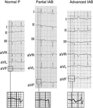
To diagnose IAB, the surface electrocardiogram should show P wave ≥ 120ms and morphology allowing the degree of blockage to be diagnosed2,16–18 (figure 2):

  • Partial IAB: positive P wave in lower leads, often bimodal (“notched”) in some leads of the frontal and horizontal planes.

  • Advanced IAB: biphasic P wave (±) in lower leads.

Figure 2.

Examples of normal P wave and partial and advanced interatrial block (IAB). Adapted with permission from Martínez-Sellés et al.18

(0.2MB).

We recently described other atypical morphologies of advanced IAB19 not discussed in the present review, as they are rare and do not currently seem to be clinically relevant.

PATHOPHYSIOLOGY AND CONSEQUENCES OF ADVANCED INTERATRIAL BLOCK

Both AF and advanced IAB have fibrotic atrial cardiomyopathy as their anatomic substrate20 which, along with atrial dyssynchrony21 in patients with advanced IAB, result in left atrial hypocontractility that favors blood stasis and encourages atrial remodeling. Under these circumstances, through thrombin activation by protease-activated receptors, a hypercoagulable state follows that further increases fibrosis and atrial remodeling and triggers the clotting cascade and the appearance of systemic embolization.22

Magnetic resonance imaging is the technique of choice to detect fibrotic atrial cardiomyopathy,23 the anatomopathologic substrate for most cases of advanced IAB and AF. Patients with advanced IAB may exhibit a high degree of fibrosis, even without documented AF.24 Speckle-tracking echocardiography also can be used to assess atrial fibrosis, with prognostic implications for AF recurrence.25–27

Until recently, AF was considered the final cause leading to systemic embolization. However, several studies in patients with an implanted Holter device have not found a temporal relationship between episodes of paroxysmal atrial fibrillation and the onset of stroke.28–30 Therefore, AF is only one risk factor, just like advanced IAB and other factors such as age, hypertension, diabetes, and obesity. Hence, the importance of atrial fibrosis and its relationship with the appearance of stasis, which encourages the formation of thrombi in the left atrium and is present in both AF and advanced IAB. In fact, the novel concept of atrial failure31 includes atrial fibrosis, often in combination with atrial dilatation, interatrial conduction disorders, and thus Bayés syndrome among them.

EPIDEMIOLOGY OF ADVANCED INTERATRIAL BLOCK

The prevalence of advanced IAB in the overall middle-aged population (45-64 years) is only 0.5%,32 but rises to 8.2% in septuagenarians and 26.3% in centenarians18 (figure 3). The age-related association is even more pronounced than suggested by these figures because, as AF also increases with age, the prevalence of advanced IAB in patients who remain in sinus rhythm is higher than the overall figures.

Figure 3.

Prevalence of advanced interatrial block (IAB) in the overall population, according to age.

(0.06MB).
ADVANCED INTERATRIAL BLOCK AND RISK OF ATRIAL FIBRILLATION, STROKE, AND DEATH

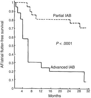
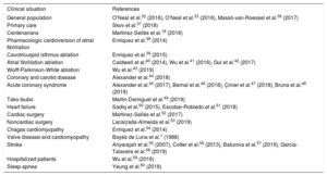
In middle-aged patients, advanced IAB results in a 3-fold risk of AF and an almost 2-fold risk of stroke,32,33 thus providing an explanation for about half the cases of AF and a quarter of the strokes occurring in this age bracket. P-wave duration is also associated with cardiovascular mortality34 and sudden cardiac death.35 At very advanced ages, the presence of IAB is also associated with total mortality.18 Since an association between advanced IAB and supraventricular arrhythmias was first described1 (figure 4), advanced IAB has been reported as associated with prognosis in various clinical situations1,18,32,33,36–60 (table 1) and AF has been confirmed as associated with stroke in a meta-analysis.61

Figure 4.

Supraventricular tachyarrhythmia (atrial fibrillation [AF]/atrial flutter)-free survival in patients with advanced interatrial block (IAB) compared with a similar group of patients with partial IAB in the first article to describe this association. Adapted with permission from Bayés de Luna et al.1

(0.06MB).
Table 1.

Studies reporting an association between advanced interatrial block and prognosis (atrial fibrillation, stroke, and mortality)

Clinical situation  References 
General population  O’Neal et al.32 (2016), O’Neal et al.33 (2016), Massó-van-Roessel et al.36 (2017) 
Primary care  Skov et al.37 (2018) 
Centenarians  Martínez-Sellés et al.18 (2016) 
Pharmacologic cardioversion of atrial fibrillation  Enriquez et al.38 (2014) 
Cavotricuspid isthmus ablation  Enriquez et al.39 (2015) 
Atrial fibrillation ablation  Caldwell et al.40 (2014), Wu et al.41 (2016), Gul et al.42 (2017) 
Wolff-Parkinson-White ablation  Wu et al.43 (2019) 
Coronary and carotid disease  Alexander et al.44 (2018) 
Acute coronary syndrome  Alexander et al.45 (2017), Bernal et al.46 (2018), Çinier et al.47 (2018), Bruna et al.48 (2019) 
Tako-tsubo  Martín-Demiguel et al.49 (2019) 
Heart failure  Sadiq et al.50 (2015), Escobar-Robledo et al.51 (2018) 
Cardiac surgery  Martínez-Sellés et al.52 (2017) 
Noncardiac surgery  Lacalzada-Almeida et al.53 (2019) 
Chagas cardiomyopathy  Enriquez et al.54 (2014) 
Valve disease and cardiomyopathy  Bayés de Luna et al.1 (1988) 
Stroke  Ariyarajah et al.55 (2007), Cotter et al.56 (2013), Baturova et al.57 (2019), García-Talavera et al.58 (2019) 
Hospitalized patients  Wu et al.59 (2016) 
Sleep apnea  Yeung et al.60 (2018) 
ADVANCED INTERATRIAL BLOCK AND DEMENTIA

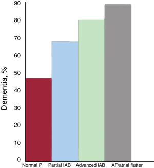
The association of AF with mild cognitive decline and dementia is no longer questioned.62 Although the pathophysiologic mechanisms explaining this association are not yet fully elucidated, they are likely to be multifactorial and to include the most obvious (eg, symptomatic ischemic stroke and silent strokes/microstrokes) as well as brain bleeds and brain hypoperfusion due to hemodynamic impairment that lowers cardiac output and reduces diastolic arterial flow to the brain. In the case of advanced IAB, the association appears to be very similar. In the Cardiac and Clinical Characterization of Centenarians (4C) study,18 the prevalence of dementia was progressively higher as normal P waves developed into partial IAB, advanced IAB, and AF (figure 5). This association is likely due to silent strokes, although other factors such as chronic cerebral hypoperfusion may play a role. This relationship between advanced IAB and dementia indicates that routine cognitive screening is needed among patients with advanced IAB. Advanced IAB should also be ruled out in patients with cognitive impairment.

Figure 5.

Prevalence of dementia in the 4C registry,18 according to the presence and type of interatrial block (IAB) and atrial fibrillation (AF)/atrial flutter.

(0.04MB).
CLINICAL IMPLICATIONS OF ADVANCED INTERATRIAL BLOCK

Patients with advanced IAB and prior episodes of documented AF (Bayés syndrome) should be treated like other patients with a history of AF. In terms of strategy, the presence of advanced IAB is an independent predictor for AF recurrence and, therefore, heart rate control could be considered in some patients.

Patients with advanced IAB and no prior episodes of documented AF also have a higher risk of stroke, particularly if there are additional risk factors, such as advanced age, diabetes, hypertension, structural heart disease, and frequent supraventricular extrasystole. However, no ongoing clinical trials support the use of anticoagulants in the absence of documented AF. Therefore, patients should be monitored for AF episodes which could require anticoagulant therapy.63 We believe it would be useful to conduct a placebo-controlled, randomized study on direct-action oral anticoagulants with patients who have advanced IAB as well as some of the other risk factors reported.64–66 It may also be useful to investigate how atrial fibrosis may be reduced with antifibrotic drugs.

CONCLUSIONS

P waves do not usually draw the attention of clinicians during electrocardiogram evaluation. However, the diagnosis of advanced IAB is not merely of academic interest, as it is associated with supraventricular arrhythmia (Bayés syndrome), stroke, mortality, and dementia.

CONFLICTS OF INTEREST

None declared.

References
[1]
A. Bayés de Luna, M. Cladellas, R. Oter, et al.
Interatrial conduction block and retrograde activation of the left atrium and paroxysmal supraventricular tchyarrhythmia.
[2]
A. Bayés de Luna, P. Platonov, F.G. Cosio, et al.
Interatrial blocks: A separate entity from left atrial enlargement: A consensus report.
J Electrocardiol., 45 (2012), pp. 445-451
[3]
V. Ariyarajh, S. Apiyasawat, J. Fernandes, M. Kranis, D.H. Spodick.
Association of atrial fibrillation in patients with interatrial block over prospectively followed controls with comparable echocardiographic parameters.
Am J Cardiol., 99 (2007), pp. 390-392
[4]
F.G. Cosío, A. Martín-Peñato, A. Pastor, et al.
Atrial activation mapping in sinus rhythm in the clinical electrophysiology laboratory: observations during Bachmann's bundle block.
J Cardiovasc Electrophysiol., 15 (2004), pp. 524-531
[5]
P.G. Platonov, L. Mitrofanova, V. Ivanov, et al.
Substrates for intra-atrial and interatrial conduction in the atrial septum: anatomical study on 84 human hearts.
Heart Rhythm., 5 (2008), pp. 1189-1195
[6]
A. Bayés de Luna, M. Cladellas, R. Oter, et al.
Interatrial conduction block with retrograde activation of the left atrium and paroxysmal supraventricular tachyarrhythmias: influence of preventive antiarrhythmic treatment.
Int J Cardiol., 22 (1989), pp. 147-150
[7]
A. Bayés de Luna, J. Guindo, X. Viñolas, et al.
Third-degree inter-atrial block and supraventricular tachyarrhythmias.
Europace., 1 (1999), pp. 43-46
[8]
D. Conde, A. Baranchuk.
Bloqueo interauricular como sustrato anatómico-eléctrico de arritmias supraventriculares: síndrome de Bayés.
Arch Cardiol Mex., 84 (2014), pp. 32-40
[9]
L. Bacharova, G.S. Wagner.
The time for naming the interatrial block syndrome: Bayes syndrome.
J Electrocardiol., 48 (2015), pp. 133-134
[10]
A. Arboix, L. Martí, S. Dorison, et al.
Bayés syndrome and acute cardioembolic ischemic stroke.
World J Clin Cases., 5 (2017), pp. 93-101
[11]
A. Baranchuk.
Interatrial block and supraventricular arrhythmias.
Clinical implications of Bayés’ Syndrome, Cardiotext Publishing, (2017),
[12]
S.Y. Ho, R.H. Anderson, D. Sánchez-Quintana.
Atrial structure and fibers: morphological basis of atrial coducton.
Cardiovasc Res., 54 (2002), pp. 325-336
[13]
S.Y. Ho, D. Sánchez-Quintana, J.A. Cabrera, et al.
Anatomy of the left atrium: Implications for radiofrequency ablation of atrial fibrillation.
J Cardiovasc Electrophysiol., 10 (1999), pp. 1525-1533
[14]
A.L. Waldo, H.L. Bush Jr., H. Gelband, G.L. Zorn Jr., K.J. Vitikainen, B.F. Hoffman.
Effects on the canine P wave of discrete lesions in the specialized atrial tracts.
Circ Res., 29 (1971), pp. 452-467
[15]
J.M. Guerra, G. Vilahur, A. Bayés de Luna, et al.
Interatrial block can occur in the absence of left atrial enlargement: New experimental model.
Pacing Clin Electrophysiol., (2020),
[16]
A. Bayés de Luna, R. Fort de Ribot, E. Trilla, et al.
Electrocardiographic and vectorcardiographic study of interatrial conduction disturbances with left atrial retrograde activation.
J Electrocardiol., 18 (1985), pp. 1-13
[17]
P. Puech.
L’activité électrique auriculaire. Normale et pathologique.
Masson & Editeurs, (1956),
[18]
M. Martinez-Selles, A. Masso-van Roessel, J. Alvarez-Garcia, et al.
Interatrial block and atrial arrhythmias in centenarians: Prevalence, associations, and clinical implications.
Heart Rhythm., 13 (2016), pp. 645-651
[19]
A. Bayés de Luna, L.A. Escobar-Robledo, D. Aristizabal, et al.
Atypical advanced interatrial block: Definition electrocardiographic recognition.
J Electrocardiol., 51 (2018), pp. 1091-1093
[20]
H. Kottkamp.
Fibrotic atrial cardiomyopathy: a specific disease/syndrome supplying substrates for atrial fibrillation, atrial tachycardia, sinus node disease, AV node disease, and thromboembolic complications.
J Cardiovasc Electrophysiol., 23 (2012), pp. 797-799
[21]
L.C. Ciuffo, H. Doria De Vasconcellos, V. Bruña, et al.
Advanced interatrialblock as an independent marker of left atrial fibrosis and intra-atrial dyssynchrony: ECG-MRI correlation.
[22]
A. Bayés de Luna, M. Martínez-Sellés, R. Elosua, et al.
Relation of advanced interatrial block to risk of atrial fibrillation and stroke.
[23]
N.F. Marrouche, D. Wilber, G. Hindricks, et al.
Association of atrial tissue fibrosis identified by delayed enhancement MRI and atrial fibrillation catheter ablation. The DECAAF study.
JAMA., 311 (2014), pp. 498-506
[24]
E.M. Benito, A. Bayés de Luna, A. Baranchuk, L. Mont.
Extensive atrial fibrosis assessed by late gadolinium enhancement cardiovascular magnetic resonance associated with advanced interatrial block electrocardiogram pattern.
Europace., 19 (2017), pp. 377
[25]
S. Montserrat, L. Gabrielli, B. Bijnens, et al.
Left atrial deformation predicts success of first and second percutaneous atrial fibrillation ablation.
Heart Rhythm., 12 (2015), pp. 11-18
[26]
J. Lacalzada-Almeida, M.M. Izquierdo-Gómez, C. Belleyo-Belkasem, et al.
Interatrial block and atrial remodeling assessed using speckle tracking echocardiography.
BMC Cardiovascular Disord., 18 (2018), pp. 38
[27]
J. Lacalzada-Almeida, M.M. Izquierdo-Gómez, J. García-Niebla, et al.
Advanced interatrial block is a surrogate for left atrial strain reduction which predicts atrial fibrillation and stroke.
Ann Noninvasive Electrocardiol., 24 (2019), pp. e12632
[28]
T.V. Glotzer, E.G. Daoud, D.G. Wyse, et al.
The relationship between daily atrial tachyarrhythmia burden from implantable device diagnostics and stroke risk. The TRENDS Study.
Circ Arrhythmia Electrophysiol., 2 (2009), pp. 474-480
[29]
J.S. Healey, S.J. Connolly, M.R. Gold, et al.
for the ASSERT Investigators. Subclinical atrial fibrillation and the risk of stroke.
N Engl J Med., 366 (2012), pp. 120-129
[30]
D.T. Martin, M.M. Bersohn, A.L. Waldo, et al.
IMPACT Investigators. Randomized trial of atrial arrhythmia monitoring to guide anticoagulation in patients with implanted defibrillator and cardiac resynchronization devices.
Eur Heart J, 36 (2015), pp. 1660-1668
[31]
F. Bisbal, A. Baranchuk, E. Braunwald, A. Bayés de Luna, A. Bayés-Genís.
Atrial failure as a clinical entity.
J Am Coll Cardiol., 75 (2020), pp. 222-232
[32]
W.T. O’Neal, Z.M. Zhang, L.R. Loehr, L.Y. Chen, A. Alonso, E.Z. Soliman.
Electrocardiographic advanced interatrial block and atrial fibrillation risk in the general population.
Am J Cardiol., 117B (2016), pp. 1755-1759
[33]
W.T. O’Neal, H. Kamel, Z.M. Zhang, L.Y. Chen, A. Alonso, E.Z. Soliman.
Advanced interatrial block and ischemic stroke. The atherosclerosis risk in communities study.
Neurology., 87 (2016), pp. 352-356
[34]
J.W. Magnani, V.M. Johnson, L.M. Sullivan, et al.
P-wave indices: derivation of reference values from the Framingham heart study.
Ann Noninvasive Electrocardiol., 15 (2010), pp. 344-352
[35]
A. Maheshwari, F.L. Norby, E.Z. Soliman, et al.
Relation of prolonged P-wave duration to risk of sudden cardiac death in the general population (from the Atherosclerosis risk in Communities Study).
Am J Cardiol., 119 (2017), pp. 1302-1306
[36]
A. Massó-van Roessel, L.A. Escobar-Robledo, I.R. Dégano, et al.
Analysis of the association between electrocardiographic P-wave characteristics and atrial fibrillation in the REGICOR Study.
Rev Esp Cardiol., 70 (2017), pp. 841-847
[37]
M.W. Skov, J. Ghouse, J.T. Kühl, et al.
Risk prediction of atrial fibrillation based on electrocardiographic interatrial block.
[38]
A. Enriquez, D. Conde, W. Hopman, et al.
Advanced interatrial block is associated with recurrence of atrial fibrillation post pharmacological cardioversion.
Cardiovasc Ther., 32 (2014), pp. 52-56
[39]
A. Enriquez, A. Sarrias, R. Villuendas, et al.
New-onset atrial fibrillation after cavotricuspid isthmus ablation: identification of advanced interatrial block is key.
Europace., 17 (2015), pp. 1289-1293
[40]
J. Caldwell, S. Koppikar, W. Barake, et al.
Prolonged P wave duration is associated with atrial fibrillation recurrence after successful pulmonary vein isolation for paroxysmal atrial fibrillation.
J Interv Card Electrophysiol., 39 (2014), pp. 131-138
[41]
J.T. Wu, D.Y. Long, J.Z. Dong, et al.
Advanced interatrial block predicts clinical recurrence of atrial fibrillation after catheter ablation.
J Cardiol., 68 (2016), pp. 352-356
[42]
E.E. Gul, R. Pal, J. Caldwell, et al.
Interatrial block and interatrial septal thickness in patients with paroxysmal atrial fibrillation undergoing catheter ablation: Long-term follow-up study.
Ann Noninvasive Electrocardiol., 22 (2017), pp. e12428
[43]
J.T. Wu, D.Q. Zhao, F.F. Li, et al.
Advanced interatrial block predicts recurrence of atrial fibrillation after accessory pathway ablation in patients with Wolff-Parkinson-White syndrome.
Clin Cardiol., 42 (2019), pp. 806-811
[44]
B. Alexander, A. Baranchuk, S. Haseeb, et al.
Interatrial block predicts atrial fibrillation in patients with carotid and coronary artery disease.
J Thorac Dis., 10 (2018), pp. 4328-4334
[45]
B. Alexander, J. MacHaalany, B. Lam, et al.
Comparison of the extent of coronary artery disease in patients with versus without interatrial block and implications for new-onset atrial fibrillation.
Am J Cardiol., 119 (2017), pp. 1162-1165
[46]
E. Bernal, A. Bayés-Genís, A. Ariza-Solé, et al.
Interatrial block, frailty and prognosis in elderly patients with myocardial infarction.
J Electrocardiol., 51 (2018), pp. 1-7
[47]
G. Çinier, Tekkeşin Aİ, D. Genç, et al.
Interatrial block as a predictor of atrial fibrillation in patients with ST-segment elevation myocardial infarction.
Clin Cardiol., 41 (2018), pp. 1232-1237
[48]
V. Bruña, J. Velásquez-Rodríguez, M.J. Valero-Masa, et al.
Prognostic of interatrial block after an acute ST-segment elevation myocardial infarction.
Cardiology., 142 (2019), pp. 109-115
[49]
I. Martín-Demiguel, I.J. Núñez-Gil, A. Pérez-Castellanos, et al.
Prevalence and significance of interatrial block in takotsubo syndrome (from the RETAKO Registry).
Am J Cardiol., 123 (2019), pp. 2039-2043
[50]
F. Sadiq Ali, A. Enriquez, D. Conde, et al.
Advanced interatrial block predicts new onset atrial fibrillation in patients with severe heart failure and cardiac resynchronization therapy.
Ann Noninvasive Electrocardiol., 20 (2015), pp. 586-591
[51]
L.A. Escobar-Robledo, A. Bayés-de-Luna, J. Lupón, et al.
Advanced interatrial block predicts new-onset atrial fibrillation and ischemic stroke in patients with heart failure: The “Bayes’ Syndrome-HF” study.
Int J Cardiol., 271 (2018), pp. 174-180
[52]
M. Martínez-Sellés, E. García-Izquierdo Jaén, I. Fernández Lozano.
Anticoagulation in elderly patients at high risk of atrial fibrillation without documented arrhythmias.
J Geriatr Cardiol., 14 (2017), pp. 166-168
[53]
J. Lacalzada-Almeida, M.M. Izquierdo-Gómez, J. García-Niebla, et al.
Advanced interatrial block is a surrogate for left atrial strain reduction which predicts atrial fibrillation and stroke.
Ann Noninvasive Electrocardiol., 24 (2019), pp. e12632
[54]
A. Enriquez, D. Conde, F. Femenia, et al.
Relation of interatrial block to new-onset atrial fibrillation in patients with Chagas cardiomyopathy and implantable cardioverter-defibrillators.
Am J Cardiol., 113 (2014), pp. 1740-1743
[55]
V. Ariyarajah, P. Puri, S. Apiyasawat, D.H. Spodick.
Interatrial block: a novel risk factor for embolic stroke?.
Ann Noninvasive Electrocardiol., 12 (2007), pp. 15-20
[56]
P.E. Cotter, P.J. Martin, L. Ring, E.A. Warburton, M. Belham, P.J. Pugh.
Incidence of atrial fibrillation detected by implantable loop recorders in unexplained stroke.
Neurology., 80 (2013), pp. 1546-1550
[57]
M.A. Baturova, A. Lindgren, Y.V. Shubik, J. Carlson, P.G. Platonov.
Interatrial block in prediction of all-cause mortality after first-ever ischemic stroke.
BMC Cardiovasc Disord., 19 (2019), pp. 37
[58]
C.S. García-Talavera, A. Aceña, A.A. López, et al.
Advanced interatrial block: An electrocardiographic marker for stroke recurrence.
J Electrocardiology., 57 (2019), pp. 1-5
[59]
J.T. Wu, S.L. Wang, Y.J. Chu, et al.
Usefulness of a combination of interatrial block and high CHADS2 score to predict new onset atrial fibrillation.
Int Heart J., 57 (2016), pp. 580-585
[60]
C. Yeung, D. Drew, S. Hammond, et al.
Extended cardiac monitoring in patients with severe sleep apnea and no history of atrial fibrillation (The Reveal XT-SA Study).
Am J Cardiol., 122 (2018), pp. 1885-1889
[61]
G. Tse, C.W. Wong, M. Gong, et al.
International Health Informatics Study (IHIS) Network. Predictive value of inter-atrial block for new onset or recurrent atrial fibrillation: A systematic review and meta-analysis.
Int J Cardiol., 250 (2018), pp. 152-156
[62]
H.C. Diener, R.G. Hart, P.J. Koudstaal, D.A. Lane, G.Y.H. Lip.
Atrial fibrillation and cognitive function: JACC review topic of the week.
J Am Coll Cardiol., 73 (2019), pp. 612-619
[63]
M. Martínez-Sellés, A. Baranchuk, R. Elosua, A.B. de Luna.
Rationale and design of the BAYES (Interatrial Block and Yearly Events) registry.
Clin Cardiol., 40 (2017), pp. 196-199
[64]
A. Bayés de Luna, M. Martínez-Sellés, A. Bayés-Genís, R. Elosua, A. Baranchuk.
Surface ECG interatrial block-guided treatment for stroke prevention: rationale for an attractive hypothesis.
BMC Cardiovasc Disord., 17 (2017), pp. 211
[65]
M. Martínez-Sellés, I. Fernández Lozano, A. Baranchuk, A. Bayes-Genis, A. Bayés de Luna.
Should we anticoagulate patients at high risk of atrial fibrillation?.
Rev Esp Cardiol., 69 (2016), pp. 374-376
[66]
A. Bayés de Luna, A. Baranchuk, M. Martínez-Sellés, P.G. Platonov.
Anticoagulation in patients at high risk of stroke without documented atrial fibrillation. Time for a paradigm shift?.
Ann Noninvasive Electrocardiol., 22 (2017), pp. e12417
Copyright © 2020. Sociedad Española de Cardiología
Idiomas
Revista Española de Cardiología (English Edition)

Subscribe to our newsletter

View newsletter history
Article options
Tools
es en

¿Es usted profesional sanitario apto para prescribir o dispensar medicamentos?

Are you a health professional able to prescribe or dispense drugs?