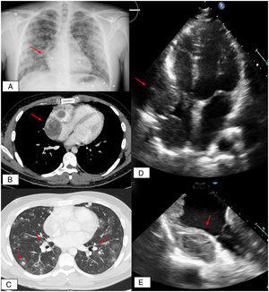
A 25-year-old male smoker with no other relevant medical history attended the emergency department with a 2-week history of persistent cough and bloody sputum. Blood analysis revealed microcytic and hypochromic anemia. A chest X-ray revealed bilateral patchy alveolar infiltrates and a “bump”-like swelling on the right atrium (figure 1A, arrow).

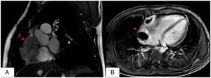
Contrast-enhanced computed tomography revealed a heterogeneous, sinuous mass containing necrotic areas in the right atrium (figure 1B, arrow). The mass was in contact with and invading the tricuspid valve and the free basal wall of the right ventricle. The X-ray also detected numerous angioinvasive sphere-like metastases dispersed in the lungs (figure 1C, arrows). These findings were confirmed by further examination by transthoracic echocardiography (figure 1D), transesophageal echocardiography (figure 1E), and cardiac magnetic resonance imaging (figure 2A and B).

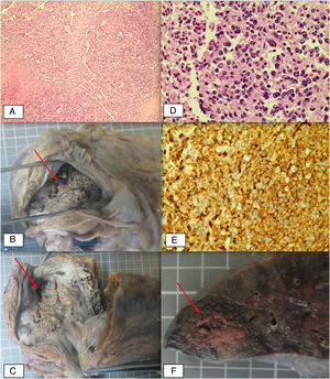
An intracardiac biopsy of the lesion was obtained via percutaneous access. Pathological analysis (figure 3A) revealed atypical mesenchymal proliferation with endothelial differentiation compatible with angiosarcoma (expression of cluster of differentiation 34 and cluster of differentiation 31). A consensus decision was taken to initiate chemotherapy with first-cycle anthracyclines followed by a schedule of etoposide, cisplatin, and isophosphamide. The initial response was encouraging; however, further clinical progress was poor, and the patient died from a massive alveolar hemorrhage.

Macroscopic autopsy (figure 3B and C) confirmed the presence of a tumor mass in the right atrium, and microscopic autopsy identified atypical endothelial cells and anaplastic fusiform cells (figure 3D). These cells were intensely immunopositive for cluster of differentiation 34 and cluster of differentiation 31 (figure 3E), consistent with angiosarcoma. An example of lesion metastasis in the lung is shown in figure 3F (arrow).