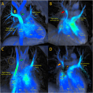

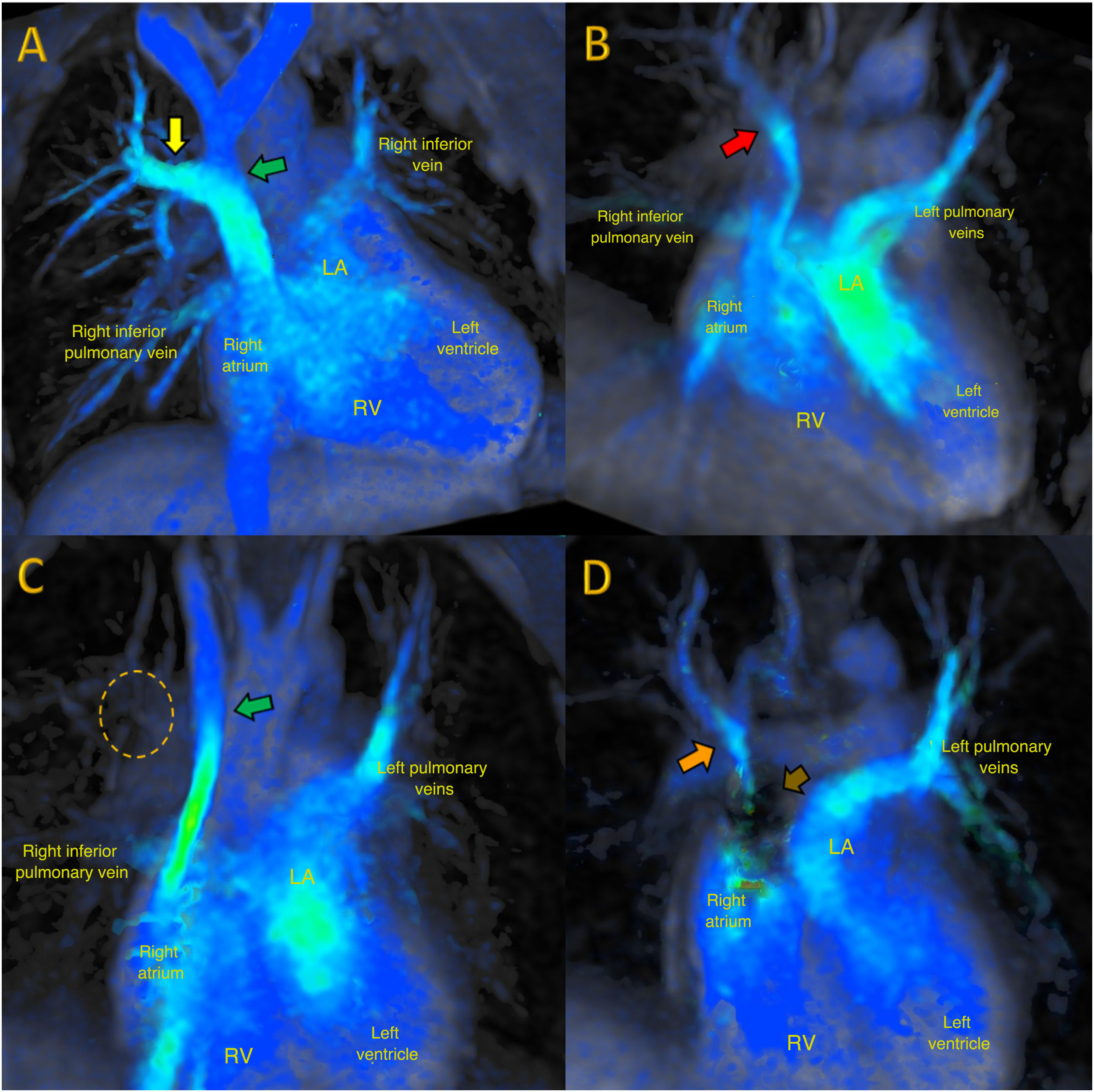
We describe the case of a 22-year-old man assessed due to heart murmur, right bundle-branch block, and right ventricle dilatation. 4D-flow cardiac magnetic resonance imaging (CMR) diagnosed partial anomalous drainage of the right and middle upper pulmonary vein (figure 1A, yellow arrow) flowing into the superior vena cava (figure 1A, green arrow, and video 1 of the supplementary data) (MRI with1.5T GE Optima 450W 4D-flow after gadolinium [Gadovist] 0.15 mmol/kg: matrix 172×172, VENC 250cm/s, voxel: 2.4×2.4×2.4mm). The right ventricle was dilatated, with Qp:Qs> 1.5. Repair surgery was performed to redirect the drainage through a bovine pericardium patch from its origin to the left atrium through an atrial septal defect artificially created in the fossa ovalis. Adequate outcome was subsequently confirmed (figure 1B, arrow) by 4D-flow CMR (figure 1B and video 2 of the supplementary data).

Three years later, echocardiographic follow-up revealed suspected stenosis of the repaired pulmonary vein and, therefore, 4D-flow CMR (figure 1C and video 3 of the supplementary data) was performed again, showing no significant flow (figure 1C, circle with dashed line), suggesting critical stenosis versus occlusion of the pulmonary vein channel. Surgery was performed to bypass the drainage through the atrial septal defect using a Gore-Tex graft (inhomogeneity artifact, figure 1D, short arrow), and was confirmed as optimal (figure 1D, long arrow) by 4D-flow CMR (figure 1D and video 4 of the supplementary data).
4D-flow CMR was used for successful multiplane qualitative and quantitative assessment of the flows of the partial anomalous pulmonary venous return treated by surgery at various stages of progression.
This figure is shown in full color only in the electronic version of the article.
FUNDINGNone.
ETHICAL CONSIDERATIONSInformed consent was obtained to perform the tests and to publish the images, and the respective international ethical recommendations were followed. Possible sex and gender biases have been taken into account.
STATEMENT ON THE USE OF ARTIFICIAL INTELLIGENCENo artificial intelligence tool was used.
AUTHORS’ CONTRIBUTIONSJ. Urmeneta Ulloa: manuscript text, CMR acquisition and interpretation, and CMR image reconstruction. A. Forteza Gil: surgical procedure and manuscript supervision. J.Á. Cabrera: manuscript supervision.
CONFLICTS OF INTERESTThe authors declare that they have no conflicts of interest.