
Native aortic regurgitation (AR) affects 8% to 13% of patients with valvular heart disease. The need for intervention is determined not only by the severity of the heart disease but also by other factors such as left ventricular (LV) function and degree of ascending aortic dilation. Aortic valve replacement is the surgical treatment of choice and has proven prognostic value; experienced surgical teams can also consider valve repair.1 Only a small percentage of patients, however, are referred for surgery as the risk is often prohibitive.1
Recent years have seen an increase in the use of percutaneous procedures to treat severe aortic stenosis in patients at high surgical risk, transforming the management of this disease and sparking growing interest in the use of less invasive treatments for AR. Until recently, valves originally designed for aortic stenosis were used to treat AR on a compassionate basis only. Outcomes in this setting are affected by technical limitations, and there have been reports of valve embolization rates of as high as 12%.3 Seeking to overcome these limitations and optimize clinical outcomes, studies of transcatheter aortic valve implantation (TAVI) in patients with AR have to date largely focused on the anatomic features of the aortic annulus.1,2 Little, however, has been published on dynamic changes and ventricular remodeling after implantation.3
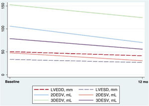
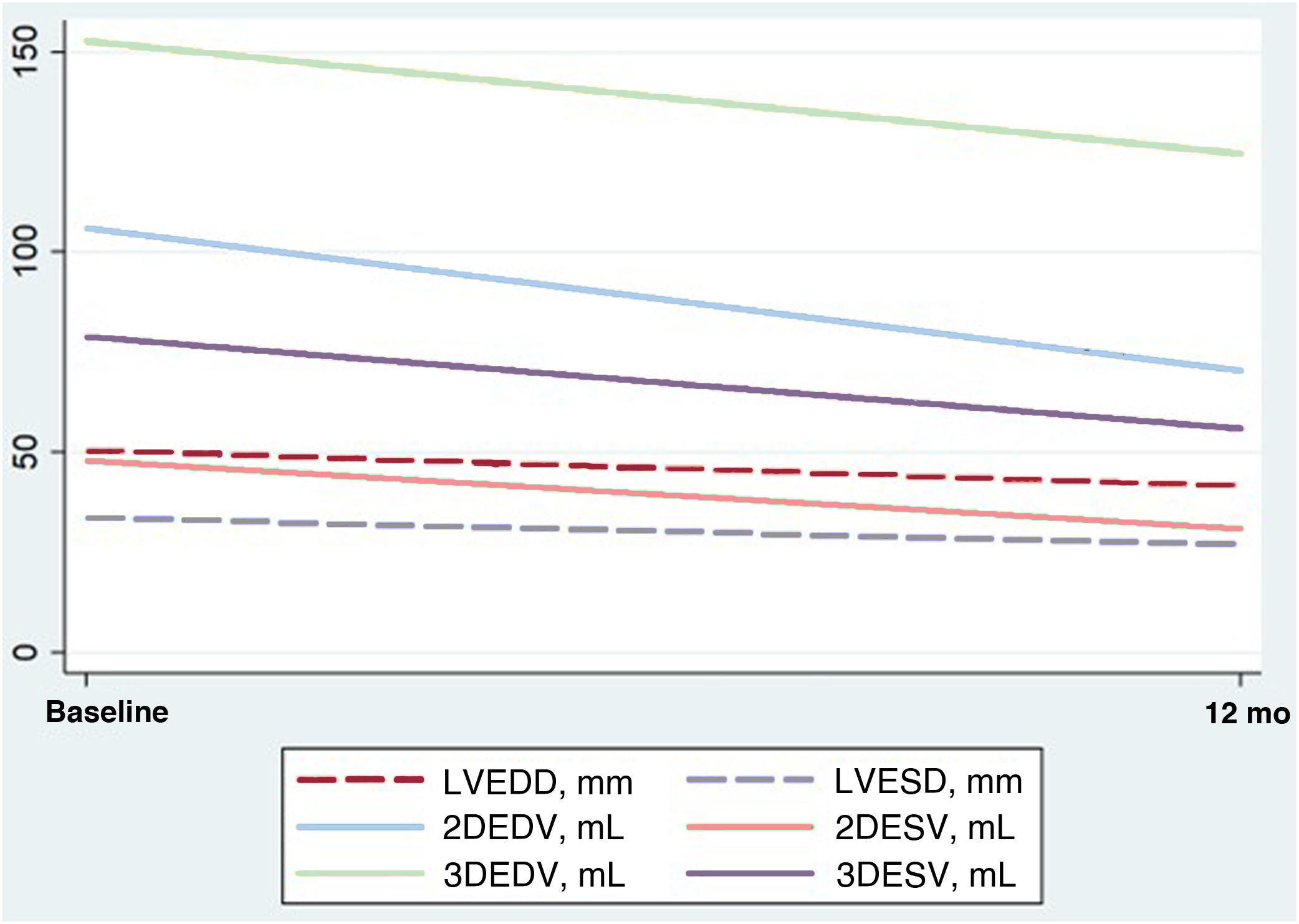
We present the results of a retrospective pilot study conducted at our hospital in which echocardiographic parameters were used to assess ventricular remodeling in patients with severe AR treated with TAVI (figure 1).
Changes in echocardiographic parameters from baseline to 12 months after transcatheter aortic valve implantation for aortic regurgitation. 3DEDV, 3-dimensional end-diastolic volume; 3DESV, 3-dimensional end-systolic volume; LVEDD, left ventricular end-diastolic diameter; LVESD, left ventricular end-systolic diameter; 2DEDV, 2-dimensional end-diastolic volume; 2DESV, 2-dimensional end-systolic volume.
Patients treated between 2013 and 2023 were included. The study was approved by the hospital's ethics committee and data anonymity was guaranteed. To be included, patients had to been followed for at least 6 months and have undergone standard transthoracic echocardiography before and after TAVI. Forty-two patients met the inclusion criteria. Patients without a comparative imaging study or with significant aortic stenosis—defined as moderate or worse—were excluded. This left 21 patients (50% of the initial sample) with a median follow-up of 12 [range, 11-15] months. Mean age at implantation was 78.8 [range, 60-89] years; 67.7% of the patients were women. Hypertension was present in 81% of cases, dyslipidemia in 62%, diabetes mellitus in 33%, chronic ischemic heart disease in 14.3%, chronic kidney disease—defined as an estimated glomerular filtration rate <45mL/min/1.73 m2—in 28.6%, and atrial fibrillation in 42.9%. The estimated risk of surgical mortality according to the Society of Thoracic Surgeons (STS) score was 5.7%; 76% of the patients had mild to moderate aortic valve calcification. Two-dimensional (2D) and 3D echocardiographic parameters were analyzed using the images obtained at baseline (pre-TAVI) and at 1 year. The parameters included end-diastolic and end-systolic diameters and volumes, 2D and 3D left ventricular ejection fraction (LVEF), and global longitudinal strain (GLS). Additional factors were valve embolization rate, significant residual paravalvular AR, and conduction disturbances requiring permanent pacemaker implantation.
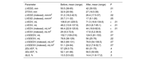
Self-expanding prostheses were implanted in 71% of patients. Pre- and post-TAVI echocardiographic parameters are shown in table 1 together with their level of statistical significance. The Wilcoxon test for paired data was used for statistical analysis.
Echocardiographic parameters before and after transcatheter aortic valve implantation
| Parameter | Before, mean (range) | After, mean (range) | P |
|---|---|---|---|
| LVEDD, mm | 50.5 (38-65) | 42 (30-53) | .01 |
| DTSVI, mm | 32.9 (20-56) | 27 (16.5-39) | .1 |
| LVEDD (indexed), mm/m2 | 31.2 (18.2-42.5) | 26.4 (17.5-37) | .02 |
| LVESD (indexed), mm/m2 | 20.7 (11-32) | 17 (9.1-26) | .05 |
| LVEDV, mL | 108.8 (41-229.5) | 71.3 (18.8-134.6) | <.01 |
| LVESV, mL | 48.2 (13.8-137.9) | 29.3 (8.7-65.1) | <.01 |
| LVEDV (indexed), mL/m2 | 66.4 (22.8-120.8) | 44 (18.8-82.6) | <.01 |
| LVESV (indexed), mL/m2 | 29 (9.3-72.6) | 17.8 (5.2-39.9) | .01 |
| LV3DEDV, mL | 152.7 (109-216) | 124.5 (81-153) | .07 |
| LV3DESV, mL | 78.8 (38-129) | 56 (25-76) | .22 |
| LV3DEDV (indexed), mL/m2 | 98.3 (69-131) | 78.9 (54-106.5) | .07 |
| LV3DESV (indexed), mL/m2 | 51.1 (24-84) | 32.2 (7.8-52.7) | .07 |
| 2DLVEF, % | 57 (29.3-73) | 60 (31-75) | .4 |
| 3DLVEF, % | 52.1 (41-66) | 56.5 (50-69) | .4 |
| GLS, % | 13.3 (5.5-22) | 14.4 (11.8-17.2) | .3 |
2D, 2-dimensional; 3D, 3-dimensional; LVEDD, left ventricular end-diastolic diameter; LVESD, left ventricular end-systolic diameter; LVEF, left ventricular ejection fraction; GLS, global longitudinal strain; LVEDV, left ventricular end-diastolic volume; LVESV, left ventricular end-systolic volume.
Implantation of a bioprosthetic valve to treat severe AR significantly reduced absolute and indexed end-diastolic and end-systolic diameters and volumes, as measured by the biplane Simpson method. A nonsignificant increase in LVEF was also observed. In line with previous findings,4 volumes estimated using 3D echocardiography were higher than those calculated using the biplane Simpson method. Analysis of changes in 3D echocardiographic parameters showed a clear numerical reduction and an increase in LVEF, although statistical significance was not reached. These values were only available for 42.8% of the patients due to the retrospective design and duration of the study (10 years). The ability to detect changes in 3D measurements was also limited by the small sample size. GLS values did not vary significantly, possibly because myocardial deformation parameters depend on preload. A sharp initial reduction in GLS, followed by normalization within 3 to 12 months, has been reported in patients with AR treated with surgical aortic valve replacement.5 Similar changes may have occurred in our series. The valve embolization rate was 4.8%; an additional 4.8% of patients experienced significant paravalvular AR and 9.5% required permanent pacemaker implantation. They had all received self-expanding valves and had no signs of significant conduction disturbances on the baseline electrocardiogram.
In this pilot study, transcatheter treatment of AR achieved left ventricular reverse remodeling and was associated with a low complication rate. The development of valves specifically designed for AR should offer even better results. Nonetheless, larger multicenter studies are needed to confirm our findings.
FUNDINGNo funding was received for the current study.
ETHICAL CONSIDERATIONSThis study was approved by the ethics committee at our hospital, and data anonymity was ensured. The Sex and Gender Equity in Research guidelines were followed.
STATEMENT ON THE USE OF ARTIFICIAL INTELLIGENCEArtificial intelligence was not used to prepare this manuscript.
AUTHORS’ CONTRIBUTIONSS. Antoñana-Ugalde: design, data acquisition and interpretation, manuscript drafting. A. García-Martín: design, data interpretation, critical review of content. C. Fernández-Golfín Lobán: design, critical review of content. L. Salido-Tahoces: design, critical review. A. Sánchez-Recalde and J.L. Zamorano-Gómez: critical review of intellectual content. All authors meet the criteria for authorship of scientific articles as defined by the International Committee of Medical Journal Editors.
CONFLICTS OF INTERESTNo conflicts of interest in relation to this article are declared.