

Sinus of Valsalva aneurysm (SVA) is a rare cardiac anomaly that is often caused by the congenital absence of muscle or elastic tissue in the aortic wall of the sinus of Valsalva. The most common coexisting cardiac lesions are ventricular septal defects (VSD) and aortic regurgitation (AR). Since the first successful surgical correction in 1957,1 surgical repair has become the primary management strategy for SVA (video 1 of the supplementary data).
Postoperative AR is a significant risk factor that affects patient prognosis. Investigating the risk factors for postoperative AR progression is important to improve outcomes, but data are lacking. Therefore, we retrospectively analyzed consecutive patients diagnosed with SVA who underwent surgical repair at our hospital between January 2006 and December 2020. The aim of our study was to determine whether there are any differences between patients with and without VSD and to estimate hazard ratios (HRs) for postoperative AR progression using multivariable Cox regression models.
Follow-up data were obtained from the outpatient department. Echocardiography was recommended at 3 months, 6 months, and annually thereafter. The severity of AR was graded using color Doppler echocardiography as none, mild, moderate, or severe. This grading was multiparametric and integrative, emphasizing both quantitative and qualitative parameters endorsed by the American Society of Echocardiography and the Valve Academic Research Consortium-2 (VARC-2) guidelines.
In the parasternal long-axis view, the ratio of the proximal regurgitation jet width to the left ventricular outflow tract was measured. A ratio of> 65% was considered severe, 25% to 65% moderate, and <25% mild. AR progression was defined as either the new onset of AR or worsening of pre-existing AR. To identify predictors of AR progression, we used a Cox proportional hazards regression model.
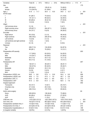
A total of 317 patients (64.0% male) were included in the study, of whom 228 (71.9%) had found ruptured aneurysms. The mean age was 31.9±11.7 years. SVA most commonly originated from the right-coronary sinus (RCS) (n=278; 87.7%), followed by the noncoronary sinus (NCS) (n=34; 10.7%) and the left coronary sinus (LCS) (n=5; 1.6%). The most frequent exit sites were the right ventricle (n=248; 78.2%) and the right atrium (n=50; 15.7%). RCS aneurysms mostly ruptured or protruded into the right ventricle (n=237; 85.3%), while NCS aneurysms most commonly ruptured or protruded into the right atrium (n=23; 67.6%) and, less frequently, the right ventricle (n=10; 29.4%).
As shown in table 1, patients with VSD had a lower mean age (30.4 vs 34.9 years; P <.001) and had a lower proportion of New York Heart Association (NYHA) class III/IV (23.8% vs 37.8%; P=.025). They also had a higher proportion of unruptured aneurysms (36.2% vs 12.1%; P <.001), a higher proportion of preoperative AR (70.0% vs 49.5%; P=.004), and a larger mean left ventricular end-diastolic diameter (LVEDD) (57.6 vs 54.4mm; P=.005).
Clinical data of patients with and without ventricular septal defect
| Variables | Total (N=317) | VSD (n=210) | Without VSD (n=117) | P |
|---|---|---|---|---|
| Sex | .175 | |||
| male | 203 (64.0) | 129 (61.4) | 74 (69.2) | |
| female | 114 (36.0%) | 81 (38.6) | 33 (30.8) | |
| Age, y | 31.9±11.7 | 30.4±10.9 | 34.9±12.7 | <.001 |
| NYHA class | .006 | |||
| I | 91 (28.7) | 70 (33.3) | 21 (19.6) | |
| II | 131 (41.1) | 89 (42.4) | 42 (39.3) | |
| III | 83 (26.2) | 46 (21.9) | 37 (34.6) | |
| IV | 12 (3.8) | 5 (2.4) | 7 (6.5) | |
| Origin | <.001 | |||
| Left coronary sinus | 5 (1.6) | 0 | 5 (4.7) | |
| Right coronary sinus | 278 (87.7) | 204 (97.1) | 74 (69.2) | |
| Noncoronary sinus | 34 (10.7) | 6 (2.9) | 28 (26.2) | |
| Exit sites | <.001 | |||
| Right atrium | 50 (15.8) | 2 (1.0) | 48 (44.9) | |
| Right ventricle | 248 (78.2) | 205 (97.6) | 43 (40.2) | |
| Left ventricle | 12 (3.8) | 2 (1.0) | 10 (9.3) | |
| Left ventricle and right ventricle | 1 (0.3) | 1 (0.5) | 0 | |
| Unclear | 6 (1.9) | 0 | 6 (5.6) | |
| Ruptured | <.001 | |||
| Yes | 228 (71.9) | 134 (63.8) | 94 (87.9) | |
| No | 89 (28.1) | 76 (36.2) | 13 (12.1) | |
| Preoperative AR severity | .003 | |||
| None | 117 (36.9) | 63 (30.0) | 54 (50.5) | |
| Mild | 110 (34.7) | 83 (39.5) | 27 (25.2) | |
| Moderate | 35 (11.0) | 23 (11.0) | 12 (11.2) | |
| Severe | 55 (17.4) | 41 (19.5) | 14 (13.1) | |
| Postoperative AR | .007 | |||
| None | 162 (51.1) | 96 (45.7) | 66 (61.7) | |
| Mild | 120 (37.9) | 85 (40.5) | 35 (32.7) | |
| Moderate | 26 (8.2) | 23 (11.0) | 2 (1.9) | |
| Severe | 9 (2.8) | 6 (2.9) | 4 (3.7) | |
| BAV | 11 (3.5) | 5 (2.4) | 6 (5.1) | .19 |
| Preoperative LVEDD, mm | 56.6±9.8 | 57.6±10.8 | 54.4±6.9 | .006 |
| Postoperative LVEDD, mm | 46.6±7.2 | 47.0±7.8 | 45.7±5.8 | .129 |
| preoperative LVEF, % | 64.6±8.4 | 64.3±8.8 | 65.2±7.7 | .375 |
| Postoperative LVEF | 61.5±8.9 | 61.0±9.4 | 62.5±7.8 | .182 |
| Surgical approach | .002 | |||
| Transaortic | 34 (10.7) | 14 (6.7) | 20 (18.7) | |
| Chamber only | 47 (14.8) | 29 (13.8) | 18 (16.8) | |
| Dual | 236 (74.4) | 167 (79.5) | 69 (64.5) | |
| Aortic valve surgery | .64 | |||
| None | 209 (65.9) | 136 (64.8) | 73 (68.2) | |
| AVR | 90 (28.4) | 63 (30.0) | 27 (25.2) | |
| AVP | 18 (5.7) | 11 (5.2) | 7 (6.5) | |
| CPB time, min | 118.0 (92.0,151.0) | 122.0 (99.3,167.0) | 103.0 (79.5,134.5) | <.001 |
| ACC time, min | 78.0 [57.0-107.0] | 86.5 [62.0-120.0] | 65.0 [49.5-92.5] | <.001 |
| Postoperative ventilator time, h | 10.0 [8.0-18.0] | 10.0 [8.0-18.0] | 11.0 [8.0-11.5] | .514 |
| Postoperative ICU time, h | 24.0 [20.0-47.0] | 24.0 [20.0-45.0] | 26.0 [21.0-58.0] | .474 |
| Postoperative hospitalization, d | 8.0 [6.0-13.0] | 8.0 [6.0-13.0] | 8.0 [7.0-13.0] | .198 |
| In-hospital mortality | 3 [0.9%] | 2 [1.0%] | 1 [0.9%] | 1.000 |
ACC, aortic cross-clamp; AR, aortic regurgitation; AVP, aortic valvuloplasty; AVR, aortic valve replacement; BAV, bicuspid aortic valve; CPB, cardiopulmonary bypass; ICU, intensive care unit; LVEDD, left ventricular end-diastolic diameter; LVEF, left ventricular ejection fraction; NYHA, New York Heart Association; VSD, ventricular septal defect.
Data are expressed as Noo. (%), mean±standard deviation, or median [interquartile range].
Patients with VSD had longer median cardiopulmonary bypass (CPB) times (122 vs 103min; P=.003) and aortic cross-clamp times (86.5 vs 65min; P <.001). However, there were no differences in the length of postoperative intensive care unit stay, mechanical ventilation duration, or hospital stay. Ninety patients underwent aortic valve replacement, while 18 underwent aortic valvuloplasty.
There were 3 in-hospital deaths. Follow-up data were available for 309 (98.4%) of the surviving patients. The mean follow-up period was 6.2 years. During follow-up, there were 3 late deaths: 1 due to heart failure 2 years postintervention and another died due to coronary heart disease. The cause of the third death was unknown. Among the surviving patients who did not undergo aortic valve replacement, 156 (75%) had late echocardiographic assessment. The mean length of follow-up was 5.8 years. AR progression occurred in 57 patients (36.5%). The mean time between the intervention and confirmation of AR progression was 3.7 years. AR progression was associated with preoperative LVEDD> 55mm (HR, 2.37; 95% confidence interval [95%CI], 1.15-4.88; P=.02) and preoperative AR (HR, 2.87; 95%CI, 1.66-4.97; P <0.01) (table 2).
Cox regression for aortic regurgitation progression
| Hazard ratio [95%CI] | P | |
|---|---|---|
| Age [per 10 y] | 1.06 [0.68-2.43] | .62 |
| NYHA III/IV | 1.28 [0.68-2.43] | .44 |
| Originating from RCS | 0.92 [0.34-2.51] | .87 |
| Ruptured | 0.58 [0.25-1.38] | .22 |
| Coexisting with VSD | 1.54 [0.74-3.19] | .24 |
| Preoperative LVEDD> 55 mm | 2.37 [1.15-4.88] | .02 |
| Preoperative LVEF | 1.00 [0.96-1.04] | .98 |
| Transaortic approach | 2.11 [0.83-5.36] | .14 |
| Preoperative AR | 2.87 [1.66-4.97] | <.01 |
95%CI, 95% confidence interval; AR, aortic regurgitation; LVEDD, left ventricular end diastolic diameter; LVEF, left ventricular ejection fraction; NYHA, New York Heart Association; RCS, right-coronary sinus; VSD, ventricular septal defect.
VSD is the most commonly associated lesion in patients with SVA. In our cohort, there were significant differences in patient characteristics and pathological anatomy between patients with and without VSD. Compared with patients without VSD, almost all patients with VSD had aneurysms originating from the RCS (97.1%), which had a strong tendency to protrude and rupture into the right ventricle. This may be due to the proximity of the RCS to the VSD and the impact of blood flow, which promotes expansion of the coronary sinus. Therefore, there were significant differences in patient characteristics and pathological anatomy between the 2 groups. Moreover, Additionally, patients with VSD may have sought medical attention earlier due to the cardiac murmur and symptoms caused by the VSD, leading to a younger age at diagnosis and a higher proportion of unruptured aneurysms. AR was more common and severe in patients with VSD, likely due to the Venturi effect and drag forces.2
AR progression was the most notable complication and is a risk factor for secondary surgery, contributing to poor prognosis. In our study, 57 patients (38.0%) experienced postoperative AR progression. Numerous potential risk factors for postoperative AR progression have been proposed in previous studies, including postoperative AR at discharge, direct suture closure, and coexisting VSD.3–6 However, no consensus has been reached. In our study, we identified larger preoperative LVEDD and preoperative AR as risk factors for postoperative AR progression.
These results suggest that AR is a progressive condition, that, once present, tends to worsen over time. In addition, a larger preoperative LVEDD may reflect more severe cardiac dysfunction associated with left heart overload. Further large-scale studies with longer follow-up periods are warranted.
FUNDINGThis work was supported by the Science and Technology Planning Project of Guangdong Province, China [No.2019B020230003], Science and Technology Projects in Guangzhou, China (202206010049), Guangdong Provincial Clinical Research Center for Cardiovascular disease (2020B1111170011), Guangdong Special Funds for Science and Technology Innovation Strategy, China (Stability Support for Scientific Research Institutions Affiliated to Guangdong Province- No. KD022023019).
ETHICAL CONSIDERATIONSThe current study was approved by the Institutional Review Board and Hospital Research Ethics Committee of Guangdong Provincial People's Hospital (GDREC2019338H). It is a retrospective study and no identifiable patient data were used. Therefore, the patients’ written consent was not required. Sex and gender issues were taken into consideration in the study design and analysis.
STATEMENT ON THE USE OF ARTIFICIAL INTELLIGENCENo artificial intelligence tool was used to prepare this work.
AUTHORS’ CONTRIBUTIONST. Chen conducted the data collection, statistical analysis, and wrote the manuscript. S. Wen, J. Cen, and J. Chen revised the manuscript. J. Zhuang and X. Liu provided the funding support and supervision. All authors have contributed to the manuscript and approved the submitted version.
CONFLICTS OF INTERESTThe authors declare no conflict of interest.
Supplementary data associated with this article can be found in the online version, at https://doi.org/10.1016/j.rec.2025.01.004