ISSN: 1885-5857 Impact factor 2024 4.9
Vol. 77. Num. 11.
Pages 885-895 (November 2024)

Original article
Systematic review and meta-analysis of randomized and nonrandomized studies on fractional flow reserve-guided revascularization

Revisión sistemática y metanálisis de estudios aleatorizados y no aleatorizados sobre revascularización guiada por reserva fraccional de flujo

Fabio MangiacapraabLuca PaolucciabNils P. JohnsoncMichele Mattia ViscusiabdGian Paolo UssiaabFrancesco GrigioniabBernard De BruynedEmanuele Barbatode
Rev Esp Cardiol. 2024;77:896-810.1016/j.rec.2024.04.012
Ramón López-Palop, Pilar Carrillo, Eduardo Pinar
https://doi.org/10.1016/j.rec.2024.02.010

Options

Supplementary data
Imagen extra
Rev Esp Cardiol. 2024;77:885-95
Abstract
Introduction and objectives

Several studies have investigated the effectiveness of fractional flow reserve (FFR) guidance in improving clinical outcomes after myocardial revascularization, yielding conflicting results. The aim of this study was to compare clinical outcomes in patients with coronary artery disease following FFR-guided or angiography-guided revascularization.

Methods

Both randomized controlled trials (RCTs) and nonrandomized intervention studies were included. Coprimary endpoints were all-cause death, myocardial infarction, and major adverse cardiovascular events (MACE). The study is registered with PROSPERO (CRD42022344765).
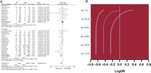
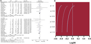
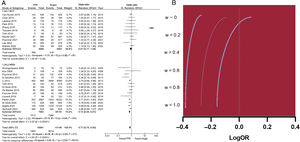
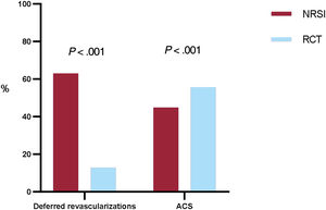
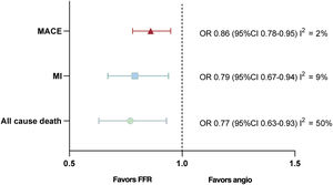
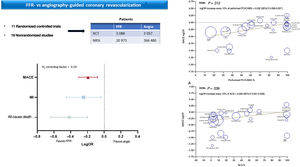
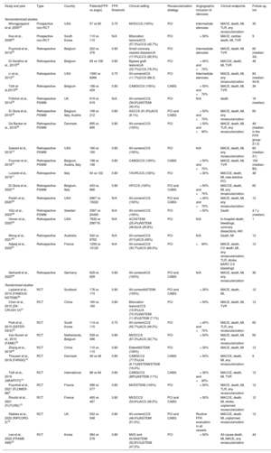
Results

A total of 30 studies enrolling 393 588 patients were included. FFR-guided revascularization was associated with significantly lower rates of all-cause death (OR, 0.63; 95%CI, 0.53-0.73), myocardial infarction (OR, 0.70; 95%CI, 0.59-0.84), and MACE (OR, 0.77; 95%CI, 0.70-0.85). When only RCTs were considered, no significant difference between the 2 strategies was observed for any endpoints. However, the use of FFR was associated with reduced rates of revascularizations and treated lesions. Metaregression suggested that the higher the rate of revascularized patients the lower the benefit of FFR guidance on MACE reduction compared with angiography guidance (P=.012). Similarly, higher rates of patients with acute coronary syndromes were associated with a lower benefit of FFR-guided revascularization (P=.039).

Conclusions

FFR-guided revascularization was associated with lower rates of all-cause death, myocardial infarction and MACE compared with angiographic guidance, with RCTs and nonrandomized intervention studies yielding conflicting data. The benefits of FFR-guidance seem to be less evident in studies with high revascularization rates and with a high prevalence of patients with acute coronary syndrome.

Keywords

Coronary artery disease
Coronary revascularization
Fractional flow reserve
INTRODUCTION

Fractional flow reserve (FFR) is an intracoronary pressure-derived index that quantifies the hemodynamic significance of coronary stenoses. The use of FFR to guide coronary revascularization is recommended by guidelines1 following the publication of several landmark clinical randomized controlled trials (RCTs) such as the “Fractional Flow Reserve Versus Angiography for Multivessel Evaluation” (FAME)2 and the “Fractional Flow Reserve guided PCI vs Medical Therapy in Stable Coronary Disease” (FAME 2).3 Following these seminal studies, the benefit of the FFR-guided revascularization strategy has been confirmed in several nonrandomized studies. However, FFR has been challenged in some clinical settings (eg, in patients with acute coronary syndromes or severe multivessel disease),4,5 raising doubts about its overall usefulness in clinical practice. This meta-analysis provides an updated assessment of the available literature to define which strategy is preferable when comparing FFR-guided vs angiography-guided revascularization according to the study type and different patient subsets.

METHODSSelection criteria and search strategy

The present analysis was conducted according to the recommendations of the Cochrane Collaboration6 and Preferred Reporting Items for Systematic reviews and Meta-Analyses (PRISMA) statements.7 Studies were considered eligible if they provided comparison between clinical endpoints following FFR-guided or angiography-guided revascularization. Both RCTs and nonrandomized intervention studies (NRSI) were included; studies with either percutaneous coronary intervention (PCI) or coronary artery bypass grafting (CABG) as revascularization strategies were considered. If an NRSI provided both propensity score matched and unmatched cohorts, only matched cohorts were included. From inception through April 2023, PubMed/MEDLINE and Cochrane/CENTRAL databases were systematically searched for studies matching the inclusion criteria. A detailed search algorithm can be found in the supplementary data. A minority of articles was selected through citation searching and from authors’ contributions. Two authors (F. Mangiacapra and L. Paolucci) independently selected eligible works at the title/abstract level and disagreements were resolved by a senior author (E. Barbato). Eligible articles were evaluated at the full-text level. Risk of bias was estimated using the Cochrane RoB 2 tool for RCTs8 and the ROBINS-I tool for NRSI.9

Clinical endpoints

Coprimary endpoints were all-cause death, myocardial infarction (MI) and major adverse cardiovascular events (MACE) at longest available follow-up. Among the 25 studies reporting MACE, 7 reported major adverse cerebral and cardiovascular events.10–16 To ensure that the analysis was sufficiently powered, we decided to merge these outcomes and refer to them simply as “MACE”. The various MACE definitions used are summarized in table 1 of the supplementary data. Secondary endpoints were any revascularization, a combination of target vessel failure/target lesion failure and the occurrence of periprocedural MI.

To compare the extent of revascularization deferral (ie, number of performed PCIs and treated lesions) between FFR- and angiography-guidance, an exploratory analysis was conducted using RCTs with only PCI as the revascularization strategy.

Data analysis

The number of events for each endpoint and the cohort size were extracted by careful evaluation of articles. If there were zero events in both groups for a certain outcome, a continuity factor of 1 was used. Heterogeneity was assessed using I2 statistics.17 Odds ratios (ORs) and 95% confidence intervals (95%CI) were calculated using a random-effects model according to the Der Simonian and Leird method.18 A fixed-effects model according to the Mantel-Haenszel method was used to assess OR heterogeneity (complete absence indicated by I2 statistic values near 0%).18 The absolute number of performed PCIs and treated lesions in the RCTs subgroup were compared using a Pearson chi-square test between the 2 groups. The rates of acute coronary syndromes (ACS) and revascularization deferral in the FFR arms of RCT and NRSI were compared in a similar fashion. Two separate analyses were conducted for the coprimary endpoints. In the primary analysis, both RCT and NRSI were included using a cross study design modelling within a Bayesian framework. With this method, studies with a suspected low level of evidence (ie, NRSI) are progressively undersized by inflating their mean effects variances by a wj factor (minimum relative weight: wj∼0; maximum relative weight: wj=1). No prior distributions of bias vectors were assumed.19 The results of the Bayesian adjustment are reported as logOR and 95%CI.

In the secondary analysis, only RCT and propensity score matching adjusted/prospective NRSI were included.20,21 Publication bias was estimated by Egger's test. Prespecified sensitivity analyses were performed by removing 1 study at a time across the groups and by using fixed-effect models for outcomes previously evaluated with random-effects models and showing low-to-moderate levels of heterogeneity. Subgroup analysis was performed between RCTs and non-RCT studies and formally evaluated with chi-square and I2 tests.22

Random-effects meta-regression analysis was performed to study the influence of revascularization rates and clinical presentation (acute vs chronic) on the coprimary endpoints. The percentage of patients undergoing revascularization and the percentage of patients with ACS enrolled in the FFR arm of each study were used as covariates. For the latter analysis, ACS was defined by the sum of patients with unstable angina (UA), non–ST-segment elevation MI, and ST-segment elevation MI.

Statistical analysis was performed with RevMan version 5.4.1 (The Cochrane Collaboration, The Nordic Cochrane Centre, Copenhagen, Denmark) and STATA 16 (Stata Corp, College Station, United States). The study is registered with PROSPERO (CRD42022344765).

RESULTSFeatures of the included studies and extent of revascularization deferral in RCTs

The systematic search led to 4520 records considered for title/abstract level evaluation and 43 articles for full-text review, of which 21 were considered eligible for inclusion. Another 8 studies were added from other sources, eventually leading to the inclusion of 30 studies.5,10–16,23–44 The PRISMA diagram summarizing the research strategy is reported in figure 1 of the supplementary data. The final analysis included 11 RCTs,5,10,12,15,16,30–33,43,45 2 prospective nonrandomized studies,23,24 8 retrospective propensity score-adjusted studies,13,14,29,35–37,39,44 and 9 retrospective unadjusted studies.11,25–28,38,40–42 Risk of bias is shown in figures 2 and 3 of the supplementary data. The analysis involved 393 588 patients: 369 527 underwent angiographic guidance and 24 061 underwent FFR-guidance. The main characteristics of the 30 studies are shown in table 1.

Table 1.

Main characteristics of the included studies

Study and year  Type  Country  Patients(FFR vs angio)  FFR threshold  Clinical setting  Revascularization strategy  Angiographic inclusion of stenoses  Clinical endpoints  Follow-up, mo 
Nonrandomized studies
Wrongpraparut et al. 200523  Prospective non-RCT  USA  57 vs 80  0.75  MVDCCS (100%)  PCI  Intermediate stenoses  MACE, death, MI, TLR, any revascularization  30 
Koo et al., 200824  Prospective non-RCT  South Korea  110 vs 110  N/A  Bifurcation lesionsACS (57.3%)CCS (42.7%)  PCI  > 50%  MACE, cardiac death, MI, TVR 
Puymirat et al., 201225  Retrospective  Belgium  222 vs 476  0.80  Small coronary vessels diseaseUA (17.5%)CCS (82.5%)  PCI  Intermediate stenoses  MACE, death, MI, TVR  60 (median: 34) 
Di Serafino et al., 201326  Retrospective  Belgium  65 vs 158  0.80  Bypass graft lesionsUA (23.7%)CCS (76.3%)  PCI  > 40% and <70%  MACCE, death, MI, TVR  48 
Li et al., 201327  Retrospective  USA  1090 vs 6268  0.75  All-comersACS (11.7%)CCS (88.3)  PCI  Intermediate stenoses  MACE, death, MI, any revascularization  84 (median: 44.9) 
Toth et al.201328  Retrospective  Belgium  198 vs 429  0.80  CABGCCS (100%)  CABG  > 50% and <70%  MACE, death, MI, TVR  36 
Fröhlich et al., 201429  Retrospective-PSWM  UK  919 vs 919  N/A  All-comersCCS (59.6%)NSTEMI (40.4%)  PCI  N/A  death  16 (median) 
Di Gioia et al., 201635  Retrospective-PSWM  Belgium, Italy, Austria  106 vs 212  0.80  ASCCS (91.9%)ACS (8.1%)  PCI and CABG  > 50% and <70%  MACE, death, MI, any revascularization  60 
De Backer et al., 201636  Retrospective-PSWM  Denmark  695 vs 695  0.80  All-comersCCS (100%)  PCI  > 50% and <90%  MACE, death, MI, TLR, any revascularization  48 (median in the FFR group: 21.3) 
Sawant et al., 201813  Retrospective-PSWM  USA  190 vs 190  0.80  All-comersCCS (100%)  PCI  N/A  MACE, death, MI, any revascularization  60 (median: 41) 
Fournier et al., 201837  Retrospective-PSWM  Belgium, Austria, Italy  198 vs 198  0.80  CABGCCS (100%)  CABG  > 50% and <70%  MACE, death, MI, TVR  104 (median: 85) 
Lunardi et al., 201911  Retrospective  Italy  94 vs 122  0.80  TAVRCCS (100%)  PCI  > 30%  MACCE, death, MI, new elective PCI  48 
Di Gioia et al., 202014  Retrospective-PSWM  Belgium, Italy  433 vs 866  0.80  HFCCS (100%)  PCI and CABG  > 50% and <70%  MACCE, death, MI, any revascularization  60 
Parikh et al., 202038  Retrospective  USA  2967 vs 15022  N/A  All-comersCCS (100%)  PCI and CABG  > 40% and <70%  MACE, death, MI, any revascularization  12 
Völz et al., 202039  Retrospective- PSWM  Sweden  3367 vs 20493  N/A  All-comersCCS (100%)  PCI  > 50%  Death  4.7 y (median) 
Omran et al., 202042  Retrospective  USA  7832 vs 296716  N/A  ACSSTEMI (25.4%)NSTEMI (48.8)UA (25.8%)  PCI  N/A  In-hospital death, bleedings, coronary dissections, AKI 
Wong et al., 202140  Retrospective  Australia  542 vs 9762  N/A  All-comersCCS (51%)ACS (49%)  PCI  N/A  Death, MI  12 
Adjedj et al., 202241  Retrospective  France  1259 vs 13125  N/A  All-comersCCS (30.7%)ACS (69.3%)  PCI  <90%  MACE, death, CV death, MI, any revascularization, TLR, stroke, BARC 3-5 bleedings  12 
Gerhardt et al., 202344  Retrospective  Germany  629 vs 629  0.80  All-comersACS (100%)  PCI and CABG  N/A  MACE, death, MI, any revascularization  36 
Randomized studies
Layland et al., 2015 (FAMOUS-NSTEMI)30  RCT  Scotland  176 vs 174  0.80  All-comersNSTEMI (100%)  PCI and CABG  > 30%  MACE, death, MI,  12 
Chen et al., 2015 (DK-CRUSH VI)31  RCT  China  160 vs 160  0.80  Bifurcation lesionsCCS (10.8%)UA (70.3%)NSTEMI (11.8%)STEMI (7.1%)  PCI  > 50%  MACE, death, MI, TVR  12 
Park et al., 2015 (DEFER-DES)32  RCT  South Korea  114 vs 115  0.75  All-comersCCS (50.7%)ACS (49.3%)  PCI  > 40% and <70%  MACE, death, MI, TLR, any revascularization  60 
Van Nunen et al., 2015 (FAME)33  RCT  Netherlands, Belgium  509 vs 496  0.80  MVDCCS (67.3%)ACS (32.7%)  PCI  > 50%  MACE, death, MI, any revascularization  60 
Zhang et al., 201645  RCT  China  110 vs 110  0.80  EldersNSTEMI (100%)  PCI  > 30%  MACE, death, MI  12 
Theusen et al., 2018 (FARGO)12  RCT  Denmark  49 vs 48  0.80  CABGCCS (77.5%)UA (6.1%)NSTEMI/STEMI (16.4%)  CABG  > 50%  MACCE, death, MI, any revascularization 
Toth et al., 2019 (GRAFFITI)10  RCT  International  88 vs 84  0.80  CABGCCS (89%)NSTEMI (11%)  CABG  > 30% and <90%  MACCE, death, MI, TVR  12 
Puymirat et al., 2021 (FLOWER-MI)5  RCT  France  586 vs 577  0.80  MVDSTEMI (100%)  PCI  > 50%  MACE, death, MI, TLR, any revascularization  12 
Rioufol et al., 2021 (FUTURE)15  RCT  France  460 vs 467  0.80  MVDCCS (53.8%)ACS (46.2%)  PCI and CABG  > 50%  MACCE, death, MI, stroke, unplanned revascularization  12 
Stables et al., 2022 (RIPCORD-2)16  RCT  UK  552 vs 548  0.80  All-comersCCS (48.4%)NSTEMI (51.6%)  PCI and CABG  Routine FFR evaluation in all vessels  MACCE, death, MI, unplanned revascularization  12 
Lee et al., 2022 (FRAME-AMI)43  RCT  Korea  284 vs 278  0.80  MVD and ACSNSTEMI (52.8%%)STEMI (47.2%)  PCI  > 50%  All-cause death, MI, MACE, any revascularization  44 

AKI, acute kidney injury; ACS, acute coronary syndrome; AS, aortic stenosis; BARC, bleedings academic research consortium; CABG, coronary artery bypass grafting; CCS, chronic coronary syndrome; FFR, fractional flow reserve; HF, heart failure; MACCE, major adverse cardiovascular and cerebral events; MACE, major adverse cardiovascular events; MI, myocardial infarction; MVD, multivessel disease; N/A, not available; NSTEMI, non-ST elevation myocardial infarction; PCI, percutaneous coronary intervention; PSWM, propensity score weighted matching; RCT, randomized clinical trial; STEMI, ST elevation myocardial infarction; TAVR, transcatheter aortic valve replacement; TLR, target lesion failure; TVR, target vessel failure; UA, unstable angina.

The baseline characteristics of the populations enrolled are summarized in table 2 of the supplementary data. High clinical heterogeneity was observed among the studies, which enrolled patients in different clinical settings including ACS, chronic coronary syndromes (CCS), bypass graft lesions, aortic stenosis, and bifurcation lesions.

The extent of revascularization deferral was reported in 9 PCI RCTs5,15,16,30–33,43,45: 2447 of 2947 (83.0%) patients in the FFR-guided group and 2586 of 2929 (88.2%) patients in angiography-guided group underwent PCI (P< .001). Data on the number of treated lesions were available in 4 studies.5,31–33 Overall, 1485 of 2632 (56.4%) lesions in the FFR-guided group and 2206 of 2606 (86.7%) lesions in the angiography-guided group were treated with PCI (P< .001). Results are shown in table 2. Data on the extent of coronary revascularization in the PCI and CABG studies according to different variables are summarized in tables 3 and 4 of the supplementary data, while periprocedural MI event rates in each study are reported in table 5 of the supplementary data.

Table 2.

Absolute number of performed PCI and treated lesions in the RCTs subgroup

Study and yearRCTs  Performed PCI FFR  Performed PCI angio  P 
Layland 201530  136/176 (77.2%)  151/174 (85.8%)  .020 
Park 201532  41/114 (35.9%)  115/115 (100%)  <.001 
Chen 201531  160/160 (100%)  160/160 (100%  .999 
Van Nunen 201533  509/509 (100%)  496/496 (100%)  .999 
Zhang 201645  95/110 (86.3%)  104/110 (94.5%)  .040 
Puymirat 20215  586/586 (100%)  577/577 (100%)  .999 
Rioufol 202115  328/460 (71.3%)  369/467 (79.0%)  .006 
Stables 202216  308/548 (56.2%)  336/552 (60.9%)  .200 
Lee 202243  284/284  278/278  .999 
Total  2447/2947 (83.0%)  2586/2929 (88.2%)  <.001 
Study and yearRCTs  Treated lesions FFR  Treated lesions angio  P 
Park 201532  29/114 (25.4%)  115/115 (100%)  <.001 
Chen 201533  91/160 (56.8%)  102/160 (63.7%)  .250 
Van Nunen 201531  819/1378 (59.4%)  1237/1350 (91.6%)  <.001 
Puymirat 20215  546/980 (55.7%)  806/981 (82.1%)  <.001 
Total  1485/2632 (56.4%)  2260/2606 (86.7%)  <.001 

FFR, fractional flow reserve; PCI, percutaneous coronary intervention; RCT, randomized clinical trial.

All-cause death

All-cause death was reported in 27 studies enrolling a total of 392 242 patients. An FFR-guided strategy was associated with a reduced risk of all-cause death (OR, 0.63; 95%CI, 0.53-0.73; I2=64%). A funnel plot is reported in figure 4 of the supplementary data. A sensitivity analysis performed by leaving out one study at a time showed no meaningful differences, and the fixed-effects model demonstrated comparable results (OR, 0.55; 95%CI, 0.51-0.59). In subgroup analysis, only NRSI FFR-guidance showed an association with reduced all-cause death (RCTs: OR, 0.99; 95%CI, 0.68-1.44, I2=31%; NRSI: OR, 0.56; 95%CI, 0.48-0.65; I2=64%), with a significant difference between the study design subgroups (P=.009). Mean effect variances adjustment of NRSI confirmed these findings up to a wj value of 0.2. The results of the primary analysis are shown in figure 1.

Figure 1.

Primary analysis for all-cause death. A: forest plot. B: mean effect variance adjustment of NRSI. 95%CI, 95% confidence intervals; FFR, fractional flow reserve; NRSI, nonrandomized intervention studies; RCT, randomized clinical trials. The bibliographic references mentioned in this figure correspond to: Puymirat et al.,5 Toth et al.,10 Lunardi et al.,11 Thuesen et al.,12 Di Gioia et al.,14 Rioufol et al.,15 Stables et al.,16 Wongpraparut et al.,23 Puymirat et al.,25 Di Serafino et al.,26 Li J et al.,27 Toth et al.,28 Fröhlich et al.,29 Layland et al.,30 Chen et al.,31 Van Nunen et al.,33 Di Gioia et al.,35 De Backer et al.,36 Fournier et al.,37 Parikh et al.,38 Völz et al.,39 Wong et al.,40 Adjedj et al.,41 Omran et al.,42 Lee et al.,43 Gerhardt et al.,44 Zhang et al.45.

(0.52MB).
Myocardial infarction

The analysis was performed in 25 studies involving 62 212 patients. FFR-guidance was associated with a reduced risk of MI (OR, 0.70; 95%CI, 0.59-0.84; I2=43%). A funnel plot can be found in figure 5 of the supplementary data. Sensitivity analysis performed by removing 1 study at a time showed no significant differences and a fixed-effects model supported the results (OR, 0.70; 95%CI, 0.62-0.79). In subgroup analysis, only NRSI FFR-guidance showed an association with reduced MI (RCTs: OR, 0.84; 95%CI, 0.64-1.10; I2=20%; NRSI: OR, 0.63; 95%CI, 0.50-0.80; I2=49%), with no difference between study design subgroups (P=.120). These findings were consistent after adjustment of NRSI mean effect variances up to a wj value of 0.2. The results are shown in figure 2.

Figure 2.

Primary analysis for MI. A: forest plot. B: mean effect variance adjustment of NRSI. 95%CI, 95% confidence intervals; FFR; fractional flow reserve; MI, myocardial infarction; NRSI, nonrandomized intervention studies; RCT; randomized clinical trials. The bibliographic references mentioned in this figure correspond to: Puymirat et al.,5 Toth et al.,10 Lunardi et al.,11 Thuesen et al.,12 Di Gioia et al.,14 Rioufol et al.,15 Stables et al.,16 Wongpraparut et al.,23 Koo et al.,24 Puymirat et al.,25 Di Serafino et al.,26 Toth et al.,28 Layland et al.,30 Chen et al.,31 Van Nunen et al.,33 Di Gioia et al.,35 De Backer et al.,36 Fournier et al.,37 Parikh et al.,38 Wong et al.,40 Adjedj et al.,41 Omran et al.,42 Lee et al.,43 Gerhardt et al.44.

(0.54MB).
Major adverse cardiovascular events

A total of 25 studies with 52 137 patients were included. FFR-guidance was associated with a reduced risk of MACE (OR, 0.77; 95%CI, 0.70-0.85; I2=41%). A funnel plot is shown in figure 6 of the supplementary data. A fixed-effects model supported this finding (OR, 0.76; 95%CI, 0.72-0.82) and sensitivity analysis did not impact the result. In subgroup analysis, only NRSI FFR-guidance showed an association with reduced MACE (RCTs: OR, 0.91; 95%CI, 0.77-1.08; I2=9%; NRSI: OR, 0.72; 95%CI, 0.64-0.81; I2=44%) and a significant difference between study design subgroups was evident (P=.030). These findings were confirmed even for high values of NRSI mean effect variances (wj=0.2). These results are reported in figure 3.

Figure 3.

Primary analysis for MACE. A: forest plot. B: mean effect variance adjustment of NRSI. 95%CI, 95% confidence intervals; FFR; fractional flow reserve; MACE, major adverse cardiovascular events; NRSI, nonrandomized intervention studies; RCT; randomized clinical trials. The bibliographic references mentioned in this figure correspond to: Puymirat et al.,5 Toth et al.,10 Lunardi et al.,11 Thuesen et al.,12 Di Gioia et al.,14 Rioufol et al.,15 Stables et al.,16 Wongpraparut et al.,23 Koo et al.,24 Puymirat et al.,25 Di Serafino et al.,26 Li J et al.,27 Toth et al.,28 Layland et al.,30 Chen et al.,31 Park et al.,32 Van Nunen et al.,33 Di Gioia et al.,35 De Backer et al.,36 Fournier et al.,37 Parikh et al.,38 Adjedj et al.,41 Lee et al.,43 Zhang et al.45.

(0.55MB).
Influence of revascularization deferral and ACS rates on clinical outcomes

The rates of PCI or CABG deferral in NRSI and RCTs were 63.0% and 12.9%, respectively (P <.001) (figure 4). The association between FFR-guided revascularization and reduced MACE was blunted with increasing numbers of patients treated with PCI or CABG. The MACE logOR increased by 0.032 for every 10% increase of the covariate (95%CI, 0.006-0.075; P=.057). A similar association was evident with all-cause death [logOR, 0.064; 95%CI, 0.027-0.102; P=.001], but not with MI (P=.167).

Figure 4.

Rates of coronary revascularization deferral and ACS in NRSI and RCTs. ACS, acute coronary syndromes; NRSI, nonrandomized intervention studies; RCT, randomized clinical trials.

(0.04MB).

The rate of ACS was lower in the NRSI compared with RCTs (respectively: 44.9% vs 55.7%; P <.001, figure 4). Meta-regression analysis suggested an inverse relationship between the percentage of patients with ACS and the association between FFR-guided revascularization and reduced MACE: logOR for MACE increased by 0.020 for every 10% increase of the covariate (95%CI, 0.001-0.039; P=.039). A tendency toward significance was found for the MI outcome (P=.050), while no association was found for all-cause death (P=.754).

Secondary analyses of the coprimary endpoints

Secondary analyses were performed including only RCTs and adjusted/prospective NRSI. The results of the primary analyses were confirmed for all coprimary endpoints (all-cause death: OR, 0.77; 95%CI, 0.63-0.93; I2=50%; MI: OR, 0.79; 95%CI, 0.67-0.94; I2=9%; MACE: OR, 0.86; 95%CI, 0.78-0.95; I2=2%). Notably, no difference was found between RCTs and adjusted/prospective NRSI for these outcomes. These results are reported in figure 5. Forest plot analyses are reported in figures 7, 8 and 9 of the supplementary data.

Figure 5.

Main results of the secondary analysis for the 3 coprimary outcomes. 95%CI, 95% confidence intervals; FFR, fractional flow reserve; MACE, major adverse cardiovascular events; MI, myocardial infarction; OR, odds ratio.

(0.06MB).
Secondary endpoints

The risk of further revascularizations (OR, 0.93; 95%CI, 0.83-1.04; I2=29%) or target vessel failure/target lesion failure (OR, 0.75; 95%CI, 0.55-1.01; I2=29%) was not increased by FFR. An exploratory analysis using a fixed-effects model showed significant benefit in the FFR-guided group for this latter outcome (OR, 0.75; 95%CI, 0.57-1; I2=29%). The risk of periprocedural MI was reduced in the FFR-guided group (OR, 0.60; 95%CI, 0.39-0.95; I2=41%). Forest plot analyses are reported in figures 10, 11 and 12 of the supplementary data.

DISCUSSION

Our meta-analysis comparing FFR-guided vs angiography-guided revascularization strategies demonstrated that: a) FFR-guided revascularization were associated with overall improved clinical outcomes, as in both primary (including all studies) and secondary analyses (including RCTs and adjusted NRSI), the rates of MI and MACE were reduced in the FFR-guided group; b) although these findings were mainly driven by data from NRSI, their relative influence can be largely undersized without compromising the final results; c) the percentage of coronary revascularization deferral and ACS was higher in NRSI than in RCTs, d) the benefits of FFR-guided revascularization in reducing MACE and all-cause death seemed to be less pronounced in studies with higher revascularization rates and a high prevalence of ACS; e) when only RCTs were analysed, an FFR-guided strategy was associated with significant deferral of lesions without compromising patient safety. The results of the present analysis are briefly synthesized in figure 6. Our findings confirm the results of previous studies investigating this topic in RCTs46,47 and identify the possible influence of data from NRSI in evaluation of the benefit of an FFR-guided strategy. Moreover, this is the first analysis investigating the role of revascularization deferral and type of coronary syndrome in determining the association between FFR and clinical outcomes at a meta-analytic level.

Figure 6.

Central illustration. FFR- vs angiography-guided coronary revascularization. 95%CI, confidence intervals; ACS, acute coronary syndromes; CABG, coronary artery bypass grafting; FFR, fractional flow reserve; MACE, major adverse cardiovascular events; MI, myocardial infarction; NRSI, nonrandomized intervention studies; OR, odds ratio; PCI, percutaneous coronary intervention; RCT, randomized clinical trials; REML, restricted maximum likelihood.

(0.34MB).
Clinical outcomes of FFR-guided revascularization

Compared with angiography, the adoption of physiology has the advantage of distinguishing epicardial coronary stenoses associated with myocardial ischemia,48 for which revascularization might be warranted, from those where revascularization could be deferred.49 The association between reduced FFR and adverse events has been widely described.50 Studies investigating the natural history of coronary lesions have shown that there is a strong relationship between lesion severity (evaluated with FFR) and rates of MACE and MI.51,52 These data are supported by evidence showing a high grade of inflammatory stress in FFR-positive lesions53 that could increase plaque instability. Consequently, the reduced rates of MI after revascularizing FFR-positive lesions could be associated with reduced mortality and MACE rates during follow-up. Compared with angiography, the addition of FFR maintains the benefits in terms of spontaneous MI reduction. At the same time, by deferring functionally insignificant lesions, FFR decreases the risks associated with procedural myocardial injury,54 which is related to impaired outcomes.55,56 Accordingly, our findings support that FFR-guided revascularization is consistently associated with a lower risk of MI and MACE. Moreover, when considering RCTs of patients treated with PCI, FFR adoption was associated with a lower number of interventions, confirming that deferring PCI in nonfunctionally significant lesions is safe.

Factors influencing the benefits of FFR-guidance

Current guidelines recommend the implementation of coronary angiography with physiologic assessment to guide revascularization of intermediate stenoses in patients without prior evidence of myocardial ischemia.1 However, clinical adoption of FFR has been modest over the years.38,57 In addition to several possible logistic reasons limiting FFR penetration, there might also have been conflicting evidence from RCTs following the results of the initial trials.16,30–34 A critical appraisal of the literature suggests that major differences in study designs affecting the ratio of performed/deferred interventions should be thoroughly accounted for when estimating the effectiveness of FFR. In the FAME trial, intermediate lesions considered for invasive functional evaluation with FFR were at least 50% diameter stenosis by visual estimation.2 In contrast, a 30% diameter stenosis was adopted in other major RCTs.30 The inclusion of lesions with less than 30% diameter stenosis can be is expected to dilute the potential clinical benefit associated with functional assessment. In fact, in a hypothetical study randomizing patients to FFR or angiography, the inclusion of mild coronary lesions could be expected to lead to a higher rate of deferred interventions even in patients not treated with physiology guidance. If we assume that the superiority of FFR is related to the proportion of safely deferred nonindicated interventions compared with angiography, this could hinder the detection of a net clinical benefit. Our analysis supports the hypothesis that the benefit of FFR-guidance is more evident when the proportion of revascularization deferral is higher compared with pure angiography-guidance, possibly due to the lower risk of late and acute complications.42,58

We found an association between the rates of patients enrolled with ACS and the benefits of an FFR-guided revascularization strategy. The effectiveness of FFR in detecting significant lesions depends on the achievement of steady-state and maximal coronary hyperemia. In the setting of ACS (especially during ST-segment elevation MI), dynamic changes in the microvascular function of both the jeopardized and nonjeopardized myocardium can lead to increased resting and reduced hyperemic coronary flow. These modifications could hinder the effectiveness of FFR in detecting significant lesions, potentially leading to underestimation.59 Currently, conflicting data are available regarding the reliability of FFR evaluation in this setting and no definitive conclusions can be drawn.60–62 Additionally, further possible explanations could be proposed to justify our finding. In the acute setting, several conditions can hamper the effectiveness of FFR, including hemodynamic instability, challenges in identifying the real nonculprit lesions,30 the need for multiple interventions,5 and the risk of endangering nonculprit vessels during FFR guidewire manipulation.16 Further studies are needed to address this topic, focusing on the relationship between ACS subtypes and the length of follow-up.

Clinical outcomes according to study type

One interesting point from our analysis is the striking difference in effect size between RCTs and NRSI. Recently published meta-analyses of RCTs have shown that FFR-guided revascularization was not superior to angiography in improving outcomes.46,47 These results highlight the inconsistency currently existing between data from RCT and NRSI.63 One of the main purposes of our study was to explore the nature of these conflicting data and to evaluate the relative contribution of observational studies in determining the benefits of FFR-guided revascularization. In our study, the influence of NRSI could be largely undersized without compromising the final evidence of a clinical benefit derived from the use of FFR. Similar findings have been confirmed including only adjusted NRSI. It is reasonable to assume that in NRSI, residual confounding from selection bias persists, especially in the absence of statistical adjustment. On the other hand, it could be argued that RCTs are highly selective, plagued by the so-called “entry bias”, and their generalizability is often questionable due to their tendency to exclude higher-risk patients.64 Among potential confounding factors, the appropriateness of physiological assessment based on clinical presentation and a differential risk profile of included patients may account for the observed divergence.63 Besides these considerations, other factors can influence the conflicting results of RCTs and NRSI, including the higher rate of patients deferred in NRSI and the limits of some RCTs in terms of sample sizes and the low number of observed events.63 Nevertheless, the interpretation of NRSI should be cautious and based on a thorough evaluation of inclusion/exclusion criteria and patient risk profiles.

Limitations

This study has several limitations. First, the lack of patient-level data prevented us from performing prespecified adjustments. This should be particularly considered when evaluating our results on the relationship between clinical outcomes, ACS rates and deferral rates, due to the possibility of significant uncontrolled collinearity between these confounders. Moreover, although we performed several statistical computations to balance the evidence from different study types, we cannot assume that these results are comparable to those from only pooled RCTs in terms of quality. In this regard, propensity score matching is only one of the available methods used to adjust for confounders, and several strategies could have led to slightly different results. Again, when including NRSI in a meta-analytic framework, positive bias related to selective reporting can be difficult to address. Eventually, including a large number of significantly different studies has surely led to a residual unavoidable heterogeneity. Although we showed that there are significant differences between RCTs and NRSI in terms of inclusion criteria (eg, angiographic severity of the lesions), ACS rates, deferral rates and others, we cannot directly assume that our methodological efforts to manage these major sources of heterogeneity have been completely effective. Therefore, we believe that our results can only be considered as hypothesis-generating.

CONCLUSIONS

Our analysis found that FFR-guided revascularization was associated with a significant reduction in all-cause death, MI, and MACE. These findings are driven by data from nonrandomized studies. Regardless of the study type, FFR effectively reduces the rates of non-beneficial revascularization without compromising clinical outcomes. The benefits of FFR-guidance may be lower when the number of patients with ACS and the revascularization rates are higher.

FUNDING

None.

ETHICAL CONSIDERATIONS

Review article not requiring ethical statement.

STATEMENT ON THE USE OF ARTIFICIAL INTELLIGENCE

Artificial intelligence was not used to draft the manuscript or perform analyses.

AUTHORS’ CONTRIBUTIONS

F. Mangiacapra, L. Paolucci, and E. Barbato conceived the idea. F. Mangiacapra, L. Paolucci, and M.M. Viscusi performed the studies’ research. F. Mangiacapra, L. Paolucci, and N.P. Johnson performed the statistical analysis. F. Mangiacapra and L. Paolucci, drafted the paper. All authors critically revised the manuscript.

CONFLICTS OF INTEREST

None.

WHAT IS KNOWN ABOUT THE TOPIC?

  • -

    FFR-guided coronary revascularization is associated increased rates of safely deferred interventions, but recent studies have questioned its benefits in terms of clinical outcomes.

WHAT DOES THIS STUDY ADD?

  • -

    Data from randomized and nonrandomized studies suggest that FFR can lead to reduced rates of all-cause death, MACE, and MI. These findings are mostly driven by the results of nonrandomized studies. The benefits of FFR-guided revascularization are lower in studies with high rates of nondeferred interventions and ACS patients.

References
[1]
J. Knuuti, W. Wijns, A. Saraste, et al.
2019 ESC Guidelines for the diagnosis and management of chronic coronary syndromes.
Eur Heart J., (2020), 41 pp. 407-477
[2]
P.A.L. Tonino, B. De Bruyne, N.H.J. Pijls, et al.
Fractional Flow Reserve versus Angiography for Guiding Percutaneous Coronary Intervention.
New Engl J Med., (2009), 360 pp. 213-224
[3]
B. De Bruyne, N.H.J. Pijls, B. Kalesan, et al.
Fractional flow reserve-guided PCI versus medical therapy in stable coronary disease.
N Engl J Med., (2012), 367 pp. 991-1001
[4]
W.F. Fearon, F.M. Zimmermann, B. De Bruyne, et al.
Fractional Flow Reserve-Guided PCI as Compared with Coronary Bypass Surgery.
N Engl J Med., (2022), 386 pp. 128-137
[5]
E. Puymirat, G. Cayla, T. Simon, et al.
Multivessel PCI Guided by FFR or Angiography for Myocardial Infarction.
N Engl J Med., (2021), 385 pp. 297-308
[6]
J.P. Higgins, J. Thomas, J. Chandler, M. Cumpston, M.J. Page, V.A. Welch.
Cochrane Handbook for Systematic Reviews of Interventions.
2nd ed., John Wiley and Sons Inc, (2019),
[7]
M.J. Page, J.E. McKenzie, P.M. Bossuyt, et al.
The PRISMA 2020 statement: an updated guideline for reporting systematic reviews.
BMJ., (2021), 372 pp. n71
[8]
J.A.C. Sterne, J. Savović, M.J. Page, et al.
RoB 2 a revised tool for assessing risk of bias in randomised trials.
BMJ., (2019), 366 pp. l4898
[9]
J.A.C. Sterne, M.A. Hernán, B.C. Reeves, et al.
ROBINS-I: a tool for assessing risk of bias in non-randomised intervention studiess.
BMJ., (2016), 355 pp. i4919
[10]
G.G. Toth, B. de Bruyne, P. Kala, et al.
Graft patency after FFR-guided versus angiography-guided coronary artery bypass grafting: The GRAFFITI trial.
EuroIntervention., (2019), 15 pp. E999-E1005
[11]
M. Lunardi, R. Scarsini, G. Venturi, et al.
Physiological Versus Angiographic Guidance for Myocardial Revascularization in Patients Undergoing Transcatheter Aortic Valve Implantation.
J Am Heart Assoc., (2019), 8 pp. e012618
[12]
A.L. Thuesen, L.P. Riber, K.T. Veien, et al.
Fractional Flow Reserve Versus Angiographically-Guided Coronary Artery Bypass Grafting.
J Am Coll Cardiol., (2018), 72 pp. 2732-2743
[13]
A.C. Sawant, A. Bhardwaj, K. Banerjee, et al.
Fractional flow reserve guided percutaneous coronary intervention results in reduced ischemic myocardium and improved outcomes.
Catheter Cardiovasc Interv., (2018), 92 pp. 692-700
[14]
G. Di Gioia, B. De Bruyne, M. Pellicano, et al.
Fractional flow reserve in patients with reduced ejection fraction.
Eur Heart J., (2020), 41 pp. 1665-1672
[15]
G. Rioufol, F. Dérimay, F. Roubille, et al.
Fractional Flow Reserve to Guide Treatment of Patients With Multivessel Coronary Artery Disease.
J Am Coll Cardiol., (2021), 78 pp. 1875-1885
[16]
R.H. Stables, L.J. Mullen, M. Elguindy, et al.
Routine Pressure Wire Assessment Versus Conventional Angiography in the Management of Patients With Coronary Artery Disease: The RIPCORD 2 Trial.
Circulation., (2022), 146 pp. 687-698
[17]
J.P.T. Higgins, S.G. Thompson, J.J. Deeks, D.G. Altman.
Measuring inconsistency in meta-analyses.
BMJ., (2003), 327 pp. 557-560
[18]
M. Borenstein, L.V. Hedges, J.P.T. Higgins, H.R. Rothstein.
A basic introduction to fixed-effect and random-effects models for meta-analysis.
Res Synth Methods., (2010), 1 pp. 97-111
[19]
O. Efthimiou, D. Mavridis, T.P.A. Debray, et al.
Combining randomized and non-randomized evidence in network meta-analysis.
Stat Med., (2017), 36 pp. 1210-1226
[20]
C. Montalto, A. Sticchi, G. Crimi, et al.
Outcomes After Transcatheter Aortic Valve Replacement in Bicuspid Versus Tricuspid Anatomy: A Systematic Review and Meta-Analysis.
JACC Cardiovasc Interv., (2021), 14 pp. 2144-2155
[21]
M.A. Kelshiker, H. Seligman, J.P. Howard, et al.
Coronary flow reserve and cardiovascular outcomes: a systematic review and meta-analysis.
Eur Heart J., (2022), 43 pp. 1582-1593
[22]
M. Richardson, P. Garner, S. Donegan.
Interpretation of subgroup analyses in systematic reviews: A tutorial.
Clin Epidemiol Glob Health., (2019), 7 pp. 192-198
[23]
N. Wongpraparut, V. Yalamanchili, V. Pasnoori, et al.
Thirty-month outcome after fractional flow reserve-guided versus conventional multivessel percutaneous coronary intervention.
Am J Cardiol., (2005), 96 pp. 877-884
[24]
B.K. Koo, K.W. Park, H.J. Kang, et al.
Physiological evaluation of the provisional side-branch intervention strategy for bifurcation lesions using fractional flow reserve.
Eur Heart J., (2008), 29 pp. 726-732
[25]
E. Puymirat, A. Peace, F. Mangiacapra, et al.
Long-term clinical outcome after fractional flow reserve-guided percutaneous coronary revascularization in patients with small-vessel disease.
Circ Cardiovasc Interv., (2012), 5 pp. 62-68
[26]
L. Di Serafino, B. De Bruyne, F. Mangiacapra, et al.
Long-term clinical outcome after fractional flow reserve- versus angio-guided percutaneous coronary intervention in patients with intermediate stenosis of coronary artery bypass grafts.
Am Heart J., (2013), 166 pp. 110-118
[27]
J. Li, M.Y. Elrashidi, A.J. Flammer, et al.
Long-term outcomes of fractional flow reserve-guided vs. angiography-guided percutaneous coronary intervention in contemporary practice.
Eur Heart J., (2013), 34 pp. 1375-1383
[28]
G. Toth, B. De Bruyne, F. Casselman, et al.
Fractional flow reserve-guided versus angiography-guided coronary artery bypass graft surgery.
Circulation., (2013), 128 pp. 1405-1411
[29]
G.M. Fröhlich, S. Redwood, R. Rakhit, et al.
Long-term survival in patients undergoing percutaneous interventions with or without intracoronary pressure wire guidance or intracoronary ultrasonographic imaging: A large cohort study. JAMA Intern Med.
[30]
J. Layland, K.G. Oldroyd, N. Curzen, et al.
Fractional flow reserve vs Angiography in guiding management to optimize outcomes in non-ST-segment elevation myocardial infarction: The British Heart Foundation FAMOUS-NSTEMI randomized trial.
Eur Heart J., (2015), 36 pp. 100-111
[31]
S.L. Chen, F. Ye, J.J. Zhang, et al.
Randomized comparison of FFR-guided and angiography-guided provisional stenting of true coronary bifurcation lesions: The DKCRUSH-VI trial (double kissing crush vs provisional stenting technique for treatment of coronary bifurcation lesions VI).
JACC Cardiovasc Interv., (2015), 8 pp. 536-546
[32]
S.H. Park, K.H. Jeon, J.M. Lee, et al.
Long-Term Clinical Outcomes of Fractional Flow Reserve-Guided Versus Routine Drug-Eluting Stent Implantation in Patients with Intermediate Coronary Stenosis: Five-Year Clinical Outcomes of DEFER-DES Trial.
Circ Cardiovasc Interv., (2015), 8 pp. 1-7
[33]
L.X. Van Nunen, F.M. Zimmermann, P.A.L. Tonino, et al.
Fractional flow reserve versus angiography for guidance of PCI in patients with multivessel coronary artery disease (FAME): 5-year follow-up of a randomised controlled trial.
The Lancet., (2015), 386 pp. 1853-1860
[34]
D. Zhang, S. Lv, X. Song, F. Yuan, F. Xu, M. Zhang.
Fractional fl ow reserve versus angiography for guiding percutaneous coronary intervention: a meta-analysis.
[35]
G. Di Gioia, M. Pellicano, G.G. Toth, et al.
Fractional Flow Reserve-Guided Revascularization in Patients with Aortic Stenosis.
Am J Cardiol., (2016), 117 pp. 1511-1515
[36]
O. De Backer, L. Biasco, J. Lønborg, et al.
Long-term outcome of FFR-guided PCI for stable coronary artery disease in daily clinical practice: A propensity scorematched landmark analysis.
EuroIntervention., (2016), 11 pp. e1257-e1266
[37]
S. Fournier, G.G. Toth, B. De Bruyne, et al.
Six-Year Follow-Up of Fractional Flow Reserve-Guided Versus Angiography-Guided Coronary Artery Bypass Graft Surgery.
Circ Cardiovasc Interv., (2018), 11 pp. 1-7
[38]
R.V. Parikh, G. Liu, M.E. Plomondon, et al.
Utilization and Outcomes of Measuring Fractional Flow Reserve in Patients With Stable Ischemic Heart Disease.
J Am Coll Cardiol., (2020), 75 pp. 409-419
[39]
S. Völz, C. Dworeck, B. Redfors, et al.
Survival of Patients With Angina Pectoris Undergoing Percutaneous Coronary Intervention With Intracoronary Pressure Wire Guidance.
J Am Coll Cardiol., (2020), 75 pp. 2785-2799
[40]
C. Wong, A. Ng, C. Ada, et al.
A real-world comparison of outcomes between fractional flow reserve-guided versus angiography-guided percutaneous coronary intervention.
PLoS One., (2021), 16 pp. e0259662
[41]
J. Adjedj, J.F. Morelle, C. Saint Etienne, et al.
Clinical impact of FFR-guided PCI compared to angio-guided PCI from the France PCI registry.
Catheter Cardiovasc Interv., (2022), 100 pp. 40-48
[42]
J. Omran, T. Enezate, O. Abdullah, et al.
Outcomes of fractional flow reserve-guided percutaneous coronary interventions in patients with acute coronary syndrome.
Catheter Cardiovasc Interv., (2020), 96 pp. E149-E154
[43]
J.M. Lee, H.K. Kim, K.H. Park, et al.
Fractional flow reserve versus angiography-guided strategy in acute myocardial infarction with multivessel disease: a randomized trial.
Eur Heart J., (2023), 44 pp. 473-484
[44]
T. Gerhardt, B.E. Stähli, T.K. Rudolph, et al.
Prognostic impact of fractional flow reserve measurements in patients with acute coronary syndromes: a subanalysis of the FLORIDA study.
Heart Vessels., (2023), 38 pp. 1009-1018
[45]
Z. Zhang, K. Li, J. Tian.
Efficacy and safety outcomes of fractional flow reserve in guiding clinical therapy of non-ST-segment elevation myocardial infarction compared with angiography alone in elderly Chinese patients.
Clin Interv Aging., (2016), 11 pp. 1751-1754
[46]
A.M. Maznyczka, C.J. Matthews, J.M. Blaxill, et al.
Fractional Flow Reserve versus Angiography-Guided Management of Coronary Artery Disease: A Meta-Analysis of Contemporary Randomised Controlled Trials.
J Clin Med., (2022), 11 pp. 7092
[47]
A. Elbadawi, R. Sedhom, A.T. Dang, et al.
Fractional flow reserve versus angiography alone in guiding myocardial revascularisation: a systematic review and meta-analysis of randomised trials.
Heart., (2022), 108 pp. 1699-1706
[48]
B. De Bruyne, J. Bartunek, S.U. Sys, G.R. Heyndrickx.
Relation between myocardial fractional flow reserve calculated from coronary pressure measurements and exercise-induced myocardial ischemia.
Circulation., (1995), 92 pp. 39-46
[49]
F.M. Zimmermann, A. Ferrara, N.P. Johnson, et al.
Deferral vs. performance of percutaneous coronary intervention of functionally non-significant coronary stenosis: 15-year follow-up of the DEFER trial.
Eur Heart J., (2015), 36 pp. 3182-3188
[50]
N.P. Johnson, G.G. Tóth, D. Lai, et al.
Prognostic value of fractional flow reserve: linking physiologic severity to clinical outcomes.
J Am Coll Cardiol., (2014), 64 pp. 1641-1654
[51]
E. Barbato, G.G. Toth, N.P. Johnson, et al.
A Prospective Natural History Study of Coronary Atherosclerosis Using Fractional Flow Reserve.
J Am Coll Cardiol., (2016), 68 pp. 2247-2255
[52]
Z. Piróth, B.M. Boxma-de Klerk, E. Omerovic, et al.
The Natural History of Nonculprit Lesions in STEMI: An FFR Substudy of the Compare-Acute Trial.
JACC Cardiovasc Interv., (2020), 13 pp. 954-961
[53]
O. Muller, A. Ntalianis, W. Wijns, et al.
Association of Biomarkers of Lipid Modification with Functional and Morphological Indices of Coronary Stenosis Severity in Stable Coronary Artery Disease.
J Cardiovasc Transl Res., (2013), 6 pp. 536-544
[54]
S.J. Park, J.M. Ahn, G.M. Park, et al.
Trends in the outcomes of percutaneous coronary intervention with the routine incorporation of fractional flow reserve in real practice.
Eur Heart J., (2013), 34 pp. 3353-3361
[55]
J. Silvain, M. Zeitouni, V. Paradies, et al.
Cardiac procedural myocardial injury, infarction, and mortality in patients undergoing elective percutaneous coronary intervention: a pooled analysis of patient-level data.
Eur Heart J., (2021), 42 pp. 323-334
[56]
M. Zeitouni, J. Silvain, P. Guedeney, et al.
Periprocedural myocardial infarction and injury in elective coronary stenting.
Eur Heart J., (2018), 39 pp. 1100-1109
[57]
G. Toth, N.P. Johnson, W. Wijns, et al.
Revascularization decisions in patients with chronic coronary syndromes: Results of the second International Survey on Interventional Strategy (ISIS-2).
Int J Cardiol., (2021), 336 pp. 38-44
[58]
R. Piccolo, A. Oliva, M. Avvedimento, et al.
Mortality after bleeding versus myocardial infarction in coronary artery disease: a systematic review and meta-analysis.
EuroIntervention., (2021), 17 pp. 550-560
[59]
T. Thim, N.W. van der Hoeven, C. Musto, et al.
Evaluation and Management of Nonculprit Lesions in STEMI.
JACC Cardiovasc Interv., (2020), 13 pp. 1145-1154
[60]
C. Musto, F. De Felice, S. Rigattieri, et al.
Instantaneous wave-free ratio and fractional flow reserve for the assessment of nonculprit lesions during the index procedure in patients with ST-segment elevation myocardial infarction: The WAVE study.
Am Heart J., (2017), 193 pp. 63-69
[61]
A. Ntalianis, J.W. Sels, G. Davidavicius, et al.
Fractional flow reserve for the assessment of nonculprit coronary artery stenoses in patients with acute myocardial infarction.
JACC Cardiovasc Interv., (2010), 3 pp. 1274-1281
[62]
N.W. van der Hoeven, G.N. Janssens, G.A. de Waard, et al.
Temporal Changes in Coronary Hyperemic and Resting Hemodynamic Indices in Nonculprit Vessels of Patients With ST-Segment Elevation Myocardial Infarction.
JAMA Cardiol., (2019), 4 pp. 736-744
[63]
L. Paolucci, F. Mangiacapra, M.M. Viscusi, et al.
Fractional Flow Reserve-Guided Coronary Revascularization: Evidence from Randomized and Non-Randomized Studies.
Diagnostics (Basel)., (2022), 12 pp. 2659
[64]
R.C. Sheldrick.
Randomized Trials vs Real-world Evidence.
Copyright © 2024. Sociedad Española de Cardiología