To the Editor,
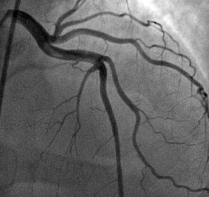
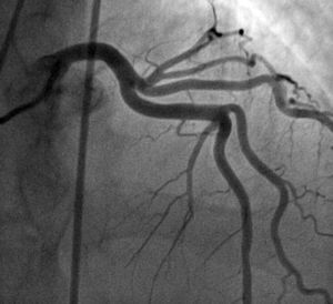
We read with interest the article in the July issue of the Revista Española de Cardiología about hematologic diseases and the heart. We believe it would be appropriate to add elevated factor VIII (FVIII) as a risk factor for venous and arterial thrombotic events. For example, we present the case of a 45-year-old man with no cardiovascular risk factors who played sports regularly and had a body mass index of 23. He attended our hospital because of severe chest pain after cycling for 20min. The electrocardiogram (ECG) showed ST elevation in leads V2 to V6, I, and aVL, and complete right bundle branch block. Fibrinolytic therapy was administered (7000 U of tenecteplase) for the first hour after onset of pain and he was transferred to our hospital. On arrival, he was asymptomatic and hemodynamically stable; the ECG showed normal ST segment with normal QS in leads V2 to V3 and negative T-waves in the precordial leads. Transthoracic echocardiography on admission revealed a dilated left ventricle, with a slightly depressed left ventricular ejection fraction (LVEF) of 38% and anteroseptal akinesia. At 23h after onset, coronary angiography was performed, showing the presence of thrombotic material in the proximal segment of the left anterior descending artery, responsible for 40% obstruction with distal TIMI III flow. The remaining vessels were free of disease (Figure 1). The physical examination was normal; laboratory tests showed troponin I levels of 57 ng/mL and peak creatine kinase of 2440 IU/L. The lipid profile, blood count, and coagulation parameters were all normal. Given the lack of cardiovascular risk factors and the presence of arterial thrombosis, a hypercoagulation study was performed 48h after admission to hospital. Fibrinogen, protein S, protein C, and antithrombin III levels were normal. Neither mutation of Leiden factor V nor lupus anticoagulant was detected. FVIII levels were elevated (234.2%; normal range, 50%-140%). The patient did not have a history of venous or arterial thrombosis. The case was discussed with members of the Hematology department, who indicated starting anticoagulation therapy with acenocumarol. Seven days after the infarction, the coronary angiography was repeated and it was confirmed that the thrombus had disappeared (Figure 2). Three months after the infarction, the patient remained asymptomatic and with improved ventricular function (LVEF, 50%) in the follow-up echocardiography but FVIII levels were still high (210%)..

Figure 1. Coronary angiography. Anteroposterior cranial view: thrombus in proximal segment of the left anterior descending artery.

Figure 2. Follow-up coronary angiogram at 7 days. The intracoronary thrombus has disappeared.
Although the main cause of myocardial infarction is atherosclerotic disease, other causes such as coronary artery thrombosis should be kept in mind, particularly in younger patients. Study of coronary artery disease in younger individuals includes patients younger than 45 years. Our patient was 45 years old, the cutoff age, with no cardiovascular risk factors, and he presented with acute anterior myocardial infarction caused by a thrombus in the left anterior descending artery, with no angiographic evidence of coronary artery disease..
Among coagulation system disorders traditionally associated with arterial thrombosis are hyperfibrinogenemia; protein C, protein S, and antithrombin factor III deficiencies; increased thrombin activity; and mutation of Leiden factor V.1, 2 For several years, FVIII elevation has been included in the list of prothrombotic factors. FVIII is a glycoprotein that acts as a cofactor for factor IX and is essential for thrombus formation. Several studies have shown that elevated FVIII is an independent risk factor for arterial thrombosis (myocardial infarction, ischemic stroke, and peripheral arterial thrombosis), venous thrombosis, and thrombotic recurrence.3, 4, 5 Most patients included in these studies had their first event at a younger age than our patient. It is also known that patients with acute myocardial infarction with elevated FVIII have a worse prognosis. However, the underlying mechanism for this FVIII elevation is not known,6 and there is little indication in the literature about management of patients who have suffered thrombotic events and how long anticoagulation therapy should be maintained in the absence of a recurrence. We followed the indications of colleagues from the Hematology department, who recommended oral anticoagulation for 6 months..
In summary, FVIII should be included in the study of hypercoagulation of a patient with myocardial infarction and no arteriosclerotic disease..
Corresponding author: veronicahernandezz@hotmail.com