
.
Propionibacterium acnes is a slow-growing Gram-positive anaerobic bacillus, which is part of the bacterial flora of the skin and is also present in mucous membranes. Isolated cases of pericarditis caused by P acnes have been reported1,2 but, even though this microorganism is a frequent cause of the disease,3 its characteristics have not been described. We report the characteristics of 5 patients with constrictive infectious pericarditis caused by P. acnes—initially manifesting as constriction in 3 and as effusive-constrictive syndrome in 2—characterized by a torpid clinical course, minimal signs of infection, much inflammatory activity and the need for surgery and prolonged treatment with antibiotics, antiinflammatory drugs, and corticosteroids.
The 5 patients were attended between 2006 and 2011. All underwent transthoracic echocardiography and 3 underwent magnetic resonance imaging studies. For diagnostic purposes, the microbiology results were combined with the echocardiogram results in 4 patients and with the magnetic resonance imaging results in 1. Pathologic analysis was performed in 4 patients.
The mean age was 44.4 years; 4 were men. Possible predisposing factors are shown in the Table. The predominant symptoms were asthenia, chest pain and symptoms of heart failure with elevated venous pressure, dyspnea and edema of the lower limbs; 3 patients had pulsus paradoxus. One patient had fever and another had chylous ascites with tension. The mean diagnostic delay was 30 weeks. One patient had hypogammaglobulinemia, adenopathy and pulmonary infiltration caused by lymphangiectasia, which disappeared once pericardial constriction had been resolved. All electrocardiograms showed low voltage in precordial leads, with ST-segment depression and T wave inversion in 3 of them.
Characteristics of the 5 Patients With Constrictive Infectious Pericarditis Caused by Propionibacterium acnes.
| Man, 55 years | Man, 26 years | Man, 31 years | Man, 72 years | Woman, 38 years | |
| Predisposing factor | Respiratory infection | Tonsillopharyngitis, dental caries | Dental infection | Not reported | 2 week-long respiratory infection |
| Symptoms and diagnostic delay | Pleuritic pain, dyspnea, heart failure, 2 months | Chest pain, dyspnea, palpitations, syncope, 2 weeks | Dyspnea, chest pain, right heart failure, fever, 9 months | Asthenia, progressive dyspnea, right heart failure, 20 months | Chest pain, asthenia, dry cough, dyspnea, right heart failure, 1 month |
| Echocardiogram | Constrictive infectious pericarditis, mild pericardial effusion, bilateral pleural effusion | Constrictive infectious pericarditis with cardiac taponade, moderate-to-severe effusion of up to 21 mm | Increased intrapericardic space, solid/fluid material causing constriction | Performed at the onset of symptoms, without changes (1 year prior to surgery) | Pericardial thickening with mild effusion, effusive-constrictive syndrome |
| Magnetic Resonance Imaging | Substantial pericardial effusion, hypointense nodular areas | Pericardial thickening, dilated inferior vena cava | Pericardial thickening with associated effusion | ||
| Pathology | |||||
| Macroscopic | Pericardial thickening, whitish colored nodule | Pericardial thickening | First surgical intervention: pericardial thickening; second intervention: adhesions, calcified nodules with caseous appearance | Pericardial thickening | Pericardial fluid with hematic material |
| Microscopic | Chronic inflammation, reactive fibrosis | Acute inflammation, fibrillin deposit, mesothelial hyperplasia | Marked fibrosis and foci oflymphocytosis | Intense pericardial fibrosis | Not performed |
| Biologic samples | P. acnes + Staphylococcus epidermidis in pericardial tissue | P. acnes in pericardial fluid and pericardial tissue | S. epidermidis in fluid in first surgical intervention; P. acnes in pericardial tissue in second intervention | P. acnes in pericardial and ascitic fluid | P. acnes in pericardial fluid |
| Surgical treatment | Subtotal pericardiectomy with pericardial patching in right cavities | Pleuropericardial pericardial window-type surgical drainage | First subtotal pericardiectomy, second patched epicardiectomy and residual pericardiectomy | Total pericardiectomy with decortication | Subxiphoid pericardial drainage without surgery |
| Medical treatment and outcome | • 1 month ceftriaxone + amoxicillin-clavulanic acid 1 month + amoxicillin and corticosteroids 6 months more• 1 relapse, resolved in 1 year | • Amoxicillin-clavulanic acid + amoxicillin 6 months + doxycycline2 months, corticosteroids 3 months• 1 relapse requiring ASA + colchicine, resolved in 21 months | • Post-second surgery, penicillin G sodium, amoxicillin 10 months + moxifloxacin 8 months, corticosteroids + colchicine + NSAID 10 months• Resolved at 3 years | • Ceftriaxone 2 weeks + minocycline 2 months• Without relapse | • Ceftriaxone + daptomycin 2 weeks, doxycycline 6 months• 2 relapses, current treatment with corticosteroids + colchicine + NSAID 12 months |
ASA, acetylsalicylic acid; NSAID, nonsteroid antiinflammatory drugs; P. acnes, Propionibacterium acnes; S. epidermidis, Staphylococcus epidermidis.
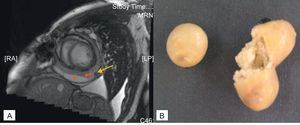
All chest X-rays showed cardiomegaly. Echocardiographic and magnetic resonance imaging studies are described in the Table. One patient had a calcified mass, with pericardial effusion and lateral myocardial hypokinesia. The Figure shows the surgical specimen, illustrating growth of P. acnes, together with an magnetic resonance imaging scan.
Medical treatment, microbiology and histology results are outlined in the Table. P. acnes grew in the samples between days 10 and 12. The results of blood cultures (3 patients) were negative. P. acnes was penicillin-sensitive in all patients. The 5 patients received intravenous beta-lactam antibiotics, followed by oral antibiotics for a mean of 5.6 months. Four received corticosteroids and 3 received nonsteroidal antiinflammatory drugs (NSAIDs) plus colchicine. Surgical treatment and outcome are described in the Table.
Pericarditis caused by P. acnes is mainly found in men and is associated with cardiac surgery and immunosuppresion.2,3 Iseki et al.1 describe a patient with comorbidity and a calcified mass containing caseous material, as found in 1 of our patients (Figure). Fever has only been described in 1 other case.1 Chest pain has also been described, as have signs of right heart failure and cardiac taponade.1,3
, pericardial thickening (**) and 1.5cm diameter hypointense nodule image (arrow), corresponding to mass full of material, in which Propionibacterium acnes was cultivated (B). LP, left posterior; MRN, magnetic resonance nuclear; RA, right anterior.")
Cardiac magnetic resonance image of a patient with constrictive infectious pericarditis. A: cine echo image of short-axis plane gradient; pericardial effusion (*), pericardial thickening (**) and 1.5cm diameter hypointense nodule image (arrow), corresponding to mass full of material, in which Propionibacterium acnes was cultivated (B). LP, left posterior; MRN, magnetic resonance nuclear; RA, right anterior.
Late growth has been described in other infections caused by P. acnes4 and, in the context of compatible symptoms, it should not be considered contamination.5 The electrocardiographic and X-ray studies showed abnormalities similar to those described elsewhere.1,2 Echocardiograms have only previously been described in 2 patients with infectious pericarditis caused by P. acnes; both had pericardial effusion.1,2 Mookadam et al.3 reported that 34 of 49 cases of pericarditis caused by P. acnes needed surgery. However, these authors did not specify the type of surgery performed. The only report specifying the type of surgery describes a partial pericardiectomy.1 Three of our patients needed a wide pericardial resection and patch epicardiectomy. Inflammatory infiltration and fibrosis confirmed that despite minimal virulence, P. acnes has an immunostimulatory effect on the mononuclear phagocyte system, which produces inflammatory mediators such as metaloproteases and tumor necrosis factor alpha.6 This microorganism has been associated with inflammatory diseases such as sarcoidosis, which would explain the need for a combined NSAID, colchicine and corticosteroid regimen. Doxycycline was selected as the maintenance antimicrobial treatment of choice, due to its ability to inhibit the metaloproteases of P. acnes.6
The antibiotic treatment was prolonged since P. acnes resists phagocytosis as an intracellular microorganism. Length of treatment has not been defined but we consider that a minimum 4 weeks are needed, which should be extended to several months in patients who relapse.
The pericardial response to infection caused by P. acnes is similar to that of tuberculous pericarditis, with a tendency to constriction. We would include P. acnes in the differential diagnosis of constrictive infectious pericarditis or idiopathic, viral and postsurgical effusive-constrictive syndrome, which has become increasingly frequent in recent years. The incubation time of surgical samples should be lengthened or polymerase chain reaction techniques be used to rule out infection caused by P. acnes.4