An understanding of the health status of animals is essential to carry out research protocols correctly. The use of genetically defined strains of rodents is widespread, and a vast number of scientific studies use consanguineous lines to prevent genetic variability. The aim of this letter is to describe a relevant finding observed in one of these lines.
Heritable pulmonary arterial hypertension (PAH) falls under group 1 of the World Health Organization classification of pulmonary hypertension (PH), along with idiopathic PAH, PAH associated with connective tissue disease, and PAH related to disorders such as drug use (methamphetamines), congenital heart disease, cirrhosis of the liver, etc. This group also includes pulmonary veno-occlusive disease of autosomal recessive inheritance pattern.1
Heritable PAH is characterized by wall thickening of the pulmonary arterioles.2 The cause is a genetic abnormality of autosomal dominant inheritance and sex-dependent incomplete penetrance with variable expression affecting 2 or more members of the same family. It is associated with a variety of mutations and is considered a serious condition that leads to right heart failure.3
Spontaneously hypertensive rats (SHR) belong to a consanguineous line developed in Japan in 1963 and are widely used in systemic hypertension research.4 Some publications associate SHR with group 2 PH, secondary to left-side heart issues. Anatomic changes have also been observed in the pulmonary veins of SHR.5
The Institute of Cellular Physiology of the National Autonomous University of Mexico acquired 34 SHR older than 20 weeks to participate in an ischemia-reperfusion protocol authorized by the ethics committee. The inclusion criteria required the performance of a transthoracic echocardiogram.
The SHR were anesthetized with ketamine (40mg/kg) and xylazine (5mg/kg), the thorax was then shaved, and conventional echocardiographic slices were acquired with a Philips CX50 ultrasound machine (Koninklijke Philips N.V., Netherlands) using high-frequency transducers (L12-4 and S12-4). As an incidental finding, 6 SHRs (17.64%) exhibited evidence of chronic PH compared with the controls: decreased ratio between the right ventricle (RV) and the left ventricle (LV) (1.04±0.12 vs 1.96±0.25), RV dilatation (4.2±0.21 vs 2.9±0.28mm), RV free wall hypertrophy (2.3±0.29 vs 1.2±0.15mm), and leftward shift of the interventricular septum. The LV ejection fraction was 80.9%±0.9% in the PAH group compared with 84.5%±4.4% in the normal group.
These findings were confirmed in tissues from 2 siblings belonging to the same litter who showed signs of PH; the tissues were fixed in formalin, embedded in paraffin, sliced at a thickness of 4μ, and stained with hematoxylin–eosin. Histology revealed pulmonary precapillary disease with considerable smooth muscle cell proliferation in the arteriole walls, resulting in a reduced vascular lumen (figure 1). Pulmonary vein and LV characteristics were normal (figure 2), and there was no evidence of postcapillary PH.
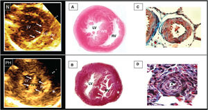
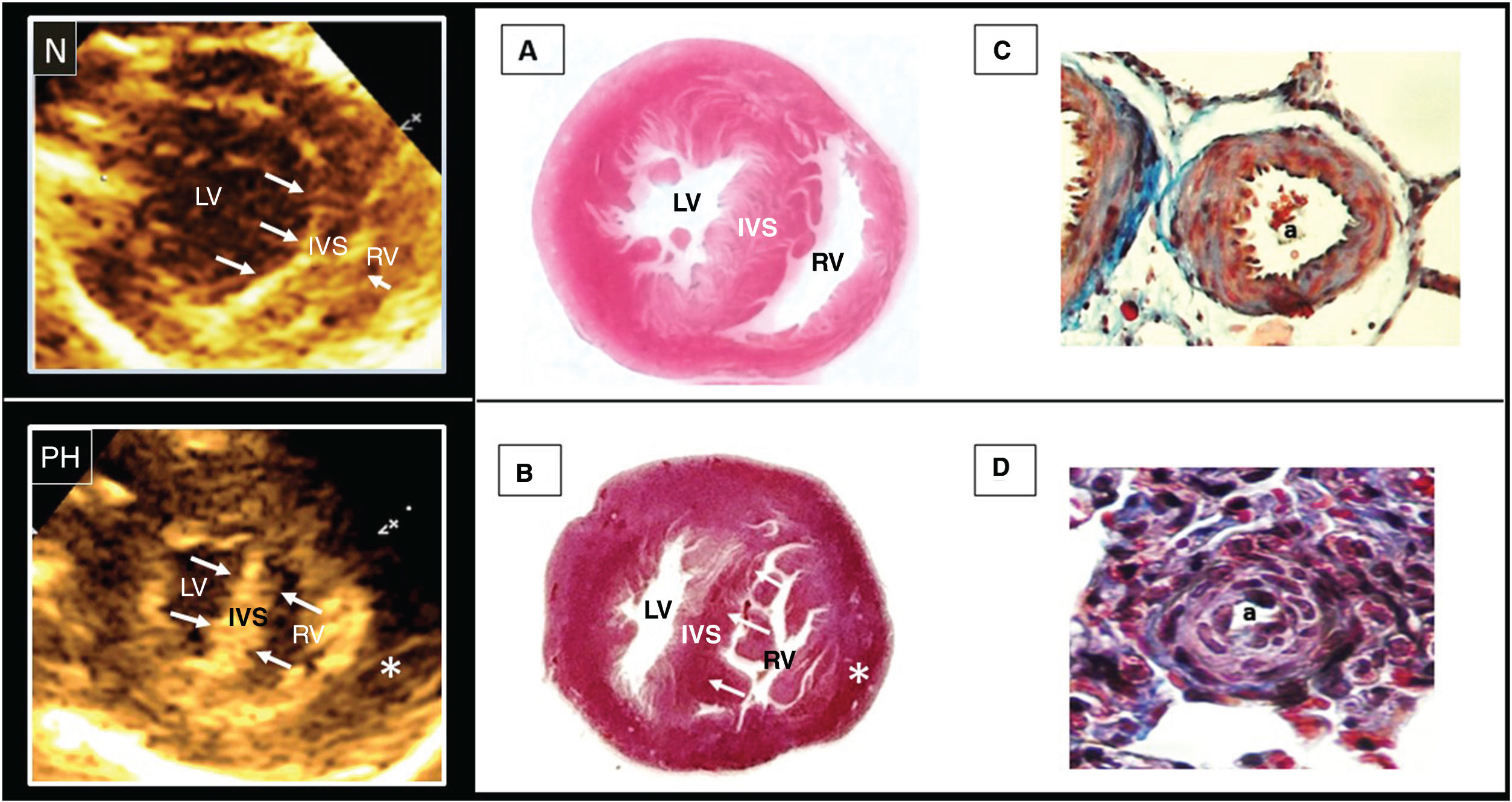
 and PH, heart with RV free wall hypertrophy (*), equalized LV/RV ratio, and leftward shift of the interventricular septum. The right side shows anatomic pathology slices corresponding to a normal heart (A) and a heart with PH (B). B shows hypertrabeculation and RV hypertrophy with rectification of the interventricular septum. C shows a pulmonary arteriole with a normal lumen (a) and a wall formed by 2 or 3 layers of smooth muscle cells. D shows smooth cell proliferation with wall thickening and a reduced lumen (a). Masson trichrome stain, ×40. IVS, interventricular septum; LV, left ventricle; PH, pulmonary hypertension; RV, right ventricle.")
The left side shows 2 echocardiographic phenotypes: N, normal heart with preserved LV/RV ratio and rightward shift of the interventricular septum (pressure forces indicated by arrows) and PH, heart with RV free wall hypertrophy (*), equalized LV/RV ratio, and leftward shift of the interventricular septum. The right side shows anatomic pathology slices corresponding to a normal heart (A) and a heart with PH (B). B shows hypertrabeculation and RV hypertrophy with rectification of the interventricular septum. C shows a pulmonary arteriole with a normal lumen (a) and a wall formed by 2 or 3 layers of smooth muscle cells. D shows smooth cell proliferation with wall thickening and a reduced lumen (a). Masson trichrome stain, ×40. IVS, interventricular septum; LV, left ventricle; PH, pulmonary hypertension; RV, right ventricle.
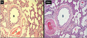
 and a rat with PAH. Both show bronchioles (B), arterioles (A), and pulmonary veins (V). The smooth muscle walls of the arterioles are thin in N, whereas noticeable thickening is seen in PAH. Both show normal vein walls. Hematoxylin–eosin stain, ×10. PAH, pulmonary arterial hypertension.")
Histologic slices of lung tissue from a normal rat (N) and a rat with PAH. Both show bronchioles (B), arterioles (A), and pulmonary veins (V). The smooth muscle walls of the arterioles are thin in N, whereas noticeable thickening is seen in PAH. Both show normal vein walls. Hematoxylin–eosin stain, ×10. PAH, pulmonary arterial hypertension.
This is the first report on hereditary PAH in SHR and warrants several comments. The first concerns the use of genetically standard lines in experimental studies, particularly consanguineous lines. These lines may exhibit spontaneous mutations arising with the introduction of new alleles, as well as unexpected medical conditions. The second refers to the need for an appropriate assessment of individuals belonging to consanguineous lines before including them in research work. Because PAH was found to be more prevalent in this group, echocardiography is recommended before including SHR in cardiovascular research protocols. Last, this finding means that an animal model may be available to study PAH with similar characteristics to those seen in the condition in humans.
Because the observations described are new, research should be done with additional genetic studies potentially related to currently known mutations.
FUNDINGThis study received no funding.
ETHICAL CONSIDERATIONSNonhuman study; all processes were carried out in compliance with Mexican Official Regulation NOM-062-ZOO-1999 (published by the Secretary of Agriculture, Livestock, Rural Development, Fisheries, and Food of the Government of Mexico), the National Institutes of Health, the European Union, and the ARRIVE (Animal Research: Reporting of In Vivo Experiments) guidelines.
The original study was approved by the research ethics committee of the institution (number 21-1262).
All documents and all histology and echocardiography records are available for the SHR.
STATEMENT ON THE USE OF ARTIFICIAL INTELLIGENCENo artificial intelligence tool was used.
AUTHORS’ CONTRIBUTIONSAll authors participated in the study and discussion.
CONFLICTS OF INTERESTAll authors declare that they have no conflicts of interest.