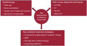
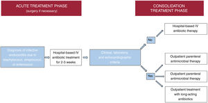
Infective endocarditis is a continually evolving disease. Present-day patients differ significantly from those treated a few decades ago: they tend to be older and have more comorbidities and health care-related episodes, while new groups of patients have emerged with new types of endocarditis, such as those affecting patients with percutaneous valve prostheses. There have also been changes in diagnostic techniques. Although transthoracic and transesophageal echocardiography are still the most commonly used imaging modalities, other techniques, such as 3-dimensional transesophageal ultrasound, cardiac computed tomography, and nuclear medicine tests (PET/CT and SPECT/CT), are increasingly used for diagnosing both the disease and its complications. In recent years, there have also been significant developments in antibiotic therapy. Currently, several treatment strategies are available to shorten the hospital phase of the disease in selected patients, which can reduce the complications associated with hospitalization, improve the quality of life of patients and their families, and reduce the health care costs of the disease. This review discusses the main recent epidemiological, diagnostic and therapeutic developments in infective endocarditis.
Keywords
Infective endocarditis (IE) has posed a challenge to the scientific community since it was first described by Sir William Osler in 1885. Despite progress in recent decades, IE continues to be associated with high mortality. The rate of approximately 20% described in relatively recent Spanish and European series1–3 is very similar to rates reported in the early 2000s.4 The aim of this review was to examine the main developments in the epidemiology, diagnosis, and treatment of primarily left-sided IE that are likely to enhance our understanding of this disease and contribute to much-needed efforts to improve its prognosis (figure 1).
Central illustration. Graphic summary of key developments in the epidemiology, diagnosis, and treatment of infective endocarditis. 3-D TEE, 3-dimensional transesophageal echocardiography; CT, computed tomography; IV, intravenous; OPAT, outpatient parenteral antimicrobial therapy; PET/CT, positron emission tomography/CT; SPECT/CT, single-photon emission computed tomography/computed tomography.
IE is a rare, complex, and serious disease that is constantly evolving due to numerous factors, including epidemiological shifts that have identified new, and in some cases, increasingly large, risk groups, such as adults with congenital heart disease5 and patients who have undergone transcatheter valve6 or cardiac device implantation.7 Other factors include changing diagnostic criteria8–10 and improvements to both diagnosis and treatment.
Epidemiological information on IE primarily comes from population-based studies and case registries. While population-based studies provide valuable insights into global trends, in particular, incidence rates, they can only be conducted in areas with established information systems, and their quality depends on the accuracy of the coded data. Case registries, while very useful for describing the natural history of a disease and detecting changes over time, are rarely exhaustive, as they typically focus on referral hospitals and provide little sociodemographic information.11 Information on the current epidemiological situation of IE should therefore be interpreted with caution as it excludes a significant proportion of the global population. Fortunately, a number of studies published in recent years have provided detailed descriptions of previously overlooked or underserved populations, such as patients diagnosed in hospitals without heart surgery units12 and those from certain regions of Africa13 or low- to middle-income countries according to the World Bank classification.14
The predominance of high-income countries in studies of IE has influenced the definition of this disease. Current diagnostic criteria are based on epidemiological and clinical data from registries managed by referral hospitals in a limited number of countries (mainly European countries and the United States).9,10 Moreover, the existence of various sets of diagnostic criteria and their lack of validation in different contexts complicates the comparison of results among studies.
IncidenceTwo population-based studies analyzing discharge coding data from hospitals in the Spanish National Health System reported a progressive increase in the incidence of IE and a higher incidence in older men. The annual incidence rose from 2.72 cases per 100 000 inhabitants in 2003 to 3.49 cases per 100 000 inhabitants in 2014.15 The estimated incidence from 2016 to 2020 was 4.1 cases per 100 000 inhabitants-year.16 The rates in Germany17 and the United States18 are even higher and are rising. Significant age, sex and regional disparities have also been reported in the United States.18
EtiologyAccording to the European Infective Endocarditis (EURO-ENDO) registry,1 the causes of IE vary according to a country's income level. In relative terms, the predominant causative microorganisms in upper and middle- to upper-income countries are staphylococci, followed by enterococci and viridans group streptococci. Staphylococci also predominate in low- to middle-income countries, but in this case, viridans group streptococci are more common than enterococci,14 as confirmed by a recent systematic review of endocarditis in African countries.13 The most striking difference, however, lies in the percentage of IE episodes with negative blood cultures (due to prior antibiotic use or difficult-to-culture causative microorganisms). Just 16% of blood cultures in high-income countries are negative compared with 48% in low- to middle-income countries.14 The main agents isolated in blood culture-negative episodes are Coxiellaburnetii in the Mediterranean basin and Bartonella species in South Africa.19 In Spain, there has been a decrease in oral streptococcal IE and an increase in enterococcal IE.1,20
Age and sexIE is more common in men than women (ratio, 2:1), a predominance that is consistent among series. Findings from various countries have shown that women are less likely to receive surgical treatment for IE and more likely to die in hospital.2,21 The reasons for these differences in management and prognosis remain to be elucidated.
IE is rare in children and adolescents. In this population, it is more common in patients with congenital heart disease, who tend to have right-sided IE and staphylococcal infections.22
There has been a progressive increase in the age of patients with IE. The median age in the EURO-ENDO registry is 63 [interquartile range, 46-73] years, and 46% are 65 years or older.1 Frailty is a significant risk factor in older adults (typically defined as an adult aged ≥ 75 years, although definitions vary) and is linked to baseline functional status, comorbidities, and contact with the health care system (more frequent in this age group). Closer contact with the health care system is associated with a higher prevalence of staphylococcal infections, while age is associated with a higher prevalence of infections caused by Enterococcus faecalis and Streptococcus gallolyticus (formerly known as Streptococcus bovis). Functional decline has been observed to have a negative effect on prognosis23; hence the importance of a baseline functional assessment, physical therapy, and early discharge to the patient's usual place of residence after clinical stabilization. Age per se is not predictive of poor surgical outcomes and should not be used in isolation to decide candidacy for surgery.24
Health care-associated IEAlthough health care-associated IE (HCIE) is preventable and has received significant attention in the past decade,25 it still accounts for one-third of all IE episodes.1,26 Its continued prevalence has been attributed to medical progress, although little is known about the burden of HCIE in low- to middle-income countries. Compared with community-acquired IE, HCIE is more likely to affect older individuals with more comorbidities and to be caused by staphylococci (in relation to catheter-related or early-onset prosthetic valve infections); patients with HCIE are also less likely to receive surgical treatment and more likely to die in hospital.27
IE following transcatheter valve implantationThe indications for transcatheter biologic valve implantation have widened in recent years, and there has been an exponential increase in the number of patients undergoing this procedure each year. Initially, the procedure was reserved for older adults with comorbidities contraindicating surgery, but this is no longer the case. It is therefore difficult to interpret data on IE in this setting unless patients are stratified by indication. The estimated incidence of IE following transcatheter aortic valve implantation (TAVI) is 5 cases per 1000 procedures-year.28 The most frequently involved microorganisms according to various studies are enterococci and staphylococcus species.6,28 Echocardiographic images can be difficult to interpret after transcatheter valve implantation, and may therefore be insufficient for establishing a definitive diagnosis. In such cases, cardiac computed tomography (CT) and positron emission tomography/CT (PET/CT) are recommended.
RECENT DEVELOPMENTS IN THE DIAGNOSIS OF IEThe diagnosis of IE has 3 key components: clinical suspicion (supported by a thorough history and physical examination focused on potential risk factors), microbiologic findings, and imaging findings.10
Echocardiography, both transthoracic and transesophageal, is the imaging modality of choice on clinical suspicion of IE as it provides valuable diagnostic and prognostic information.10 Because it is widely available, it is also the tool of choice for monitoring disease course and follow-up.
Transesophageal echocardiography (TEE) has very high sensitivity and specificity for native valve IE and also performs well in the diagnosis of prosthetic valve and intracardiac device infections.29 Acoustic shadowing by prosthetic material, however, can limit visualization of periannular complications.30
A number of newly developed imaging techniques have proven particularly useful for establishing a definitive diagnosis in patients with a strong clinical suspicion of IE but inconclusive echocardiographic findings (figure 2). These new techniques, supported by a growing body of evidence, have been incorporated into clinical practice guidelines,10 which highlight their usefulness for detecting septic emboli and valvular and perivalvular lesions.
Diagnostic utility of imaging techniques in the diagnosis of infective endocarditis. A and B: transesophageal echocardiogram (TEE) in a patient with prosthetic mitral valve endocarditis; conventional 2-dimensional TEE (A) with color Doppler imaging showing prosthetic dehiscence and paravalvular regurgitation; 3-dimensional TEE allows more precise localization of the dehiscence and assessment of the severity of the paravalvular regurgitation. B and C: prosthetic endocarditis involving an aortic valve tube, with PET/CT showing a circumferential abscess. C: CT image showing the location of the abscess (arrowheads). D: PET/CT fusion image showing intense focal fluorodeoxyglucose uptake at the site of the abscess. PET/CT, positron emission tomography/computed tomography.
The European Society of Cardiology (ESC)10 and the Duke-International Society for Cardiovascular Infectious Diseases (Duke-ISCVID)9 both updated their diagnostic criteria for IE in 2023.9,10 The criteria recommended by each of these societies are compared in table 1.
Comparison of 2023 Duke-ISCIVD and 2023 ESC criteria for the diagnosis of IE
| 2023 Duke-ISCVID criteria | 2023 ESC criteria |
|---|---|
| Major criteria | Major criteria |
| 1. Microbiologic major criteria• Positive blood cultures for typical microorganismsa that cause IE isolated from 2 separate blood culture sets• Positive blood cultures for microorganisms that occasionally or rarely cause IE isolated from ≥ 3 separate blood cultures• Positive PCR or other nucleic acid-based technique for detecting Coxiella burnetii, Bartonella species, or Tropheryma whipplei from blood• Detection of IgG or IgM antibodies by indirect immunofluorescence assays for Bartonella henselae or Bartonella quintana with an IgG titer > 1:800.2. Imaging major criteria• Echocardiography or cardiac CT showing vegetation, valve perforation, valve aneurysm, abscess, pseudoaneurysm, or intracardiac fistula, or new valvular regurgitation or new dehiscence of prosthetic valve as compared with previous studies• FDG-PET/CT showing abnormal metabolic activity involving a native or prosthetic valve, ascending aortic grafts (with concomitant evidence of valve involvement), intracardiac device leads, or other prosthetic material (beyond the first 3 mo after surgery).3. Surgical major criteria: evidence of IE documented by direct inspection during heart surgery and neither histologic nor microbiologic confirmation | 1. Blood cultures positive for IE• Typical microorganisms consistent with IE from 2 separate blood cultures: Streptococcus viridans, Streptococcus gallolyticus, HACEK group, Staphylococcusaureus, Enterococcus faecalis• Microorganisms consistent with IE from continuously positive blood cultures:- At least 2 positive blood cultures of blood samples drawn > 12 h apart- All of 3 or a majority of ≥ 4 separate cultures of blood (with first and last samples drawn ≥ 1 h apart)- Single positive blood culture for Coxiella burnetii or IgG phase I antibody titer > 1:800.2. Imaging positive for IE• Valvular, perivalvular, or periprosthetic lesions or metabolic lesions characteristic of IE detected by any of the following imaging techniques:- Echocardiography (TTE or TEE)- Cardiac CT- FDG-PET- SPECT/CT with labeled leukocytes |
| Minor criteria | Minor criteria |
| 1. Predisposition (previous history of IE, prosthetic valve, previous valve repair, congenital heart disease, moderate or severe valve disease, intracardiac implantable electronic device, hypertrophic obstructive cardiomyopathy, injection drug use)2. Fever, defined as temperature > 38̊C3. Vascular phenomena (including those detected by imaging only): arterial or pulmonary emboli and infarcts, cerebral or splenic abscesses, mycotic aneurysm, intracranial hemorrhage, conjunctival hemorrhage, Janeway lesions, purulent purpura4. Immunologic phenomena: glomerulonephritis, Osler nodes, Roth spots, rheumatoid factor5. Microbiologic evidence: positive blood culture for a microorganism consistent with IE but not meeting the requirements for major criterion, or positive blood culture, PCR, or other nucleic acid-based test for a microorganism consistent with IE from a sterile body site other than cardiac tissue, cardiac prosthesis, or arterial embolus, or a single finding of skin bacterium by PCR on a valve or wire without additional clinical or microbiologic evidence6. Imaging criteria: abnormal metabolic activity as detected by PET/CT within 3 mo of implantation of a prosthetic valve, ascending aortic graft (with concomitant evidence of valve involvement), intracardiac device leads, or other prosthetic material7. Physical examination criteria: new valvular regurgitation or worsening or changing of preexisting murmur (when echocardiography is not available) | 1. Predisposing conditions (predisposing heart condition at intermediate or high risk of IE or history of injection drug use)2. Fever, defined as temperature> 38̊C3. Vascular phenomena (including those detected by imaging only): arterial or pulmonary emboli and infarcts abscesses, hematogenous osteoarticular septic complications (spondylodiscitis), mycotic aneurysms, intracranial ischemic or hemorrhagic lesions, conjunctival hemorrhages, and Janeway lesions4. Immunologic phenomena: glomerulonephritis, Osler nodes, Roth spots, rheumatoid factor5. Microbiologic evidence: positive blood culture not meeting a major criterion as noted above or serologic evidence of active infection with microorganism consistent with IE |
CT, computed tomography; ESC, European Society of Cardiology; FDG-PET/CT, fluoro-[18F]-deoxy-2-D-glucose positron emission tomography/computed tomography ; HACEK: Haemophilus, Aggregatibacter, Cardiobacterium, Eikenella, and Kingella; IE, infective endocarditis; ISCVID, International Society of Cardiovascular Infectious Diseases; SPECT/CT, single-photon emission computed tomography/computed tomography; TEE, transesophageal echocardiography; TTE, transthoracic echocardiography.
*Typical microorganisms include S aureus, Staphylococcuslugdunensis, Enterococcus faecalis, all species of streptococci (except Streptococcuspneumoniae and Streptococcus pyogenes), Granulicatella and Abiotrophia spp., Gemella spp., and HACEK group microorganisms. In the setting of intracardiac prosthetic material, the following bacteria should also be included: coagulase-negative staphylococci, Corynebacterium striatum, and Corynebacterium jeikeium, Serratia marcescens, Pseudomonas aeruginosa, Cutibacterium acnes, nontuberculous mycobacteria (especially Mycobacterium chimaera), and Candida spp.
An expert panel recently undertook an external validation of the 2023 Duke-ISCVID and ESC criteria, using clinical diagnosis as the reference standard. They found that the Duke-ISCVID criteria had higher specificity than the ESC criteria (94% vs 82%) and similar sensitivity (85% vs 84%).31
Three-dimensional TEEThree-dimensional (3-D) TEE, when used in combination with conventional 2-D TEE, is useful for assessing periannular complications (location, size, extension) and detecting and quantifying the severity of periprosthetic regurgitation. It can also enable more precise detection and localization of valve perforations32 because it produces full-volume images that can subsequently be processed to produce planes that are not feasible with 2D-TEE alone.
Three-dimensional TEE is also a useful surgical planning tool, as it provides an accurate visualization of the surgical field and clearly depicts the relationship between periannular complications and adjacent structures.33
The procedure enables a more accurate assessment of the size and morphology of vegetations, particularly in the case of irregular masses or clusters, potentially improving the prediction of embolic risk.34
A limitation of 3-D TEE is its lower temporal and spatial resolution, potentially complicating the detection of small vegetations.35 Three-dimensional TEE can also produce dropout artifacts (reduced ultrasound signal strength) that may be mistaken for valve perforations or periprosthetic leaks. These effects can be minimized by careful adjustment of gain, compression, and smoothing settings. Concomitant color Doppler imaging is useful for confirming the presence or absence of a suspected tissue defect.36
In conclusion, the value of 3-D TEE lies not in its use as an initial diagnostic tool but as an adjunct to 2-D TEE to provide a more detailed and precise assessment of certain endocardial lesions.
Cardiac CTElectrocardiogram-gated cardiac CT, in addition to ruling out significant coronary artery stenosis in patients with IE who require surgery as an alternative to invasive angiography, can be used to investigate suspected IE when TEE is not feasible or yields inconclusive results. The superior diagnostic yield of cardiac CT in prosthetic IE lies in its ability to detect periannular complications, which are more difficult to detect with TEE.33 Cardiac CT is less sensitive than TEE for the detection of small vegetations, but it is useful for diagnosing valve perforations, fistulas, and, as mentioned, periannular complications; it also has excellent spatial resolution.37
Overall, compared with TEE, cardiac CT is more effective at detecting and providing accurate visualization of abscesses and pseudoaneurysms (extent and exact location) and less effective at detecting valve lesions such as small vegetations (< 4 mm) and leaflet perforations36-39 and at evaluating periprosthetic leaks.40
Nuclear imagingTwo particularly useful nuclear imaging techniques for investigating prosthetic IE in patients with inconclusive TEE findings are fluoro-[18F]-deoxy-2-D-glucose PET/CT (FDG-PET/CT) and single-photon emission CT (SPECT) combined with low-dose CT (SPECT/CT) using labeled leukocytes.
PET/CT combines CT-based anatomic information with PET-based metabolic information on FDG uptake in areas of higher metabolic activity. It has high sensitivity and specificity (≈85%) in prosthetic IE.36,41 Its diagnostic performance is enhanced when combined with electrocardiogram gating.41,42
SPECT/CT has a sensitivity ranging from 64% to 90% and a specificity ranging from 36% to 100%.43 It is more specific than PET/CT,43 but has several limitations, including the need to handle blood during preparation of the FDG radiotracer, the duration of the procedure, and its lower spatial resolution.44 The current recommendation is therefore to use SPECT/CT when PET/CT is unavailable.10
PET/CT has low sensitivity for the detection of native valve IE,45,46 probably because infections involving native valves tend to be associated with less intense inflammatory responses and fewer periannular complications. It could, however, be useful for diagnosing emboli and metastatic infections in this setting,10,45,46 It would not be suitable for identifying cerebral emboli or mycotic aneurysms because of physiological FDG uptake in the brain.
PET/CT has demonstrated high sensitivity for pocket and extracardiac lead infections,47 but low sensitivity for lead-related IE.48 In addition, negative results do not exclude an intracardiac infection.
Guidelines used to advise against using PET/CT in the first 3 months after surgical valve implantation, as it was considered that the associated inflammatory response could affect diagnostic performance. It has since been shown, however, that the 3-month cutoff was arbitrary and that inflammation can last for longer. In addition, it is possible to distinguish between inflammatory and infective uptake patterns after recent surgery.49,50 In its updated 2023 guidelines, the ESC no longer recommends delaying imaging tests for 3 months, highlighting instead the importance of adequate patient preparation49,51 and accurate imaging interpretation, with consideration of uptake types and patterns (heterogeneous and intense focal patterns, for example, are associated with infection),52-54 as well as factors that can lead to false positives, such as surgical adhesives.51,55,56
Artificial intelligenceThe use of artificial intelligence in diagnostic imaging can be expected to increase in the coming years. A recent study analyzed the ability of a machine learning method based on PET/CT images to diagnose prosthetic valve IE.57 The performance observed matched that of expert human interpreters, as reported in the literature, especially when findings were integrated with other diagnostic criteria for IE. Machine learning tools could be particularly useful in hospitals lacking expertise. However, the study analyzed a small sample and used radiomic texture analysis, which can be affected by the image acquisition process. The ability of this and other artificial intelligence tools to reliably diagnose IE using images from a range of diagnostic tools will undoubtedly be analyzed in larger studies.
RECENT DEVELOPMENTS IN THE TREATMENT OF IEAntibiotic therapy and surgery are the mainstays of treatment for IE.
Antibiotic therapyOne of the main developments relating to the use of antibiotics in the treatment of IE in recent years is the questioning of the dogma that antibiotics should be administered intravenously in a hospital setting until completion of treatment. A number of studies have provided evidence supporting the use of 3 strategies that have successfully achieved clinical stabilization after a period of hospital-based intravenous antibiotic therapy. These strategies are: a) a switch to outpatient oral antibiotic therapy; b) a switch to outpatient parenteral antibiotic therapy (OPAT) (completion of intravenous therapy at home); and c) use of antibiotics with pharmacokinetic and pharmacodynamic properties that allow weekly or biweekly dosing. The aim of all 3 strategies is to shorten hospital stays, ultimately reducing the risk of complications, improving patient quality of life, and reducing costs.
In the POET (Partial Oral Treatment of Endocarditis) trial, published in 2019, 400 patients with stable IE caused by the most common gram-positive microorganisms (staphylococci, streptococci, and E faecalis) were randomized to receive conventional intravenous treatment as recommended in clinical practice guidelines or oral treatment with prespecified combinations of antibiotics.58 To be randomized, patients had to have received intravenous antibiotic treatment for at least 10 days or, in the case of heart surgery, at least 7 days since the intervention. They also had to meet a series of strict clinical, laboratory, and echocardiographic criteria defining stable IE. The trial findings showed that switching to oral antibiotic treatment was noninferior to continued intravenous antibiotic treatment for the composite primary outcome of all-cause mortality, relapse of bacteremia, unplanned cardiac surgery, and new embolic events. Despite its limitations—low inclusion rate (just 20% of patients evaluated were randomized), few coagulase-negative staphylococcal infections, no methicillin-resistant Staphylococcus aureus infections, and use of antibiotics with complex dosing regimens or a high risk of interactions59—the POET trial brought about a paradigm shift in the treatment of IE, including modifications to the ESC guidelines.10
The recent publication on the 5-year outcomes of the POET trial reported lower all-cause mortality in patients who were switched to oral antibiotics compared with those who continued with intravenous antibiotics in hospital. No significant differences were observed for the other primary outcome components (relapse of bacteremia, unplanned cardiac surgery, or new embolic events).60
The authors of a recent review published in JAMA Internal Medicine concluded that there is sufficient evidence showing that oral step-down antibiotic therapy in patients with stable IE is as effective as continued intravenous treatment in hospital.61 There were, however, concerns about the feasibility and safety of such a strategy and recognition of a need for corroboration in real-world studies. Following the publication of the POET trial, the Danish IE guidelines were updated to include the recommendation to switch stable patients to oral antibiotics. Shortly after the publication of this update, a nationwide multicenter study was conducted to assess how widely the new recommendation had been implemented.62 Of the 55% of patients who met the inclusion and exclusion criteria for the POET trial, 42.7% had received oral step-down antibiotic therapy and 57.3% had completed intravenous therapy in hospital. IE episodes due to S. aureus, intracardiac abscesses, preexisting endocardial devices (pacemakers and defibrillators), and surgical treatment of IE were more common in the intravenous group. After 6 months of follow-up, patients in the step-down group had shorter hospital stays and a lower incidence of all-cause mortality. They also had a lower incidence of the composite primary outcome used in the POET trial, although the difference with the intravenous group was not significant.
Enrolment for the POET II trial began in February 2019. This trial is investigating the efficacy and safety of a shorter course of antibiotics for stable patients with the 3 main types of left-sided IE (staphylococcal, streptococcal, and enterococcal).63 Confirmation that accelerated antibiotic therapy is noninferior to standard-duration therapy could lead to a reduction in complications associated with long hospital stays (in particular catheter-related infections), as well as reduced costs, fewer antibiotic-related adverse effects, and improved quality of life for patients and their families.
OPAT, which allows patients to continue intravenous antibiotic treatment at home, has gained increasing acceptance in recent years. One notable Spanish study by researchers from the GAMES group analyzed characteristics and outcomes in 429 patients who received OPAT for IE (30% of all IE cases in nonintravenous drug users who survived the initial episode).64 Although the study was not a randomized trial, its results showed that OPAT was both effective and safe in selected patients, as 93% of patients were still alive at 1 year, just 10.9% were readmitted within 3 months of discharge, and just 1.4% experienced recurrence.
The criteria for selecting suitable candidates for oral step-down antibiotic therapy or OPAT are summarized in table 2.
Criteria for selecting patients for outpatient (home-based) oral or intravenous antibiotic therapy
| Diagnosis | Infective endocarditis involving native or prosthetic valve caused by streptococci, Staphylococcus aureus, or Enterococcus faecalis |
| Antibiotic treatment | Adequate intravenous antibiotic treatment administered for at least 10 d or at least 7 d after heart surgery |
| Clinical course | Satisfactory response to treatment: without fever for at least 2 d, C-reactive protein <20mg/L or <25% of peak value, and leukocytes <109/L |
| Echocardiogram | Transesophageal echocardiogram ruling out abscesses or other reasons for surgery in past 48 h |
Several studies of novel long-acting antibiotics with activity against most gram-positive microorganisms, such as dalbavancin, have reported noteworthy findings, with clinical cure rates in the range of 90% to 96%.65,66 The lipoglycopeptide dalbavancin can be administered weekly or biweekly, potentially allowing earlier discharge for patients with IE who achieve clinical stabilization.
There is thus real-world evidence supporting the use of various antibiotic strategies for selected patients with left-sided IE (figure 3). The choice of strategy depends on a range of factors, including clinical and logistical factors, local resources, and the experience of multidisciplinary teams.
Surgical treatment of IEApproximately 50% of patients with left-sided IE undergo surgery before completing antibiotic treatment.67 A recent study showed that patients who benefit most from surgery are also those at highest risk of mortality, calculated using the ENDOVAL risk scale developed by 3 Spanish hospitals.68 The study also found that surgery was particularly beneficial in patients with an uncontrolled infection. Timing of surgery is probably one of the most difficult decisions to make in IE, in part because robust scientific evidence is lacking. The decision regarding if and when to perform surgery should always be taken by a multidisciplinary team with expertise in the management and treatment of IE.10 Guideline recommendations on the indication for and timing of surgery in IE are based on observational studies, which are prone to systematic survivor bias (patients who live for longer are more likely to undergo surgery). As reported by Mack and Lancellotti,69 the correlation between increased survival and urgent surgery could be misconstrued as evidence that surgery improves prognosis, but studies adjusting for survivor bias have shown that early surgery may result in a loss of benefit.
Generally, when determining the most appropriate time for surgery, it is necessary to balance the urgency of surgery against the risks of postsurgical complications. Clinical, microbiological, and echocardiographic factors supporting early surgery are shown in table 3.
Factors supporting early surgery for left-sided infective endocarditis
| Clinical factors | Heart failure |
| Recurrent embolic events | |
| Atrioventricular block | |
| Microbiologic factors | Fungal endocarditis |
| Vancomycin-resistant enterococci | |
| Staphylococci | |
| Multidrug-resistant gram-negative bacilli | |
| Echocardiographic factors | Vegetation >10mm |
| Growth of vegetations during antibiotic treatment | |
| Periannular complications | |
| Severe valve regurgitation | |
| Involvement of anterior mitral valve leaflet |
Gisler et al.70 conducted an interesting study evaluating the impact of preoperative antibiotic treatment duration on valve culture results, with adjustment for causative microorganisms. These authors studied 231 episodes of surgically treated IE, 66% of which involved native valves. Positive valve cultures were detected in 25% of cases. Staphylococcal and enterococcal infections were associated with a high risk of positive valve cultures, regardless of preoperative antibiotic treatment duration. The greatest impact of treatment was observed in the first few days. The results reported by Gisler et al. help guide decisions on the timing of surgery and suggest that the decision should not be overly influenced by the goal of a negative valve culture. Rather, they suggest that early surgery should be considered when the indication is urgent.
FUNDINGThis study was partially funded by grants from Instituto de Salud Carlos III (PI22/00459) and the Gerencia Regional de Salud de Castilla y León (GRS 2657/A/22).
STATEMENT ON THE USE OF ARTIFICIAL INTELLIGENCEArtificial intelligence was not used to prepare this manuscript.
AUTHORS’ CONTRIBUTIONSAll the authors (J. López, C. Olmos, N. Fernández-Hidalgo) were involved in conceiving and designing the study, analyzing and interpreting the data, drafting the article, and approving the final version. They also take responsibility for all aspects of the research and are committed to investigating and resolving any issues regarding the accuracy and integrity of any part of the work.
CONFLICTS OF INTERESTWe have no conflicts of interest to declare.
