Nonsyndromic thoracic aortic aneurysms and dissections (ns-TAADs)1 are characterized by the silent formation of aortic aneurysms and dissections without other external manifestations that facilitate their diagnosis. Familial ns-TAADs are reported to display autosomal dominant inheritance, as well as incomplete penetrance and variable expression.2 Mutations have been found in various genes, but predominantly ACTA2.3–5 Here, we present the case of a family with a novel mutation in ACTA2 causing ns-TAADs.
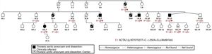
The index case was a man (II.9) who was treated at the age of 50 years for type A aortic dissection (AD) and had several relatives who died of sudden cardiac death (figure 1 and table 1). His mother (I.5) had a 38-mm dilatation of the aortic root (AR), whereas his brother (II.8) died of abdominal AD. The other 4 siblings were apparently healthy. The complete pedigree additionally included 2 cousins (sisters) who died of sudden cardiac death, one (II.6) at the age of 45 years without autopsy (one of her sons died at the age of 17 years of autopsy-confirmed AD), the other (II.4) at the age of 47 years from ascending AD.

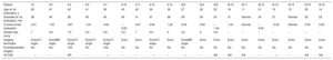
Characteristics of the family members studied
| Patient | I.5 | II.3 | II.4 | II.5 | II.7 | II.10 | II.11 | II.12 | II.13 | III.3 | III.4 | III.6 | III.10 | III.11 | III.12 | III.13 | III.14 | III.15 | III.16 |
|---|---|---|---|---|---|---|---|---|---|---|---|---|---|---|---|---|---|---|---|
| Age at 1st evaluation, y | 83 | 47 | 43 | 41 | 36 | 45 | 42 | 39 | 39 | 27 | 29 | 20 | 16 | 11 | 21 | 15 | 12 | 20 | 14 |
| Diameter at 1st evaluation, mm | 38 | 36 | 38 | 39 | 40 | 29 | 31 | 37 | 38 | 39 | 30 | 23 | 31 | Normal | 24 | 27 | Normal | 32 | 23 |
| Z-score at last evaluation | 2.31 | 1.55 | 3.67 | 1.24 | 4.42 | –0.93 | 0.61 | 2.64 | 1.22 | 2.43 | 0.64 | –1.04 | 1.44 | Normal | –0.93 | –0.23 | Normal | 0.33 | 0.45 |
| Growth rate, mm/y | 1 | 0.6 | 1.5 | 0.2 | 0.8 | 0.2 | 1 | 0.6 | 0 | 0.3 | 0 | 0.8 | – | – | – | – | – | – | – |
| Imaging technique | EchoCT-angio | EchoMR-angio | EchoCT-angio | EchoCT-angio | EchoCT-angio | Echo | Echo | EchoCT-angio | Echo | EchoMR-angio | Echo | Echo | Echo | Echo | Echo | Echo | Echo | Echo | Echo |
| Event/preventive surgery | No | No | Yes | No | Yes | No | No | No | No | No | No | No | No | No | No | No | No | No | No |
| ACTA2 | + | – | NP | – | + | – | – | + | – | + | – | NA | NA | NA | – | + | – | NA | NA |
CT-angio, computed tomography angiography; MR-angio, magnetic resonance angiography; NA, not applicable; NP, not performed.
A genetic study was performed using next-generation sequencing of a panel including 41 genes related to aortic disease (ACTA2, ADAMTSL4, B3GAT3, CBS, COL1A1-2, COL3A1, COL5A1-2, EFEMP2, ELN, FBN1-2, FLNA, GAA, GATA5, HRAS, KCNJ8, MED12, MYH11, MYLK, NKX2-5, NOTCH1, PLOD1, PRKG1, PTPN11, SKI, SLC2A10, SMAD3-4, TGFB2-3, TGFBR12, ZDHHC9, ATP7A, CHST14, ADAMTS2*, B4GALT7*, FKBP14*, and SLC39A13*). A novel heterozygous mutation was identified in exon 3 of the ACTA2 gene, which encodes alpha-smooth muscle actin: Met84Val (NC_000010.10:g.90707023T>C). Subsequently, 19 relatives were examined; 13 underwent a genetic study. AR dilatation was found in 4, all of whom had a positive genetic screening result. All family members were studied using echocardiography, and the screening of the affected carriers was completed using computed tomography or magnetic resonance angiography. The median age at presentation was 47 years and only 1 family member had early presentation (at 17 years). In our series, the predominant location of the dilatation was the AR and there was only 1 case of abdominal aneurysm.
The patients were recommended strict control of cardiovascular risk factors and to avoid isometric exercises and competitive sports. All patients were treated with angiotensin receptor blockers and 1 was managed with prophylactic surgery.
AD in young patients is often the result of a genetic aortopathy. The paradigmatic example is Marfan syndrome. However, there are other genetic aortopathies lacking external signs, characterized by the silent formation of thoracic aortic aneurysms and dissections, known as ns-TAADs.1 Mutations identified in the ACTA2 gene would explain 14% of these patients.3–5 The ACTA2 gene, located on chromosome 10 (10q23.31), encodes alpha-actin, the most abundant protein in the smooth muscle of vascular cells. Its deficiency decreases the contractibility of these cells and can cause type A and B AD.4,5 The variant identified in this family has not been described in the literature or in public genotyping databases of the general population and also involves a highly conserved residue. Variants associated with the development of thoracic aortic disease have been identified in nearby amino acids, such as p.Asp82Glu and p.Glu85Lys. Family members with ns-TAAD exhibit variable expression.2 In this family, the median age at presentation was 47 years, similar to that described in the literature. Another characteristic of the phenotypic variability of ns-TAADs is the site of aortic involvement; in our family, the AR predominated. Penetrance has been reported to be incomplete; in our case, of the 6 patients with a positive genetic study, 5 had AR dilatation, indicating that the variant identified probably has elevated penetrance. Although the penetrance has been reported to be lower in women, cases were found in both sexes in this family. Other mutations described in ACTA2 are associated with livedo reticularis, iris flocculi, and bicuspid aortic valve.4 However, these conditions were not present in any of our patients, which may indicate a low frequency of association, as suggested in previous studies.4
Treatment consists of hemodynamic stress management with medication to prevent complications or surgical repair. The indications for prophylactic surgery are not well-established,6 although such surgery seems appropriate for aortic diameters > 5cm or even lower (> 4.5cm) or growth rates exceeding 0.5cm/y. Only 1 member of our family had an AR dissection (42mm on echocardiography performed 9 months before). Preventive measures, such as hypertension management, smoking cessation, and avoidance of isometric exercise and competitive sports, may have an impact on disease progression.6
In conclusion, ACTA2 mutations are the most frequent cause of familial ns-TAAD. We describe a new mutation in the ACTA2 gene that cosegregates in a large family. In this mutation, the dilatation mainly appears at the AR and may cause events with slightly larger diameters. Important factors are the early diagnosis of family members (clinical, genetic, and imaging studies), their monitoring (clinical and imaging studies), and the prevention of complications (cardiovascular risk factor control).
CONFLICTS OF INTERESTL. Montserrat is a shareholder in Health in Code S.L.