Fractional flow reserve (FFR) has firmly proven clinical value in randomized clinical trials.1 Coronary flow impairment can alternatively be assessed by coronary flow reserve (CFR), which has also been shown to provide significant prognostic information of adverse outcomes when reduced, regardless of the presence or absence of epicardial stenosis.2,3 Patients with preserved FFR with reduced CFR have been reported to exhibit worse outcomes than those with preserved FFR and CFR. Conversely, compared with patients with concordantly ischemic FFR and CFR, those with ischemic FFR with preserved CFR showed better outcomes.4 Although we previously reported that lower FFR and microvascular dysfunction were independent predictors of the presence of optical coherence tomography (OCT)-defined thin-cap fibroatheroma (TCFA),5 no comparisons have been reported on the basis of FFR/CFR quadrants regarding OCT-derived high-risk plaque features.
We retrospectively investigated 473 de novo intermediate coronary artery lesions (30%-80% visual stenosis) from 419 patients who underwent OCT and physiological assessments during the same procedure. OCT images were acquired with the frequency domain OCT systems (ILUMIEN, Abbott Vascular, Santa Clara, California, United States; or LUNAWAVE, Terumo, Tokyo, Japan). Physiological assessments were performed using a Pressure Wire Certus as previously described.2 The prevalence of TCFA and plaque rupture (PR) were evaluated. Established cutoff values of pressure-derived physiologic indices (FFR ≤ 0.80 and CFR ≤ 2.0) were used to divide FFR/CFR into quadrants in 2 concordantly classified [FFR+/CFR+(173 vessels) and FFR-/CFR- (103 vessels)] and 2 discordantly classified [FFR+/CFR- (171 vessels) and FFR-/CFR+(26 vessels)] groups. OCT-derived plaque characteristics were compared among FFR/CFR quadrants.
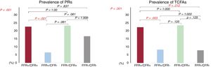
CFR was correlated to FFR (r=0.454, P<.001) and FFR-based decision making disagreed with that of CFR in 41.7% (197 vessels). TCFAs and PRs were detected in 66 vessels (14.0%) and 73 vessels (15.4%) in the total cohort. The patient and lesion characteristics of the FFR/CFR quadrants are summarized in table 1. Compared with the FFR-/CFR+group, patients in the FFR+/CFR- group were significantly younger. There were no significant differences in the prevalence of nonculprit lesions of acute coronary syndrome among the 4 quadrants. The prevalence of TCFAs and PRs were significantly different among the 4 quadrants (figure 1A,B). Compared with FFR+/CFR-, the prevalence of both TCFAs and PRs tended to be higher, although, in the post hoc analysis, this difference was not significant in the FFR-/CFR+group. The net reclassification index and integrated discrimination improvement index were both significantly improved when CFR was added to the FFR-based classification for predicting PR and TCFA (PR; net reclassification index 0.462, P <.001, integrated discrimination improvement 0.031, P <.001, TCFA; net reclassification index 0.320, P=.012, integrated discrimination improvement 0.017, P=.002).
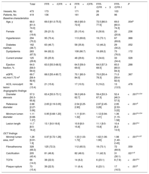
Patient and lesion characteristics
| Total | FFR+/CFR+4 | FFR+/CFR-2 | FFR-/CFR+3 | FFR-/CFR-1 | P | |
|---|---|---|---|---|---|---|
| Vessels, No. | 473 | 173 | 171 | 26 | 103 | |
| Patients, No. | 419 | 136 | 161 | 26 | 96 | |
| Baseline characteristics | ||||||
| Age, y | 68.0 [61.0-74.0] | 69.0 [61.0-75.5] | 66.0 [60.0-72.0] | 72.5 [66.0-77.5] | 69.0 [60.0-74.3] | .004a |
| Female | 82 (19.6) | 29 (21.5) | 25 (15.4) | 8 (30.8) | 20 (20.8) | .236 |
| Hypertension | 294 (70.2) | 95 (70.4) | 113 (69.8) | 19 (73.1) | 67 (69.8) | .988 |
| Diabetes mellitus | 162 (38.7) | 63 (46.7) | 58 (35.8) | 12 (46.2) | 29 (30.2) | .052 |
| Dyslipidemia | 283 (67.5) | 84 (62.2) | 108 (66.7) | 18 (69.2) | 73 (76.0) | .172 |
| Current smoker | 125 (29.8) | 35 (25.9) | 48 (29.6) | 9 (34.6) | 33 (34.4) | .528 |
| Ejection fraction, % | 64.0 [58.0-69.0] | 63.0 [55.5-68.5] | 64.5 [59.0-69.0] | 64.0 [57.3-68.5] | 65.0 [60.0-69.3] | .288 |
| eGFR, mL/min/1.73 m2 | 69.7 [58.4-82.6] | 68.0 [55.4-80.7] | 70.1 [60.0-84.0] | 70.0 [55.4-78.3] | 71.0 [59.4-82.0] | .367 |
| ACS, nonculprit lesion | 56 (13.4) | 21 (15.6) | 17 (10.5) | 5 (19.2) | 13 (13.5) | .478 |
| Angiographic findings | ||||||
| Diameter stenosis | 57.3 [50.3-65.8] | 63.4 [55.5-73.1] | 56.3 [49.6-62.7] | 59.4 [53.3-67.3] | 52.4 [46.3-57.5] | <.001b,c,d,e |
| Reference diameter | 2.68 [2.27-3.06] | 2.65 [2.19-3.05] | 2.54 [2.25-2.90] | 2.97 [2.45-3.26] | 2.95 [2.53-3.22] | <.001d |
| Minimum lumen diameter | 1.11 [0.88-1.37] | 0.95 [0.68-1.20] | 1.11 [0.91-1.32] | 1.12 [0.94-1.41] | 1.36 [1.15-1.58] | <.001b,c,d,e |
| Lesion length | 11.7 [8.5-16.2] | 13.1 [9.0-18.6] | 10.9 [8.0-15.8] | 11.1 [9.5-15.8] | 10.4 [8.2-13.5] | <.001b,c |
| OCT findings | ||||||
| Minimal lumen area, mm2 | 1.28 [0.91-1.9] | 0.97 [0.72-1.28] | 1.33 [1.03-1.72] | 1.62 [1.06-1.90] | 1.98 [1.40-2.45] | <.001b,c,d,f |
| Fibroatheroma | 328 (69.3) | 125 (72.3) | 112 (65.5) | 19 (73.1) | 72 (69.9) | .559 |
| Calcification | 205 (43.4) | 81 (46.8) | 82 (48.0) | 11 (42.3) | 31 (30.1) | .021c,d |
| TCFA | 66 (14.0) | 38 (22.0) | 14 (8.2) | 6 (23.1) | 8 (7.8) | <.001b,c |
| Plaque rupture | 73 (15.4) | 39 (22.5) | 11 (6.4) | 6 (23.1) | 17 (16.5) | <.001b |
ACS, acute coronary syndrome; CFR, coronary flow reserve; eGFR, estimated glomerular filtration rate; FFR, fractional flow reserve; OCT, optical coherence tomography; TCFA, thin-cap fibroatheroma.
Unless otherwise indicated, values are expressed as No. (%) or median [interquartile range].

Our results indicate that physiological classifications of coronary stenosis evaluated by FFR and CFR are associated with the difference in plaque instability. Even in patients with lesions showing preserved FFR, CFR may add incremental information on plaque instability, which might be associated with worse outcomes. In the present study, we would like to address the importance of CFR in addition to FFR for evaluating plaque vulnerability. This differs from our previous report5 in which we evaluated the significance of microvascular dysfunction in addition to FFR. Further studies are needed to test the hypothesis of the possible link between physiological lesion assessment and lesion instability, and its impact on subsequent adverse cardiac events.