Publish in this journal
Journal Information
Vol. 69. Issue 4.
Pages 374-376 (April 2016)
Vol. 69. Issue 4.
Pages 374-376 (April 2016)
Editorial
Full text access
Should We Anticoagulate Patients at High Risk of Atrial Fibrillation?
¿Debemos anticoagular a pacientes en alto riesgo de sufrir fibrilación auricular?
Visits
11699
Manuel Martínez-Sellésa,
Corresponding author
mmselles@secardiologia.es

Corresponding author: Servicio de Cardiología, Hospital General Universitario Gregorio Marañón, Dr. Esquerdo 46, 28007 Madrid, España.
, Ignacio Fernández Lozanob, Adrian Baranchukc, Antoni Bayes-Genisd, Antonio Bayés de Lunae
a Servicio de Cardiología, Hospital General Universitario Gregorio Marañón, Universidad Europea y Universidad Complutense, Madrid, Spain
b Servicio de Cardiología, Hospital Universitario Puerta de Hierro, Majadahonda, Madrid, Spain
c Division of Cardiology, Queen's University, Kingston, Ontario, Canada
d Servicio de Cardiología, Hospital Universitari Germans Trias i Pujol, Universitat Autònoma de Barcelona, Badalona, Barcelona, Spain
e Fundació Investigació Cardiovascular, ICCC, Barcelona, Spain
This item has received
Article information
Full Text
Bibliography
Download PDF
Statistics
Figures (2)
Tables (1)
Table. Characteristics Associated With Higher Thromboembolic Risk in Patients With Advanced Interatrial Block
Full Text

The rapidly progressive aging of Western countries suggests that about one-third of the population will be older than 65 years in 2050.1 Thus, one of the biggest challenges in modern cardiology involves determining the risk of atrial fibrillation (AF) and identifying which patients should receive anticoagulation therapy.2,3 Treatment of AF with anticoagulants, performed since the late 1980s,4 has significantly reduced the incidence of stroke. However, their exact impact on cognitive impairment is still unknown, although recent evidence has been published on the relationship between AF and cognitive decline.5 In addition, there are new data6,7 on the identification of at-risk patients and the importance of AF as a causal factor for embolisms. The information provided by continuous cardiac monitoring devices (implantable loop recorders, pacemakers, and defibrillators) has indicated that there is no temporal relationship between AF and stroke. In fact, almost no patients with paroxysmal AF have AF at the time of stroke onset, although AF is indeed a marker of endothelial dysfunction and another risk factor for stroke.8–11 Recent work has also strengthened the value of surface ECG, specifically ECG of interatrial block (IAB) disorders, which can help to determine the risk of AF.

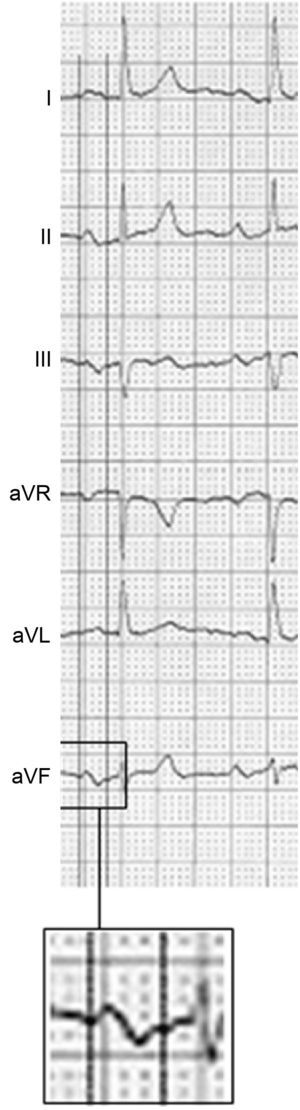
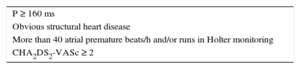
The diagnosis and classification of IABs (partial and advanced) was published in the 1980s by Bayés de Luna12,13 and later by the group of Spodick,14 among others. Their diagnostic criteria were finally defined in a consensus document in 2012.15 Partial IAB is diagnosed in the presence of a P wave ≥ 120ms, whereas the more advanced form is diagnosed if there is also a ± pattern in leads II, III, and aVF (Figure 1). In addition, ± P from V1 to V3 is almost always present, but this criterion can also be found in isolated left atrial enlargement. When advanced IAB shows a very long P wave (≥ 160 ms) and the patient has obvious structural heart disease or elevated CHA2DS2-VASc11 (heart failure or systolic dysfunction, hypertension, age ≥ 75 years, diabetes mellitus, stroke, vascular disease, age 65-74 years, female sex) and ambient atrial arrhythmias (more than 40 atrial premature beats/h on Holter monitoring), there is a very high possibility of AD/atrial flutter development in the next 2 to 3 years. Various studies have confirmed the usefulness of advanced IAB as a predictor of AF.16–22 Recently, this condition was named Bayés syndrome.23–26 Finally, IABs and ambient arrhythmias are also frequently associated with stroke and cognitive impairment.26–31

Figure 1.

Electrocardiogram showing advanced interatrial block: P wave ≥ 120ms plus a ± pattern in the II, III, and aVF leads.

(0.2MB).

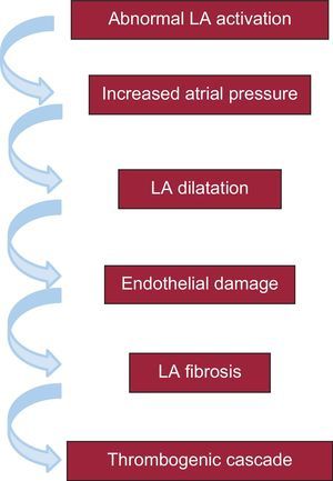
The pathophysiology of the association between advanced IAB and AF and stroke probably depends on a series of sequential electromechanical changes that, starting with an abnormal and delayed activation of the left atrium, culminates in a thrombogenic cascade, primarily in the left atrial appendage32 (Figure 2). It is interesting to note the important role played by fibrosis in this mechanism, because fibrosis has already been closely related to endothelial damage and promotes the development of AF and thrombogenic cascades.33–38

Figure 2.

Electromechanical changes possibly explaining the association of advanced interatrial block with atrial fibrillation and the risk of cerebral embolic events (stroke and cognitive impairment associated or not with the presence of atrial fibrillation). LA, left atrial.

(0.1MB).

This sequence of events has led to the conclusion that it would be pertinent to identify patients with advanced IAB who have the characteristics included in the Table, because these patients have high risk of AF and stroke. They would thus benefit from the early use of anticoagulation therapy, even without evidence of AF. This approach has been strengthened6,7 by the recent demonstration that, although patients with a high CHA2DS2-VASc score show an increased prevalence of AF, thromboembolic complications appear to be independent of the presence of AF when this score is very high. If advanced IAB is also present, it would give rise to a subgroup of patients who will probably show an even higher risk of short-term embolic complications with or without the development of AF. This strategy might at least partly avoid the cognitive impairment that appears to be associated with AF, but that surely should be considered linked to the microembolic processes frequently occurring in these patients before a stroke.5

Table.

Characteristics Associated With Higher Thromboembolic Risk in Patients With Advanced Interatrial Block

P ≥ 160 ms 
Obvious structural heart disease 
More than 40 atrial premature beats/h and/or runs in Holter monitoring 
CHA2DS2-VASc ≥ 2 

The hypothesis of anticoagulating patients in sinus rhythm showing the characteristics of the Table but without confirmed AF seems to us to be obvious and appealing. In addition, the clinical application of this approach could already be considered in certain patients. Moreover, the diagnosis of IAB is simple and only requires electrocardiographic testing.39 However, 2 prerequisites must be met before this approach can be generally recommended. The first involves the creation of a registry including patients with advanced IAB and the characteristics listed in the Table and a control group comprising patients with a similar clinical profile but with partial IAB. The second is that, if this registry shows that patients with advanced IAB have a higher risk of stroke regardless of whether they have documented AF or not, a clinical trial should be performed with anticoagulants (possibly using the “new” oral anticoagulants) and a control group to determine if anticoagulation therapy can reduce the incidence of stroke and cognitive decline in these patients. If so, anticoagulant therapy would be advisable for these patients because, although it would not avoid the development of AF or atrial flutter, it could reduce the collateral effects and sequelae (stroke and cognitive impairment). As Fabritz has recently noted, a small biphasic P wave in the elderly could have a considerable impact.40

CONFLICTS OF INTEREST

None declared.

References
[1]
W.C. Sanderson, S. Scherbov.
Faster increases in human life expectancy could lead to slower population aging.
PLoS One., 10 (2015), pp. e0121922
[2]
A. Jahangir, V. Lee, P.A. Friedman, J.M. Trusty, D.O. Hodge, S.L. Kopecky, et al.
Long-term progression and outcomes with aging in patients with lone atrial fibrillation: a 30-year follow-up study.
Circulation., 115 (2007), pp. 3050-3056
[3]
W.B. Kannel, R.D. Abbott, D.D. Savage, P.M. McNamara.
Epidemiologic features of chronic atrial fibrillation.
N Engl J Med., 306 (1982), pp. 1018-1022
[4]
Atrial Fibrillation Investigators.
Risk factors for stroke and efficacy of antithrombotic therapy in atrial fibrillation.
Arch Intern Med., 154 (1994), pp. 1449-1457
[5]
E. Ammirati, I. Scotti, P.G. Camici.
Can silent brain lesions be a target to guide anticoagulation treatment in patients with low-risk atrial fibrillation to reduce cognitive impairment?.
J Am Coll Cardiol., 63 (2014), pp. 2174-2175
[6]
G.F. Polenz, T.L. Leiria, V. Essebag, M.L. Kruse, L.M. Pires, T.B. Nogueira, et al.
CHA2DS2-VASc score as a predictor of cardiovascular events in ambulatory patients without atrial fibrillation.
PACE., 38 (2015), pp. 1412-1417
[7]
Ts Tischer, R. Schneider, J. Lauschke, C. Nesselmann, A. Klemm, D. Diedrich, et al.
Prevalence of atrial fibrillation in patients with high CHADS2- and CHA2DS2-VASc scores: anticoagulate or monitor high-risk patients?.
PACE., 37 (2014), pp. 1651
[8]
T.V. Glotzer, A.S. Hellkamp, J. Zimmerman, M.O. Sweeney, R. Yee, R. Marinchak, et al.
Atrial high rate episodes detected by pacemaker diagnostics predict death and stroke. Report of the atrial diagnostics Ancillary Study of the Mode Selection Trial (MOST).
Circulation., 107 (2003), pp. 1614-1619
[9]
T.V. Glotzer, E.G. Daoud, D.G. Wyse, D.E. Singer, M.D. Ezekowitz, C. Hilker, et al.
The relationship between daily atrial tachyarrhythmia burden from implantable device diagnostics and stroke risk. The TRENDS Study.
Circ Arrhythmia Electrophysiol., 2 (2009), pp. 474-480
[10]
S.H. Hohnloser, A. Capucci, E. Fain, M.R. Gold, I.C. van Gelder, J. Healey, ASSERT Investigators and Committees, et al.
Asymptomatic atrial fibrillation and stroke evaluation in pacemaker patients and the atrial fibrillation reduction atrial pacing trial (ASSERT).
Am Heart J., 152 (2006), pp. 442-447
[11]
D.T. Martin, M.M. Bersohn, A.L. Waldo, M.S. Wathen, W.K. Choucair, G.Y. Lip, IMPACT Investigators, et al.
Randomized trial of atrial arrhythmia monitoring to guide anticoagulation in patients with implanted defibrillator and cardiac resynchronization devices.
Eur Heart J., 36 (2015), pp. 1660-1668
[12]
A. Bayes de Luna, R. Fort de Ribot, E. Trilla, J. Julia, J. Garcia, J. Sadurni, et al.
Electrocardiographic and vectorcardiographic study of interatrial conduction disturbances with left atrial retrograde activation.
J Electrocardiol., 18 (1985), pp. 1
[13]
A. Bayés de Luna, M. Cladellas, R. Oter, P. Torner, J. Guindo, V. Martí, et al.
Interatrial conduction block and retrograde activation of the left atrium and paroxysmal supraventricular tachyarrhythmia.
Eur Heart J., 9 (1988), pp. 1112-1118
[14]
D.H. Spodick.
Interatrial block and atrial arrhythmias.
Am J Cardiol., 77 (1996), pp. 326
[15]
A. Bayés de Luna, P. Platonov, F.G. Cosio, I. Cygankiewicz, C. Pastore, R. Baranowski, Interatrial blocks, et al.
A separate entity from left atrial enlargement: a consensus report.
J Electrocardiol., 45 (2012), pp. 445-451
[16]
A. Enriquez, D. Conde, F. Femenia, A. Bayés de Luna, A. Ribeiro, C. Muratore, et al.
Relation of interatrial block to new-onset atrial fibrillation in patients with Chagas cardiomyopathy and implantable cardioverter defibrillators.
Am J Cardiol., 113 (2014), pp. 1740-1743
[17]
F. Holmqvist, P.G. Platonov, S. McNitt, S. Polonsky, J. Carlson, W. Zareba, et al.
Abnormal P-wave morphology is a predictor of atrial fibrillation development and cardiac death.
Ann Noninv Electrocardiol., 15 (2010), pp. 63-72
[18]
F. Holmqvist, P.G. Platonov, J. Carlson, W. Zareba, A.J. Moss, MADIT II Investigators.
Altered interatrial conduction detected in MADIT II patients bound to develop atrial fibrillation.
Ann Noninv Electrocardiol., 14 (2009), pp. 268-275
[19]
J. Caldwell, S. Koppikar, W. Barake, D. Redfearn, K. Michael, C. Simpson, et al.
Prolonged P wave duration is associated with atrial fibrillation recurrence after successful pulmonary vein isolation for paroxysmal atrial fibrillation.
J Interv Card Electrophysiol., 39 (2014), pp. 131-138
[20]
A. Enriquez, A. Sarrias, R. Villuendas, F.S. Ali, D. Conde, W.M. Hopman, et al.
New-onset atrial fibrillation after cavotricuspid isthmus ablation: Identification of advanced interatrial block is key.
Europace., 17 (2015), pp. 1289-1293
[21]
A. Enriquez, D. Conde, W. Hopman, I. Mondragon, P.A. Chiale, A.B. de Luna, et al.
Advanced interatrial block is associated with recurrence of atrial fibrillation post pharmacological cardioversion.
Cardiovasc Ther., 32 (2014), pp. 52-56
[22]
A. Enriquez, D. Conde, W. Hopman, I. Mondragon, P.A. Chiale, A.B. de Luna, et al.
Advanced interatrial block is a predictor of new onset atrial fibrillation in patients with severe heart failure and cardiac resynchronization therapy.
Ann Noninv Electrophysiol., 20 (2015), pp. 586-591
[23]
D. Conde, A. Baranchuk.
Bloqueo interauricular como sustrato anatómico-eléctrico de arritmias supraventriculares: Sindrome de Bayés.
Arch Cardiol Mex., 84 (2014), pp. 32-40
[24]
D. Conde, A. Baranchuk.
[What a cardiologist must know about Bayés’ Syndrome].
Rev Argent Cardiol., 82 (2014), pp. 220-222
[25]
L. Bacharova, G.S. Wagner.
The time for naming the interatrial block syndrome: Bayes Syndrome.
J Electrocardiol., 48 (2015), pp. 133-134
[26]
M. Martínez-Sellés, A. Massó-van Roessel, J. Álvarez-García, B. García de la Villa, A.J. Cruz-Jentoft, M.T. Vidán, et al.
Interatrial block and atrial arrhythmias in centenarians: prevalence, associations, and clinical implications.
[Epub ahead of print]
[27]
M. Loibar, R. Labranet, D. Spodick.
Interatrial block as a predictor of embolic stroke.
Am J Cardiol., 95 (2005), pp. 667-668
[28]
V. Ariyarajah, P. Puri, S. Apiyasawat, D.H. Spodick.
Interatrial block: a novel risk factor for embolic stroke?.
Ann Noninv Electrocardiol., 12 (2007), pp. 15-20
[29]
H. Kamel, M. Hunter, Y.P. Moon, S. Yaghi, K. Cheung, M.R. Di Tullio, et al.
Electrocardiographic left atria abnormality and risk of stroke. Nothern Manhattan Study.
Stroke., 46 (2015), pp. 3208-3212
[30]
B.S. Larsen, P. Kumarathurai, J. Falkenberg, O.W. Nielsen, A. Sajadieh.
Excessive atrial ectopy and short atrial runs increase the risk of stroke beyond incident atrial fibrillation.
J Am Coll Cardiol., 66 (2015), pp. 232-241
[31]
G. Engström, B. Hedblad, S. Juul-Möller, P. Tydén, L. Janzon.
Cardiac arrhythmias and stroke. Increased risk in men with high frecuency of atrial ectopic beats.
Stroke., 31 (2000), pp. 2925-2929
[32]
B.J. Hirsh, R.S. Copeland-Halperin, J.L. Halperin.
Fibrotic atrial cardiomyopathy, atrial fibrillation, and thromboembolism: mechanistic links and clinical inferences.
J Am Coll Cardiol., 65 (2015), pp. 2239-2251
[33]
M.S. Dzeshka, G.Y. Lip, V. Snezhitskiy, E. Shantsila.
Cardiac fibrosis in patients with atrial fibrillation: mechanisms and clinical implications.
J Am Coll Cardiol., 66 (2015), pp. 943-959
[34]
E.P. Anyukhovsky, E.A. Sosunov, P. Chandra, T.S. Rosen, P.A. Boyden, P. Danilo Jr., et al.
Age-associated changes in electrophysiologic remodeling: a potential contributor to initiation of atrial fibrillation.
Cardiovasc Res., 66 (2005), pp. 353-363
[35]
H. Kottkamp.
Human atrial fibrillation substrate: towards a specific fibrotic atrial cardiomyopathy.
Eur Heart J., 34 (2013), pp. 2731-2738
[36]
M.S. Dzeshka, G.Y. Lip, V. Snezhitskiy, E. Shantsila.
Cardiac fibrosis in patients with atrial fibrillation. Mechanisms and clinical implications.
J Am Coll Cardiol., 66 (2015), pp. 943-959
[37]
N.F. Marrouche, D. Wilber, G. Hindricks, P. Jais, N. Akoum, F. Marchlinski, et al.
Association of atrial tissue fibrosis identified by delayed enhancement MRI and atrial fibrillation catheter ablation. The DECAAF study.
JAMA., 311 (2014), pp. 498-506
[38]
P.G. Platonov, L.B. Mitrofanova, V. Orshanskaya, S.Y. Ho.
Structural abnormalities in atrial walls are associated with presence and persistency of atrial fibrillation but not with age.
J Am Coll Cardiol., 58 (2011), pp. 2225-2232
[39]
A. Baranchuk, A. Bayes-Genis.
Síndrome de Bayés.
[40]
L. Fabritz.
The power of P in the elderly: Small biphasic wave, big impact.
[Epub ahead of print]
Copyright © 2016. Sociedad Española de Cardiología
Idiomas
Revista Española de Cardiología (English Edition)

Subscribe to our newsletter

View newsletter history
Article options
Tools
es en

¿Es usted profesional sanitario apto para prescribir o dispensar medicamentos?

Are you a health professional able to prescribe or dispense drugs?