Diffuse homogeneous hypoechoic leaflet thickening, with a wavy leaflet motion documented by transesophageal echocardiography (TEE), has been described in some cases of prosthetic valve endocarditis (PVE) involving aortic bioprosthesis (AoBio-PVE). This echocardiographic finding has been termed valvulitis. We aimed to estimate the prevalence of valvulitis, precisely describe its echocardiographic characteristics, and determine their clinical significance in patients with AoBio-PVE.
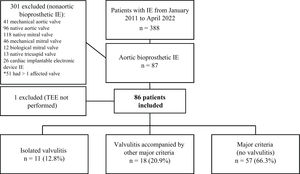
MethodsFrom 2011 to 2022, 388 consecutive patients with infective endocarditis (IE) admitted to a tertiary care hospital were prospectively included in a multipurpose database. For this study, all patients with AoBio-PVE (n=86) were selected, and their TEE images were thoroughly evaluated by 3 independent cardiologists to identify all cases of valvulitis.
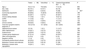
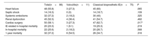
ResultsThe prevalence of isolated valvulitis was 12.8%, and 20.9% of patients had valvulitis accompanied by other classic echocardiographic findings of IE. A total of 9 out of 11 patients with isolated valvulitis had significant valve stenosis, whereas significant aortic valve regurgitation was documented in only 1 patient. Compared with the other patients with AoBio-PVE, cardiac surgery was less frequently performed in patients with isolated valvulitis (27.3% vs 62.7%, P=.017). In 4 out of 5 patients with valve stenosis who did not undergo surgery but underwent follow-up TEE, valve gradients significantly improved with appropriate antibiotic therapy.
ConclusionsValvulitis can be the only echocardiographic finding in infected AoBio and needs to be identified by imaging specialists for early diagnosis. However, this entity is a diagnostic challenge and additional imaging techniques might be required to confirm the diagnosis. Larger series are needed.
Keywords
Prosthetic valve endocarditis (PVE) is the most severe form of infective endocarditis (IE), accounting for 20% to 30% of all IE cases. In recent years, there has been a notable increase in the incidence of PVE.1-8 This form of IE is associated with poor prognosis, partly attributed to the difficulties of early identification of infectious prosthetic valvular involvement, among other factors.1-6
Transesophageal echocardiography (TEE) is the imaging technique of choice to assess structural and functional damage to prosthetic cardiac valves, playing a crucial role in the diagnosis of PVE. Currently, 7 echocardiographic findings—vegetation, abscess, pseudoaneurysm, intracardiac fistula, leaflet perforation, valvular aneurysm, and new prosthetic valve dehiscence—are considered major criteria for diagnosing PVE.1 However, it is well known that a negative TEE does not exclude the diagnosis of PVE.9-12
While other imaging modalities, such as cardiac computed tomography (CT), and nuclear techniques, particularly 18F-fluorodeoxyglucose positron emission tomography-computed tomography (18F-FDG-PET/CT) are now integral to the diagnostic strategy for IE,1 TEE continues to be mandatory when PVE is suspected. Therefore, it is crucial to optimize its utility.
Generalized valve thickening has been recognized as a potential sign of infection, even in the absence of other echocardiographic signs of IE.9 This specific echocardiographic finding has been termed valvulitis and is mainly observed in patients with bioprosthetic aortic valves.10 The echocardiographic characteristics of this lesion are not well established, and its clinical relevance is unknown.
The aim of our study was to estimate the prevalence of valvulitis, precisely describe its echocardiographic characteristics, and determine their clinical significance in patients with bioprosthetic aortic valve IE.
METHODSStudy design and patient populationThis retrospective single-center cohort study analyzed prospectively collected data. From January 2011 to April 2022, 388 consecutive patients with IE admitted to our tertiary care hospital were prospectively included in an ongoing multipurpose database.
Inclusion criteria were a diagnosis of definite or possible IE, considered and treated as IE, based on the Modified Duke criteria until 2015,13 and the European Society of Cardiology (ESC) 2015 IE diagnostic criteria thereafter.2 Early prosthetic valve IE was defined as occurring within the first year after surgery, and late prosthetic valve IE as occurring beyond 1 year.2,13
Surgery was performed when any of the current guidelines’ criteria for urgent surgery (heart failure, uncontrolled infection or prevention of embolisms) were met.2,13
Of the 388 episodes of IE, 86 involved aortic valve bioprosthesis, and they comprised our study group.
Prosthetic aortic valve stenosis and regurgitation were assessed and classified as recommended by the current imaging guidelines.14 Moderate prosthetic valve stenosis was diagnosed when the mean gradient ranged from 20 to 34mmHg and the effective orifice area was between 0.8 and 1.1 cm2. Severe stenosis was diagnosed when the mean gradient was 35mmHg or higher and the effective orifice area was less than 0.8 cm2. Epidemiological, clinical, and microbiological characteristics, imaging features, and outcome were entered in a standardized case report form that has been previously described elsewhere.15
The study was approved by the local ethics committee and informed consent was obtained from all patients.
Definition of valvulitis as detected by transesophageal echocardiographyWe propose the following definition of valvulitis: diffuse leaflet thickening (1 or more leaflets) with homogeneous and hypoechoic appearance without clearcut vegetations. Notably, a characteristic motion of the leaflets with wavy edges is particularly seen in short-axis view (figure 1, video 1 of the supplementary data). In contrast to prosthetic valve degeneration, extensive calcification and significant aortic regurgitation is usually absent.
Images of a normal bioprosthetic aortic valve and of bioprosthetic valvulitis. A: short-axis view, 45° and long-axis view, 135° of a normal bioprosthetic aortic valve. B: patient with bioprosthetic aortic valvulitis: a marked thickening (blue arrows) of the valve leaflets is seen on TEE.
The histopathological correlation of this lesion shows thickening of the prosthetic valve leaflets, due to polymorphonuclear cell infiltration (figure 2).
Histological correlation of valvulitis. Histological slide from a bioprosthetic aortic valve leaflet (surgical specimen, patient #3 from table 1 of the supplementary data). Hematoxylin and eosin stain (panel A: 2x; panel B: 4x; panel C: 10x). Normal thickness of the aortic leaflet (panel A, large bracket). A layer of early granulation tissue (small bracket) is seen covering the aortic leaflet (panels A and B). This granulation tissue is mainly characterized by the presence of fibroblasts, fibrin, new thin-walled capillaries, and inflammatory cells (panel B). The inflammatory infiltrate (asterisk) is mainly composed of polymorphonuclear cells and it is especially located in the interface between the leaflet and the granulation tissue (panel C). The normal 3 distinctive layers of the aortic valve, fibrosa (F), spongiosa (S) and ventricularis (V) are well delineated in panel A. Fibrosa (F) and spongiosa (S) are seen in panel B.
When patients with suspected bioprosthetic IE are assessed, it may be challenging to differentiate them from patients with common prosthetic degenerative changes.
In this study, we defined bioprosthetic valve degeneration as leaflet thickening and increased echogenicity suggestive of fibrosis or calcification with limited, delayed, or asymmetrical leaflet opening or closure by TEE. Tearing, or disruption of the prosthetic valve leaflets eventually resulting in valvular stenosis or regurgitation, were also considered degenerative bioprosthetic valve lesions.16
Several meta-analyses have shown that structural valve degeneration commonly begins 8 years after implantation. Mid-term failure rates of porcine and bovine aortic bioprosthesis are fairly low: <1% before 5 years for patients aged >65 years.17 In addition, the process of bioprosthetic degeneration is typically gradual, taking place over years; however, certain prostheses, including Mitroflow (Sorin Group, Canada) and Trifecta (Abbott, United States), are known to degenerate more rapidly.17 Thus, comparison with previous TEE and transthoracic echocardiography (TTE) studies was also performed, when available.
Identification of patients with valvulitisFor this study, all episodes of bioprosthetic aortic valve IE after surgical aortic valve replacement (SAVR) or transcatheter aortic valve replacement (TAVR) were thoroughly evaluated (n=86). TEE images from all patients were retrospectively reviewed by 3 cardiologists to identify all potential cases of valvulitis. The diagnosis of valvulitis was present when all 3 cardiologists were in agreement. In 6 of the 29 cases that were finally classified as valvulitis, 2 out of the 3 reviewers made the diagnosis of valvulitis; images were jointly reevaluated, and a consensus was reached.
Of the 86 patients with bioprosthetic aortic valve IE, 11 patients met the TEE criteria of valvulitis and had no other echocardiographic findings of IE, 18 had leaflet thickening and other signs of prosthetic aortic valve IE (vegetations or perivalvular complications), and 57 had classic signs of IE without diffuse leaflet thickening. The study flowchart is illustrated in figure 3.
Study flowchart. All patients with IE (n=388) treated in our tertiary care center were included in a prospective registry. Patients with IE involving aortic bioprostheses (n=87) were retrospectively analyzed. One patient was excluded as TEE was not performed. IE: infective endocarditis; TEE: transesophageal echocardiography.
Clinical features and outcomes of patients with bioprosthetic valvulitis were compared with those of the remaining patients with bioprosthetic aortic valve IE.
Statistical analysisContinuous variables are expressed as mean ±standard deviation (SD) or median and interquartile range (IQR). Assessment of normality and homogeneity of variances for continuous data was performed using the Shapiro-Wilk test and Levene's test, respectively. The Student t-test and Mann-Whitney U test were used for comparison.
Categorical variables are expressed as frequency and percentage. Hypothesis testing was performed with the Pearson chi-square test and Fisher exact test, when appropriate.
Interobserver variability was assessed by the Kappa index. All tests were 2-sided, and differences were considered statistically significant at P <.05. Statistical analysis was performed with Stata V.16.0 (Stata Corp, College Station, United States).
RESULTSDescription of patients with bioprosthetic valvulitisThe prevalence of isolated valvulitis in the total cohort of patients with bioprosthetic aortic valve IE was 12.8% (11 patients). Eighteen patients (20.9%) had features of valvulitis accompanying other classic echocardiographic findings of IE.
The main epidemiological, clinical, and echocardiographic findings of the 11 patients with isolated valvulitis are shown in table 1 of the supplementary data. Mean age was 73.6±8.6 years and 8 were male. Median time from prosthetic valve implantation to IE diagnosis was 40 [IQR 40] months. Ten patients had a SAVR and 1 had a TAVR. Ten cases were classified as late PVE (90.9%) and 1 as early PVE. The most common symptom at presentation was fever, and 2 patients had systemic embolic events during hospitalization. Blood cultures were positive in all patients. The causative microorganisms were viridans streptococci in 4 cases, staphylococci in 4, and enterococci in 3.
Regarding imaging findings, valvulitis involved a mean of 2.5±0.7 leaflets. The mean leaflet thickness measured by TEE was 7±2.9mm. Unlike typical findings of prosthetic valve degeneration, extensive calcification was rare. Six patients had no calcification, 4 had mild calcification on TEE and 1 had moderate calcification. Prosthetic valve stenosis was present in 9 patients, whereas significant prosthetic valve regurgitation was documented in only 1 patient.
Of the 9 patients with prosthetic aortic valve stenosis, 5 had a normal TTE during the previous year of IE diagnosis, 2 had a normal TTE 2 years before IE diagnosis, and 2 had no recent prior echocardiograms.
Follow-up echocardiograms were performed in 7 patients, including 5 patients with valve stenosis. Four of these 5 patients experienced a significant reduction in the severity of valvular stenosis (figure 4), and 1 showed no improvement in prosthetic valvular gradient. Four patients had no follow-up echocardiograms due to early surgery or death.
Changes in transesophageal echocardiography (TEE) in a patient with aortic bioprosthetic valvulitis. A: TEE short-axis view, 45° and long-axis view, 135° of a patient with valvulitis. Leaflet thickening is clearly shown (arrows). B: follow-up TEE after completion of antibiotic treatment, in which valve thickening has resolved.
Maximum leaflet thickness on the follow-up TEE performed prior to discharge is described at table 1 of the supplementary data.
18F-FDG-PET/CT was carried out in 4 patients. Two of them had 18F-FDG uptake at the level of the prosthesis (figure 5), 1 had a spondylodiscitis, and the other had no pathological 18F-FDG uptake. None of the patients underwent cardiac-gated CT.
Central illustration. A: Transesophageal echocardiography long-axis and short-axis views of a patient with aortic bioprosthetic valvulitis (homogeneous and hypoechoic thickening of the aortic leaflets) resulting in severe aortic stenosis. B: abnormal 18F-FDG uptake (arrow) at the prosthetic valve is seen on PET/CT scan. C: hematoxylin and eosin stain of the aortic leaflet; granulation tissue is well demonstrated (bracket) covering the aortic fibrosa (asterisk).
18F-FDG, 18F-fluorodeoxyglucose; PET/CT, positron emission tomography-computed tomography.
Four patients met a surgical indication, of which 3 (27.3%) underwent surgery due to persistent signs of infection (persistently positive blood cultures and fever). Valve culture was positive in 1 patient, negative in another, and not done in the third. Patient number 8 had uncontrolled heart failure as an indication for urgent surgery but was not operated on due to high surgical risk.
Regarding patient outcomes, 2 patients (18.2%) died during hospitalization, 1 of them due to COVID-19 bilateral pneumonia and the other due to an intracranial hemorrhage secondary to a cerebral embolism. At 1 year of follow-up, 4 patients had died (none related to IE), and 5 remained alive. The only patient with a TAVR had a relapse a few months after the index episode and was medically managed with full recovery. Interestingly, the TEE findings of this new IE episode again met the criteria for valvulitis.
Comparison of patients with isolated valvulitis with those with classic bioprosthetic aortic valve infective endocarditisComparison of clinical, microbiological, and echocardiographic findings of patients with isolated valvulitis with those with classic TEE findings of bioprosthetic aortic valve IE are shown in table 1. No significant differences were found in age or sex among the 2 groups.
Comparison of epidemiological, microbiological, and echocardiographic findings between patients with isolated valvulitis and those with classic bioprosthetic aortic valve infective endocarditis
| Total(n=86) | Valvulitis(n=11) | Classical bioprosthetic IE(n=75) | P | |
|---|---|---|---|---|
| Age, y | 72.3 (11.3) | 73.6 (8.6) | 71.9 (12) | .652 |
| Sex, male | 62 (72.1) | 8 (72.7) | 54 (72) | .982 |
| Definite IE | 78 (90.7) | 6 (54.5) | 72 (96) | <.001* |
| Community-acquired IE | 59 (68.6) | 10 (90.9) | 49 (65.3) | .189 |
| Diabetes | 23 (26.7) | 3 (27.3) | 20 (26.7) | .716 |
| Chronic kidney disease | 17 (19.8) | 1 (9.1) | 16 (21.3) | .280 |
| COPD | 11 (12.8) | 0 (0) | 11 (14.7) | .123 |
| Cancer | 14 (16.3) | 4 (36.4) | 10 (13.3) | .102 |
| Chronic anemia | 28 (32.6) | 4 (36.4) | 24 (32) | .956 |
| Time from symptom onset to diagnosis, d | 19.4 (31.8) | 14.7 (25.2) | 20.1 (32.8) | .5837 |
| Staphylococcus aureus | 12 (14) | 1 (9.1) | 11 (14.7) | .325 |
| Other staphylococci | 19 (22.1) | 3 (27.3) | 16 (21.3) | .796 |
| Enterococcus | 18 (20.9) | 3 (27.3) | 15 (20) | .643 |
| Viridans group streptococci | 27 (31.4) | 4 (36.4) | 23 (30.7) | .905 |
| Other microorganisms | 9 (10.5) | 0 (0) | 9 (12) | .231 |
| Negative cultures | 1 (1.2) | 0 (0) | 1 (1.3) | .115 |
| Persistently positive blood cultures | 26 (30.2) | 3 (27.3) | 23 (30.7) | .975 |
| Moderate or severe valve stenosis | 30 (34.9) | 9 (81.8) | 21 (28) | .001* |
| Moderate or severe valve regurgitation | 19 (22.1) | 1 (9.1) | 18 (24.1) | .231 |
COPD, chronic obstructive pulmonary disease; IE, infective endocarditis; SD, standard deviation.
“Valvulitis” group (n=11) refers to patients with valvulitis as the only echocardiographic sign of IE, and “Classical bioprosthetic IE” group (n=75) refers to patients presenting with vegetations, abscesses, or pseudoaneurysms.
Data are expressed as No. (%) or mean±standard deviation.
Moderate or severe bioprosthetic valve stenosis was more frequent in patients with valvulitis (81.8% vs 28.0%, P=.001), whereas moderate or severe aortic regurgitation was more common in patients with classic prosthetic valve IE (9.1% vs 24.1%, P=.568), although this difference was not statistically significant. In-hospital outcomes, including heart failure, septic shock, systemic embolisms, acute renal failure, all-cause mortality, and 1-year mortality are shown in table 2. Cardiac surgery was less frequently performed in patients with valvulitis (27.3% vs 62.7%, P=.017).
In-hospital events and 1-year mortality in patients with valvulitis vs classic bioprosthetic infective endocarditis
| Total(n=86) | Valvulitis(n=11) | Classical bioprosthetic IE(n=75) | P | |
|---|---|---|---|---|
| Heart failure | 48 (55.8) | 3 (27.3) | 45 (60) | .065 |
| Septic shock | 14 (16.3) | 0 (0) | 14 (18.7) | .109 |
| Systemic embolisms | 32 (37.2) | 2 (18.2) | 30 (40) | .325 |
| Renal dysfunction | 45 (52.3) | 4 (36.4) | 41 (54.7) | .462 |
| Cardiac surgery | 50 (58.1) | 3 (27.3) | 47 (62.7) | .017* |
| IE-related in-hospital mortality | 20 (23.3) | 1 (9.1) | 19 (25.3) | .219 |
| In-hospital mortality | 22 (25.6) | 2 (18.2) | 20 (26.7) | .568 |
| 1-year mortality | 32 (37.2) | 6 (54.5) | 26 (34.7) | .316 |
IE, infective endocarditis.
Data are expressed as No. (%).
During hospitalization and before surgery, 4 patients met the criteria for definite IE and 7 for possible IE. Major and minor criteria for each patient are detailed in table 1 of the supplementary data. If valvulitis were considered as a major criterion for bioprosthetic aortic valve IE, these 7 patients with possible IE would have been reclassified as definite IE (figure 1 of the supplementary data).
According to the current ESC diagnostic criteria for IE, 6 patients finally met the criteria for definite IE: 4 had pathological criteria (3 patients with prosthetic valves obtained at surgery and 1 patient with a coronary embolic vegetation) and 2 patients without a pathological analysis met the clinical criteria (1 of them had 2 major criteria: positive blood cultures and positive PET/CT; the other had 1 major (positive blood cultures) and 3 minor criteria: fever, predisposing lesion and emboli). Five patients remained classified as possible IE (1 major and 2 minor criteria).
Interobserver agreementTo evaluate the external applicability of this proposed echocardiographic new criterion in patients with prosthetic valvulitis and bioprosthetic degeneration, interobserver variability was assessed. We selected 10 patients among those with valvulitis and 10 participants with bioprosthetic aortic valve degeneration as assessed by TEE. These 20 cases were then blindly and randomly presented to an experienced cardiology-trained imaging specialist, who was external to the study and who had been instructed on the imaging features of bioprosthetic valvulitis. Overall agreement with the expert consensus was 80% (95% confidence interval (CI): 58.4%-91.9%), and the Kappa value was 0.60 (95%CI: 0.2429-0.9506).
DISCUSSIONThis is the first study to focus on a poorly described form of IE in patients with bioprosthetic aortic valve infection: bioprosthetic valvulitis. A diffuse and homogeneous leaflet thickening with undulating edges is characteristically seen on TEE. This leaflet swelling is typically the result of inflammation and granulation tissue proliferation. This echocardiographic finding should be incorporated into the major echocardiographic criteria to help reach a diagnosis of IE.
This form of infectious leaflet involvement in bioprosthetic aortic valves is not rare. In our cohort of patients with bioprosthetic IE, the prevalence of isolated valvulitis was 12.8%. Patients with typical findings of valvulitis plus classic echocardiographic findings of IE were more prevalent (20.9%). Although time from symptom onset to diagnosis was not statistically different among groups, patients with valvulitis had a shorter time: 14.7 days±25.2 vs 20.1 days±32.8). Our hypothesis is that valvulitis could be more frequently seen at earlier stages of the disease.
This study highlights substantial differences in hemodynamics between patients with bioprosthetic aortic valvulitis and those with classic bioprosthetic aortic valve IE. In the latter, as is commonly reported, the most characteristic valve lesion was valvular regurgitation, while in patients with valvulitis the most frequent lesion was aortic stenosis. In fact, no patient with valvulitis had severe aortic regurgitation. Valvulitis causes leaflet stiffening, impeding full leaflet excursion and resulting in significant valve stenosis in a high percentage of cases.
Cardiac surgery was less frequently performed in patients with valvulitis than in the other participants with bioprosthetic aortic valve IE. In 4 patients with valvulitis, the severity of prosthetic valvular stenosis decreased during appropriate antibiotic therapy. This leads us to believe that an initially conservative approach could be selected in some patients with bioprosthetic valvulitis. However, this study is underpowered to detect differences among groups, and we cannot definitively recommend conservative treatment in these patients.
Although no significant differences were found regarding the causative microorganisms between the 2 groups (valvulitis vs no valvulitis), Staphylococcus aureus was the causative microorganism in 14.7% of patients with classic signs of IE whereas only 1 patient with valvulitis had an infection due to this microorganism. We hypothesize that due to the type of morphological lesion left by the infection, most virulent microorganisms, such as S. aureus, may cause more valve destruction and may be less prone to valvulitis. In contrast, less virulent microorganisms, such as streptococci, will likely be more commonly isolated in patients with valvulitis. However, this hypothesis should be confirmed in a larger cohort.
TEE is the imaging technique of choice in the diagnosis of PVE.1 The differential diagnosis of valvulitis by TEE with other pathologic conditions that may have a similar echocardiographic appearance is of paramount importance. Bioprosthetic structural valve degeneration may also cause leaflet thickening and mimic valvulitis, posing a challenge in diagnosis. In this study, the level of agreement among different observers was moderate. Therefore, we suggest that the evaluation of TEE images should be carefully performed by expert cardiac imaging cardiologists.
The following clues may help differentiate between the 2 conditions: extensive calcification, significant regurgitation, and very restricted leaflet motion are more characteristic of bioprosthetic structural valve degeneration.16,17 On the other hand, homogeneous and hypoechoic leaflet thickening, the absence of calcification, and a typical wavy motion of the leaflets, particularly in the short-axis view, are more representative of valvulitis. Additionally, the absence of leaflet thickening in a recent echocardiogram (within the last 6 months) favors the diagnosis of valvulitis over prosthetic valve degeneration. Finally, and most importantly, the clinical scenario remains crucial for guiding the final diagnosis. However, distinguishing between structural valve degeneration and valvulitis remains a challenge.9 If there are discrepancies or when the diagnosis is unclear, our recommendation would be to perform serial TEE and rely on other imaging techniques such as PET/CT, which can be useful in demonstrating both pathological prosthetic FDG uptake and systemic emboli. Information from clinical history, physical findings, blood tests and cultures is also of the utmost importance.
The incorporation of PET/CT into the diagnostic criteria for IE in the 2015 ESC guidelines for the management of IE was shown to increase the sensitivity of the Duke criteria, at the cost of a relative decrease in specificity.18 A recent meta-analysis showed 86% sensitivity and 84% specificity for PET/CT in PVE.19 The main contribution of 18F-FDG-PET/CT is in cases with a high clinical suspicion of PVE, where, despite a proper clinical and imaging diagnostic approach, IE diagnosis remains possible by traditional criteria.1 Accordingly, PET/CT could be a potentially useful technique to confirm a diagnosis of IE in patients with valvulitis. However, in this clinicopathologic context (absence of annular complications and fewer embolic events), this technique might not be as effective as in classic PVE, as infection along the prosthetic valvular plane, a mechanistic explanation for 18F-FDG uptake in classic PVE,20 is not present. Unfortunately, we did not have enough patients with valvulitis and PET/CT to reach a conclusion.
LimitationsAn important limitation of the study is its single-center nature, which may reduce the external validity required to consider this echocardiographic finding as a major diagnostic criterion for IE. The sensitivity and specificity of TEE for diagnosing bioprosthetic aortic valvulitis in a setting other than that of our group remain to be determined.
Five patients did not meet the criteria for definite IE and therefore so we cannot be completely sure that they all had endocarditis. However, given that 4 of these patients had a normal TTE during the year prior to IE diagnosis and that 4 of them experienced a reduction in the severity of aortic bioprosthesis stenosis during antibiotic treatment, diagnoses other than IE are unlikely.
Another limitation of this study is the low number of patients with isolated valvulitis. Consequently, this study is underpowered to detect differences among groups. A large, multicenter study might provide definitive data and more insights into this new type of IE lesion.
Additionally, the study was not designed to address the sensitivity and specificity of including valvulitis as a criterion of IE in patients with suspected bioprosthetic aortic valve IE, and dedicated studies are necessary.
Finally, only patients with known IE were studied. Thus, the occurrence of diffuse valve thickening with a similar echocardiographic appearance to valvulitis as a result of hypoattenuated leaflet thickening or early structural valve degeneration has not been addressed.
CONCLUSIONSInfection of aortic bioprosthetic valves may give rise to a characteristic echocardiographic picture: diffuse leaflet thickening with a wavy leaflet motion, which can be present as the only echocardiographic IE finding and needs to be recognized by imaging specialists for early patient diagnosis. The identification of this type of lesion by TEE is important since the clinical profile and management of these patients may differ from that of patients with classic PVE. However, these lesions represent a diagnostic challenge, and additional imaging techniques might be required. Larger series are needed to accurately describe this entity and to guide the clinical management of these patients.
- -
Prosthetic valve endocarditis is the most severe form of infective endocarditis, accounting for 20% to 30% of all infective endocarditis cases.
- -
This form of infective endocarditis has been associated with poor prognosis, which has been attributed to difficulties in the early identification of infectious prosthetic valvular involvement, among other factors.
- -
Transesophageal echocardiography is the imaging technique of choice to assess the structural and functional damage of prosthetic cardiac valves and is mandatory when prosthetic valve endocarditis is suspected.
- -
This is the first study to focus on a new form of infective endocarditis in patients with bioprosthetic aortic valve infection: valvulitis.
- -
Valvulitis presents as diffuse and homogeneous leaflet thickening with a wavy leaflet motion. Valve stenosis is frequent, whereas valve regurgitation is uncommon.
- -
Larger series are needed to better understand this clinical entity, and to evaluate the diagnostic performance of our proposed echocardiographic criterion in patients with suspected bioprosthetic aortic valve endocarditis.
This research did not receive a specific grant from funding agencies in the public, commercial, or not-for-profit sectors.
ETHICAL CONSIDERATIONSThe study was approved by the local ethics committee and informed consent was obtained from all participants.
STATEMENT ON THE USE OF ARTIFICIAL INTELLIGENCENo artificial intelligence tools were used in the preparation of this work.
AUTHORS’ CONTRIBUTIONSThe study conception and design were done by E. Pozo, I. Vilacosta and C. Olmos. Material preparation and data collection were carried out by D. García-Arribas, C.N. Pérez-García, A. Jerónimo, D. Gómez, P. Zulet, C. Olmos, M. Carnero, D. Pérez-Camargo, L. Montero, M. Saiz-Pardo, P. Mahía, F. Islas, and J.A. San Román. P. Zulet and C. Olmos analyzed and interpreted the data, wrote the manuscript, and generated the associated figures and tables. I. Vilacosta and J.A. San Román contributed to the interpretation and discussion, and reviewed and edited the manuscript. I. Vilacosta and C. Olmos supervised the study. All authors have read and approved the final version of the manuscript.
CONFLICTS OF INTERESTNone.
