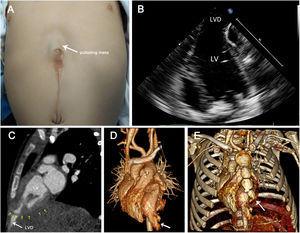

A 2-month-old infant with an asymptomatic epigastric hernia at birth was admitted for further diagnosis. Physical examination revealed a small pulsating mass covered by a layer of skin on the upper abdomen, synchronizing with the heartbeat (figure 1A, arrow). There was no murmur on auscultation. Echocardiography indicated left ventricular (LV) diverticulum (figure 1B). Contrast-enhanced computed tomography angiography showed that contrast (figure 1C, big arrow) leaked to the subxiphoid region through defects of the diaphragm and pericardium (figure 1C, small arrows). The LV apex was finger-like (figure 1D, arrow). The lower part of the sternum was short and misshapen (figure 1E, arrow). Pentalogy of Cantrell (PC) was suspected. To avoid lethal complications, such as thrombogenesis, spontaneous rupture, and sudden cardiac death, excision of the LV diverticulum (LVD) was performed. During the intervention, the diagnosis of PC was confirmed. Six months later, the patient recovered well with normal growth. Informed consent was obtained from the patient's guardian.

PC, with an incidence of 1 in 65 000 to 200 000 live newborns, is composed of 5 congenital defects of the midline abdominal wall, diaphragm, sternum, pericardium and intracardiac abnormalities. Prenatal ultrasound identifying omphalocele might be useful to suspect PC, but prenatal ultrasound is insufficient to exclude diagnosis. The treatment strategy varies depending on the size and type of the defects. Most cases have typical omphalocele and ectopia cordis combined with different degrees of intracardiac abnormalities, which are easily associated with PC. Our patient had all the defects but had mild intracardiac anomalies and epigastric hernia. A pulsating mass on the upper abdomen may be the first finding in PC and should be comprehensively evaluated by multimodality imaging.
FUNDINGNot applicable.
AUTHORS’ CONTRIBUTIONSF. Huang collected the clinical data and wrote this paper. L. Rao contributed to interpretation of imaging. W. Bai revised the manuscript.
CONFLICTS OF INTERESTThe authors declare that they have no competing interests.