Cardiac amyloidosis (CA) is caused by amyloid deposits in the heart and is usually secondary to systemic amyloidosis. Echocardiography and cardiac magnetic resonance imaging are useful for the diagnosis of CA but they do not specifically distinguish it from other infiltrative heart diseases.
Positron emission tomography/computed tomography (PET/CT) with 18F-florbetair is an accurate nuclear medicine imaging tool for the detection of cerebral amyloid and diagnosis of Alzheimer disease and is currently a promising imaging modality for the detection of CA and extracardiac amyloidosis.
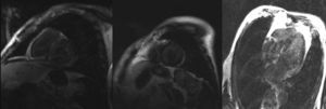
We present the case of a 75-year-old man with a history of multiple myeloma and progressive heart failure symptoms. Echocardiography showed left ventricular hypertrophy while cardiac magnetic resonance imaging, to rule out infiltrative disease, revealed mild left ventricular dysfunction (), altered gadolinium kinetics, and the presence of global subendocardial late enhancement in the left ventricle and atrium (Figure 1), suggestive of CA.

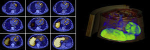
Axial images and 3-dimensional cardiac reconstruction with 18F-florbetapir PET/CT 40minutes after intravenous administration of 381 MBq revealed an intense and heterogeneous uptake of the amyloid tracer (Figure 2), confirming the diagnosis of CA.

Imaging with 18F-florbetapir PET/CT permits the early and noninvasive detection of cardiac amyloid, precluding the need for myocardial biopsy. In addition, because this modality avoids the risk of complications associated with invasive procedures, it is more cost-effective than cardiac biopsy for the diagnosis of CA.
FUNDINGThis work was supported by a grant from the 2014 Program of ERESA Funding Group to Mariano Linares.