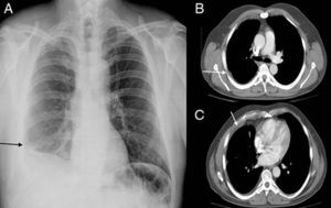
A 46-year-old man was diagnosed with right pleural epithelioid epithelioma, with partial response to chemotherapy and underwent extended pleuropneumonectomy to the pericardium and diaphragm. The preoperative x-ray (Figure 1A) and computed tomography (CT) scan (Figures 1B and 1C, of the supplementary material) showed the correct cardiac position and pleural thickening (arrows in Figure 1) corresponding to epithelioid epithelioma. The preoperative electrocardiogram () showed no changes.

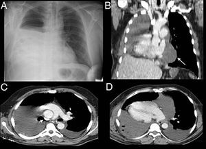
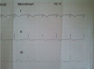
Postoperatively, the patient developed hypotension with oliguria; therefore, a chest radiograph (Figure 2A) was requested; it was interpreted as normal. An electrocardiogram (Figure 3) showed a deviation from the electric axis to the right. Given the clinical-radiological discordance, a chest CT scan was performed (Figures 2B-D, ), which showed right heart rotation about the craniocaudal axis and slight bending of the superior vena cava (Figure 2C, arrow). The occupation resulting from the remaining pericardial sac effusion (Figure 2B, arrow) resembled the left cardiac contour in the x-ray. After treatment of the pleural and pericardial effusion, the patient improved clinically; therefore, we decided on conventional rather than surgical treatment.


A cardiac hernia is a very rare episode, consisting of the protrusion of the heart through a pericardial defect, mostly secondary to lung cancer surgery.
At 62 months after cardiac herniation (), the patient was asymptomatic. This is the most extraordinary aspect of our case, since the most common treatment is emergency surgery, given the high mortality rate resulting from this type of episode.
Supplementary material associated with this article can be found in the online version available at doi:10.1016/j.rec.2014.05.021.