A 16-year-old male handball player, seen for a routine medical check-up, was found to have an electrocardiography alteration (right frontal plane QRS axis), and heartbeat radiating toward the left axillary area.
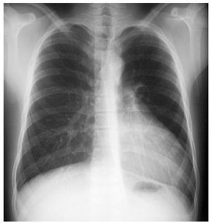
Posterior-anterior chest x-rays (Figure 1) showed displacement of the cardiac silhouette toward the left, with no deviation of the trachea. At the left cardiac border, 3 convexities were visualized: the aortic knob, pulmonary artery, and convexity of the left atrium. In addition, 2 radiotransparent bands were seen, one between the aortic knob and the pulmonary artery, and another between the cardiac base and the left hemidiaphragm.

Figure 1.
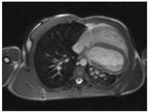
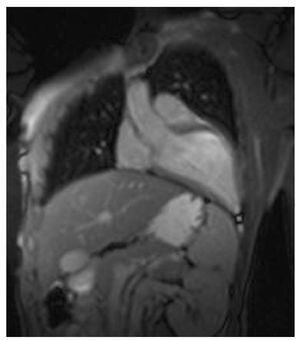
On magnetic resonance imaging, T2-weighted gradient-echo (*) axial (Figure 2), and coronal (Figure 3) views showed left posterolateral rotation and displacement of the heart, with the apex located in a posterior position. The pericardium could not be visualized, and the recess between the aorta and the root of the left pulmonary artery was seen to be filled. A diagnosis of complete agenesis of the pericardium was established and no treatment was required.

Figure 2.

Figure 3.
In the posterior-anterior view of the chest, partial left pericardial agenesis is manifested as an extremely prominent cardiac border at the level of the left atrial appendage and the pulmonary tract, caused by protrusion of these structures through the pericardial defect. Symptomatic cases of partial agenesis and asymptomatic cases with a risk of ventricular strangulation shown imaging studies should be treated surgically.