Cor triatriatum sinister is a rare congenital cardiac malformation, characterized by anomalous septation of the left atrium, leading to obstruction of the left ventricular inflow. We report an unusual case of cor triatriatum in which 3D ultrasound with the new 6Vc-D pediatric transducer from GE-Healthcare (Spain) was essential to confirm the preoperative diagnosis.
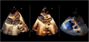
A late preterm neonate (36+3/7 weeks’ gestation, 2.84kg) had a prenatal diagnosis of possible tricuspid dysplasia on the third-trimester ultrasound study. The pertinent consent was obtained. On the first day of life, 2D echocardiography showed a membrane that crossed the entire left atrium (LA) to the roof of the appendage, dividing the atrium into 2 chambers: a high-pressure posterosuperior chamber (PSC) where the pulmonary veins arrived, and a low-pressure anteroinferior chamber (AIC) that included the appendage. In addition to defining the dysplastic tricuspid valve (TV) anatomy, ultrasound using a 3D pediatric transducer (figure 1) was able to delineate the connection between the 2 chambers. The high-pressure chamber drained to the coronary sinus (CS) through a dilated orifice (asterisk) with no obstruction. A persistent left superior vena cava was not observed. An interatrial communication (IAC) was seen between the right atrium (RA) and the lower chamber, and there was no significant pressure gradient. Direct visualization during surgery confirmed the diagnosis.

This type of malformation, which is not included in the Lucas-Schmidt classification, would be similar to type IB, with the difference that the coronary sinus was involved in draining the posterosuperior cavity to the RA. 3D echocardiography was a key noninvasive diagnostic tool for preoperative confirmation of the diagnosis.
FundingNo funding was received for this study.
AUTHORS’ CONTRIBUTIONSAll authors participated equally in the design and preparation of the manuscript.
CONFLICTS OF INTERESTNo conflicts of interest to declare.