
A 34-year-old woman with a history of a large apical ventricular septal defect (VSD) underwent palliative surgery involving pulmonary artery banding during the lactation period. Four years later, pulmonary debanding and surgical closure of the VSD were performed.
Currently, although the patient is asymptomatic, her echocardiogram did not completely rule out the presence of a small residual VSD due to a poor acoustic window. Consequently, her cardiologist requested a cardiac magnetic resonance (CMR) examination.
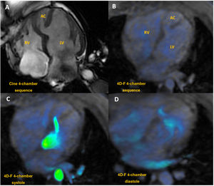
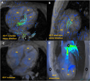
The CMR showed, in cine sequences, a nondilated left ventricle (LV) with normal systolic function. In the mid-apical segment, the LV exhibited a double chamber configulation (DCLV) with a smaller accessory chamber (AC) separated from the main ventricular cavity by a prominent moderator band, also known as septomarginal trabeculae, and a remnant of a pericardial patch (figure 1A, videos 1 and 2 of the supplementary data).

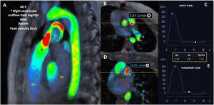
To assess pulmonary flow and rule out the presence of a hidden small residual VSD, we obtained a 4D-flow CMR sequence, which revealed flow through the interventricular septum from the AC to the LV, without any communication with the right ventricle (RV) (figure 1 and figure 2, video 3 of the supplementary data). This flow was attributed to high pressure in the accessory chamber (concomitant moderate supravalvular pulmonary stenosis, peak velocity 3m/seg, [figure 3], and ventricular conduction disorder).


VSDs located in the apical portion of the septum can be challenging to asses via echocardiography. In this case, CMR clearly demonstrated that the apical portion of the RV was isolated from the rest of the RV circulation, leaving the right apex incorporated into the LV chamber, with a good long-term surgical result. Written informed consent was obtained from the patient.
FUNDINGNo funding was received for the preparation of this manuscript.
ETHICAL CONSIDERATIONSAs this is a single clinical case report, evaluation by the ethics committee of our institution was not necessary. Written Informed consent was obtained from the patient. As this is a single clinical case report, it was not necessary to take the SAGER guidelines into account. In fact, this case concerned a female patient.
STATEMENT ON THE USE OF ARTIFICIAL INTELLIGENCENo artificial intelligence tools were used in the preparation of this manuscript.
AUTHORS’ CONTRIBUTIONSThe authors contributed equally and consensually to the completion of this manuscript.
CONFLICTS OF INTERESTThe authors of this manuscript have no conflicts of interest in relation to this manuscript.