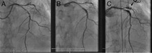
A 71-year-old man with chronic ischemic heart disease was hospitalized for heart failure. On diagnostic coronary angiography, two severe lesions with considerable calcification were detected in the left anterior descending artery (Fig. 1A). Predilation of the entire proximal and medial segment of the descending artery was performed using nondistensible balloons at high-pressure inflation. Subsequently, a 3.5×20-mm drug-eluting stent at 18 atm was implanted, with a good angiographic outcome in 3 postprocedure views (Fig. 1B).

At 60 min following the procedure, the patient showed hypotension, and echocardiography detected a large pericardial effusion. Pericardiocentesis was performed, and 600mL of hematic pericardial fluid was obtained.
A decision was made to review the previous procedure. At the first contrast injection, coronary perforation with contrast extravasation was seen, with a visible leak in the stent implanted 2h previously (type III coronary perforation with delayed presentation) (Fig. 1C). Correction of the perforation with prolonged inflation failed; hence, we decided to seal the perforation with a polytetrafluoroethylene-covered stent, which was successfully carried out.
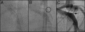
On comparison of the image of the empty stent at completion of the first procedure (Fig. 2A) with its morphology before the first injection in the second procedure, a small gap is seen in the struts at the superior portion (Fig. 2B, circle), where contrast is leaking (Fig. 2C, arrow). Therefore, the possible mechanism of the delayed coronary perforation (an unusual complication rarely described in the literature) may have been partial stent fracture.
