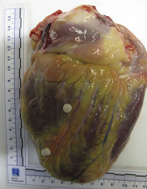
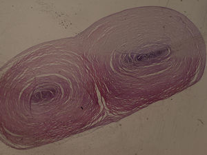
A 44-year-old man with a history of depressive syndrome on pharmacological treatment died from suicide by hanging. At autopsy, in addition to the characteristic signs of the cause of death, 3 pearly pedunculated nodules were found, measuring 0.5cm in diameter in the posterior wall of the left ventricle (Figure 1) and 2 with the same characteristics at the root of the lungs. A histopathological study found that the nodules consisted of hyaline tissue with trace amounts of fibrous cells in the periphery. In the central area, there were small microcalcifications and some red blood cells. The histopathological diagnosis was of nonspecific hyaline nodules (Figure 2).


There was no evidence that these epicardial nodules caused any symptoms, so there had been no previous examinations such as echocardiography or magnetic resonance imaging. If it had been necessary to study these nodules, the first exploratory option would have been ultrasound due to its availability and low cost. However, ultrasound is considerably limited by poor image quality and a relatively small field of vision. Magnetic resonance imaging provides high-quality images with an excellent tissue contrast and a wider field of vision, as well as more complete anatomical information that may be critical to patient care, such as any infiltration of heart and mediastinal structures, vascularization, and the anchor point of the mass. Magnetic resonance imaging also allows the functional alterations caused by masses to be studied and often their tissue characterization.